Inside all living cells, loosely formed assemblies known as biomolecular condensates perform many critical functions. However, it is not well understood how proteins and other biomolecules come together to form these assemblies within cells.
MIT biologists have now discovered that a single scaffolding protein is responsible for the formation of one of these condensates, which forms within a cell organelle called the nucleolus. Without this protein, known as TCOF1, this condensate cannot form.
The findings could help to explain a major evolutionary shift, which took place around 300 million years ago, in how the nucleolus is organized. Until that point, the nucleolus, whose role is to help build ribosomes, was divided into two compartments. However, in amniotes (which include reptiles, birds, and mammals), the nucleolus developed a condensate that acts as a third compartment. Biologists do not yet fully understand why this shift happened.
“If you look across the tree of life, the basic structure and function of the ribosome has remained quite static; however, the process of making it keeps evolving. Our hypothesis for why this process keeps evolving is that it might make it easier to assemble ribosomes by compartmentalizing the different biochemical reactions,” says Eliezer Calo, an associate professor of biology at MIT and the senior author of the study.
Now that the researchers know how this condensate, known as the fibrillar center, forms, they may be able to more easily study its function in cells. The findings also offer insight into how other condensates may have originally evolved in cells, the researchers say.
Former MIT graduate students Nima Jaberi-Lashkari PhD ’23 and Byron Lee PhD ’23 are the lead authors of the paper, which appears today in Cell Reports. Former MIT research associate Fardin Aryan is also an author of the paper.
Condensate formation
Many cell functions are carried out by membrane-bound organelles, such as lysosomes and mitochondria, but membraneless condensates also perform critical tasks such as gene regulation and stress response. In some cases, these condensates form when needed and then dissolve when they are finished with their task.
“Almost every cellular process that is essential for the functioning of the cell has been associated somehow with condensate formation and activity,” Calo says. “However, it’s not very well sorted out how these condensates form.”
In a 2022 study, Calo and his colleagues identified a protein region that seemed to be involved in forming condensates. In that study, the researchers used computational methods to identify and compare stretches of proteins known as low-complexity regions (LCRs), from many different species. LCRs are sequences of a single amino acid repeated many times, with a few other amino acids sprinkled in.
That work also revealed that a nucleolar protein known as TCOF1 contains many glutamate-rich LCRs that can help scaffold biomolecular assemblies.
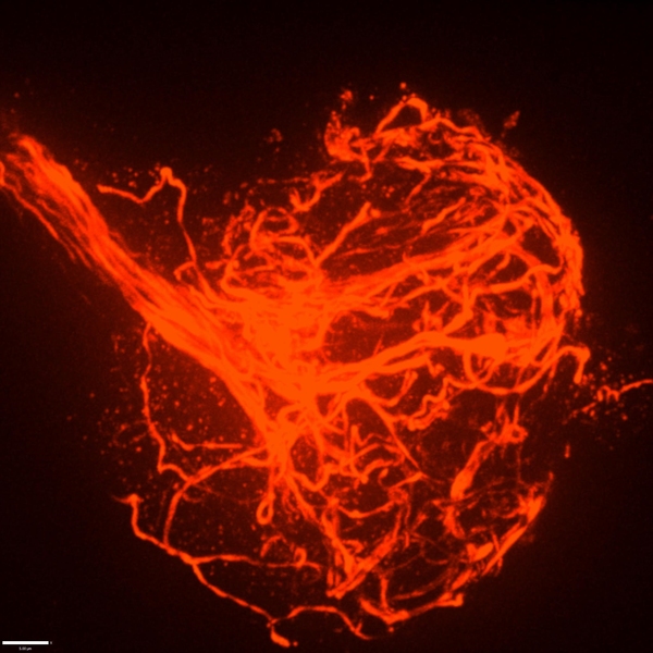
In the new study, the researchers found that whenever TCOF1 is expressed in cells, condensates form. These condensates always include proteins usually found within a particular condensate known as the fibrillar center (FC) of the nucleolus. The FC is known to be involved in the production of ribosomal RNA, a key component of ribosomes, the cell complex responsible for building all cellular proteins.
However, despite its importance in assembling ribosomes, the fibrillar center appeared only around 300 million years ago; single-celled organisms, invertebrates, and the earliest vertebrates (fish) do not have it.
The new study suggests that TCOF1 was essential for this transition from a “bipartite” to “tripartite” nucleolus. The researchers found without TCOF1, cells form only two nucleolar compartments. Furthermore, when the researchers added TCOF1 to zebrafish embryos, which normally have bipartite nucleoli, they could induce the formation of a third compartment.
“More than just creating that condensate, TCOF1 reorganized the nucleolus to acquire tripartite properties, which indicates that whatever chemistry that condensate was bringing to the nucleolus was enough to change the composition of the organelle,” Calo says.
Scaffold evolution
The researchers also found that the essential region of TCOF1 that helps it form scaffolds is the glutamate-rich low-complexity regions. These LCRs appear to interact with other glutamate-rich regions of neighboring TCOF1 molecules, helping the proteins assemble into a scaffold that can then attract other proteins and biomolecules that help form the fibrillar center.
“What’s really exciting about this work is that it gives us a molecular handle to control a condensate, introduce it into a species that doesn’t have it, and also get rid of it in a species that does have it. That could really help us unlock the structure-to-function relationship and figure out what is the role of the third compartment,” Jaberi-Lashkari says.
Based on the findings of this study, the researchers hypothesize that cellular condensates that emerged earlier in evolutionary history may have originally been scaffolded by a single protein, as TCOF1 scaffolds the fibrillar center, but gradually evolved to become more complex.
“Our hypothesis, which is supported by the data in the paper, is that these condensates might originate from one scaffold protein that behaves as a single component, and over time, they become multicomponent,” Calo says.
The formation of certain types of biomolecular condensates has also been linked to disorders such as ALS, Huntington’s disease, and cancer.
“In all of these situations, what our work poses is this question of why are these assemblies forming, and what is the scaffold in these assemblies? And if we can better understand that, then I think we have a better handle on how we could treat these diseases,” Lee says.
The research was funded by the National Institutes of Health, the National Institute of General Medical Sciences, the Pew Charitable Trusts, and the National Cancer Institute.
Sipping a beer on a warm summer evening, one might not consider that humans and yeast have been inextricably linked for thousands of years; winemaking, baking, and brewing all depend on budding yeast. Outside of baking and fermentation, researchers also use Saccharomyces cerevisiae, classified as a fungus, to study fundamental questions of cell biology.
Budding yeast gets its name from the way it multiplies. A daughter cell forms first as a swelling, protruding growth on the mother cell. The daughter cell projects further and further from the mother cell until it detaches as an independent yeast cell.
How do cells decide on a front and back? How do cells decode concentration gradients of chemical signals to orient in useful directions, or sense and navigate around physical obstacles? New Department of Biology faculty member Daniel “Danny” Lew uses the model yeast S. cerevisiae, and a non-model yeast with an unusual pattern of cell division, to explore these questions.
Q: Why is it useful to study yeast, and how do you approach the questions you hope to answer?
A: Humans and yeast are descended from a common ancestor, and some molecular mechanisms developed by that ancestor have been around for so long that yeast and mammals often use the same mechanisms. Many cells develop a front and migrate or grow in a particular direction, like the axons in our nervous system, using similar molecular mechanisms to those of yeast cells orienting growth towards the bud.
When I started my lab, I was working on cell cycle control, but I’ve always been interested in morphogenesis and the cell biology of how cells change shape and decide to do different things with different parts of themselves. Those mechanisms turn out to be conserved between yeast and humans.
But some things are very different about fungal and animal cells. One of the differences is the cell wall and what fungal cells have to do to deal with the fact that they have a cell wall.
Fungi are inflated by turgor pressure, which pushes their membranes against the rigid cell wall. This means they’ll die if there is any hole in the cell wall, which would be expected to happen often as cells remodel the wall in order to grow. We’re interested in understanding how fungi sense when any weak spots appear in the wall and repair them before those weak spots become dangerous.
Yeast cells, like most fungi, also mate by fusing with a partner. To succeed, they must do the most dangerous thing in the fungal lifecycle: get rid of the cell wall at the point of contact to allow fusion. That means they must be precise about where and when they remove the wall. We’re fascinated to understand how they know it is safe to remove the wall there, and nowhere else.
We take an interdisciplinary approach. We’ve used genetics, biochemistry, cell biology, and computational biology to try and solve problems in the past. There’s a natural progression: observation and genetic approaches tend to be the first line of attack when you know nothing about how something works. As you learn more, you need biochemical approaches and, eventually, computational approaches to understand exactly what mechanism you’re looking at.
I’m also passionate about mentoring, and I love working with trainees and getting them fascinated by the same problems that fascinate me. I’m looking to work with curious trainees who love addressing fundamental problems.
Q: How does yeast decide to orient a certain way—towards a mating partner, for example?
A: We are still working on questions of how cells analyze the surrounding environment to pick a direction. Yeast cells have receptors that sense pheromones that a mating partner releases. What is amazing about that is that these cells are incredibly small, and pheromones are released by several potential partners in the neighborhood. That means yeast cells must interpret a very confusing landscape of pheromone concentrations. It’s not apparent how they manage to orient accurately toward a single partner.
That got me interested in related questions. Suppose the cell is oriented toward something that isn’t a mating partner. The cell seems to recognize that there’s an obstacle in the way, and it can change direction to go around that obstacle. This is how fungi get so good at growing into things that look very solid, like wood, and some fungi can even penetrate Kevlar vests.
If they recognize an obstacle, they have to change directions and go around it. If they recognize a mating partner, they have to stick with that direction and allow the cell wall to get degraded. How do they know they’ve hit an obstacle? How do they know a mating partner is different from an obstacle? These are the questions we’d like to understand.
Q: For the last couple of years, you’ve also been studying a budding yeast that forms multiple buds when it reproduces instead of just one. How did you come across it, and what questions are you hoping to explore?
A: I spent several years trying to figure out why most yeasts make one bud and only one bud, which I think is related to the question of why migrating cells make one and only one front. We had what we thought was a persuasive answer to that, so seeing a yeast completely disobey that and make as many buds as it felt like was a shock, which got me intrigued.
We started working on it because my colleague,Amy Gladfelter, had sampled the waters around Woods Hole, Massachusetts. When she saw this specimen under a microscope, she immediately called me and said, “You have to look at this.”
A question we’re very intrigued by is if the cell makes five, seven, or 12 buds simultaneously, how do they divide the mother cell’s material and growth capacity five, seven, or 12 ways? It looks like all of the buds grow at the same rate and reach about the same size. One of our short-term goals is to check whether all the buds really get to exactly the same size or whether they are born unequal.
And we’re interested in more than just growth rate. What about organelles? Do you give each bud the same number of mitochondria, nuclei, peroxisomes, and vacuoles? That question will inevitably lead to follow-up questions. If each bud has the same number of mitochondria, how does the cell measure mitochondrial inheritance to do that? If they don’t have the same amount, then buds are each born with a different complement and ratio of organelles. What happens to buds if they have very different numbers of organelles?
As far as we can tell, every bud gets at least one nucleus. How the cell ensures that each bud gets a nucleus is a question we’d also very much like to understand.
We have molecular candidates because we know a lot about how model yeasts deliver nuclei, organelles, and growth materials from the mother to the single bud. We can mutate candidate genes and see if similar molecular pathways are involved in the multi-budding yeast and, if so, how they are working.
It turns out that this unconventional yeast has yet to be studied from the point of view of basic cell biology. The other thing that intrigues me is that it’s a poly-extremophile. This yeast can survive under many rather harsh conditions: it’s been isolated in Antarctica, from jet engines, from all kinds of plants, and of course from the ocean as well. An advantage of working with something so ubiquitous is we already know it’s not toxic to us under almost any circumstances. We come into contact with it all the time. If we learn enough about its cell biology to begin to manipulate it, then there are many potential applications, from human health to agriculture.

B lymphocytes are the fulcrum of our immunological memory, the source of antibodies, and the focus of vaccine development. My lab has investigated how, where, and when B cell responses take shape. In recent years, my group has expanded into preclinical vaccinology, developing cutting-edge humanized mouse models for diseases including malaria, HIV, and SARS-CoV-2.
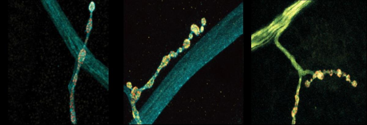
Perhaps the most obvious feature of a neuron is the long branch called an axon that ventures far from the cell body to connect with other neurons or muscles. If that long, thin projection ever seems like it could be vulnerable, a new MIT study shows that its structural integrity may indeed require the support of a surrounding protein called perlecan. Without that protein in Drosophila fruit flies, researchers at The Picower Institute for Learning and Memory found axonal segments can break apart during development and the connections, or synapses, that they form end up dying away.
Perlecan helps make the extracellular matrix, the proteins and other molecules that surround cells, stable and flexible so that cells can develop and function in an environment that is supportive without being rigid.
“What we found was that the extracellular matrix around nerves was being altered and essentially causing the nerves to break completely. Broken nerves eventually led to the synapses retracting,” says study senior author Troy Littleton, the Menicon Professor in MIT’s departments of Biology and Brain and Cognitive Sciences.
Humans need at least some perlecan to survive after birth. Mutations that reduce, but don’t eliminate, perlecan can cause Schwartz-Jampel syndrome, in which patients experience neuromuscular problems and skeletal abnormalities. The new study may help explain how neurons are affected in the condition, Littleton says, and also deepen scientists’ understanding of how the extracellular matrix supports axon and neural circuit development.
Ellen Guss PhD ’23, who recently defended her doctoral thesis on the work, led the research published June 8 in eLife.
At first she and Littleton didn’t expect the study to yield a new discovery about the durability of developing axons. Instead, they were investigating a hypothesis that perlecan might help organize some of the protein components in synapses that fly nerves develop to connect with muscles. But when they knocked out the gene called “trol” that encodes perlecan in flies, they saw that the neurons appeared to “retract” many synapses at a late stage of larval development. Proteins on the muscle side of the synaptic connection remained, but the neuron side of the connection withered away. That suggested that perlecan had a bigger role than they first thought.
Indeed, the authors found that the perlecan wasn’t particularly enriched around synapses. Where it was pronounced was in a structure called the neural lamella, which surrounds axon bundles and acts a bit like the rubbery cladding around a TV cable to keep the structure intact. That suggested that a lack of perlecan might not be a problem at the synapse, but instead causes trouble along axons due to its absence in the extracellular matrix surrounding nerve bundles.
Littleton’s lab had developed a technique for daily imaging of fly neural development called serial intravital imaging. They applied it to watch what happened to the fly axons and synapses over a four-day span. They observed that while fly axons and synapses developed normally at first, not only synapses but also whole segments of axons faded away.
They also saw that the farther an axon segment was from the fly’s brain, the more likely it was to break apart, suggesting that the axon segments became more vulnerable the further out they extended. Looking segment by segment, they found that where axons were breaking down, synapse loss would soon follow, suggesting that axon breakage was the cause of the synapse retraction.
“The breakages were happening in a segment-wide manner,” Littleton says. “In some segments the nerves would break and in some they wouldn’t. Whenever there was a breakage event, you would see all the neuromuscular junctions (synapses) across all the muscles in that segment retract.”
When they compared the structure of the lamella in mutant versus healthy flies, they found that the lamella was thinner and defective in the mutants. Moreover, where the lamella was weakened, axons were prone to break and the microtubule structures that run the length of the axon would become misdirected, protruding outward and becoming tangled up in dramatic bundles at sites of severed axons.
In one other key finding, the team showed that perlecan’s critical role depended on its secretion from many cells, not just neurons. Blocking the protein in just one cell type or another did not cause the problems that total knockdown did, and enhancing secretion from just neurons was not enough to overcome its deficiency from other sources.
Altogether, the evidence pointed to a scenario where lack of perlecan secretion caused the neural lamella to be thin and defective, with the extracellular matrix becoming too rigid. The further from the brain nerve bundles extended, the more likely movement stresses would cause the axons to break where the lamella had broken down. The microtubule structure within the axons then became disorganized. That ultimately led to synapses downstream of those breakages dying away because the disruption of the microtubules means the cells could no longer support the synapses.
“When you don’t have that flexibility, although the extracellular matrix is still there, it becomes very rigid and tight and that basically leads to this breakage as the animal moves and pulls on those nerves over time,” Littleton says. “It argues that the extracellular matrix is functional early on and can support development, but doesn’t have the right properties to sustain some key functions over time as the animal begins to move and navigate around. The loss of flexibility becomes really critical.”
In addition to Littleton and Guss, the paper’s other authors are Yulia Akbergenova and Karen Cunningham.
Support for the study came from the National Institutes of Health. The Littleton Lab is also supported by The Picower Institute for Learning and Memory and The JPB Foundation.
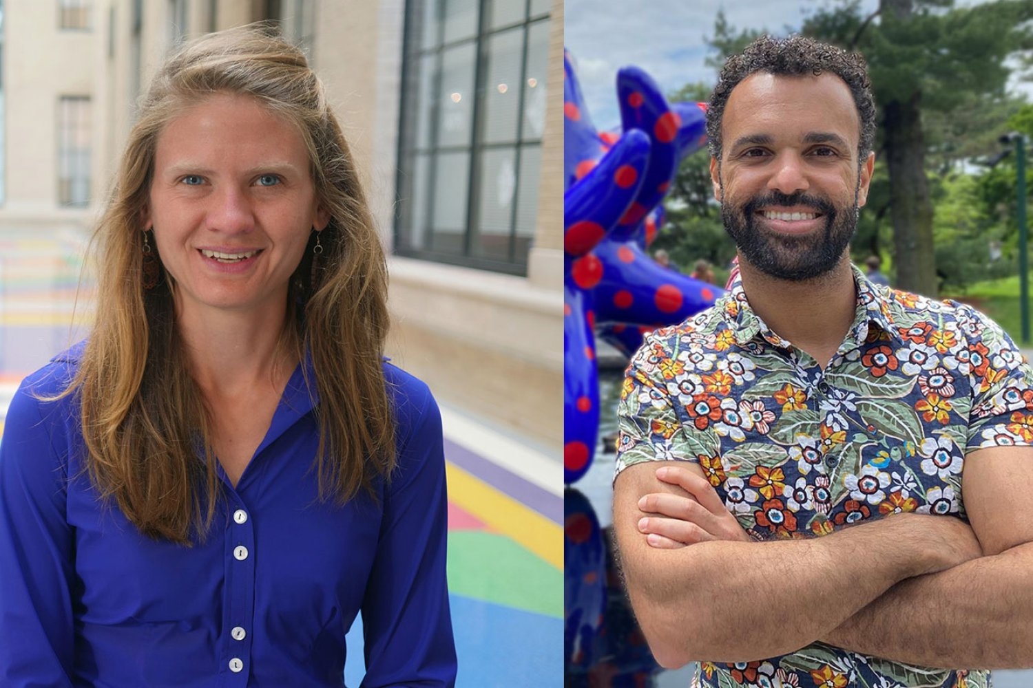
Two faculty members from the MIT Department of Biology have been selected by the Howard Hughes Medical Institute (HHMI) for the inaugural cohort of HHMI Freeman Hrabowski Scholars.
Seychelle Vos, the Robert A. Swanson Career Development Professor of Life Sciences, and Hernandez Moura Silva, an assistant professor of biology and core member of the Ragon Institute of MGH, MIT and Harvard, are among 31 early-career faculty selected for their potential to become leaders in their research fields and to create diverse and inclusive lab environments in which everyone can thrive, according to a press release.
Freeman Hrabowski Scholars are appointed to a five-year term, renewable for a second five-year term after a successful progress evaluation. Each scholar will receive up to $8.6 million over 10 years, including full salary, benefits, a research budget, and scientific equipment. In addition, they will participate in professional development to advance their leadership and mentorship skills.
The Freeman Hrabowski Scholars Program represents a key component of HHMI’s diversity, equity, and inclusion goals. Over the next 20 years, HHMI expects to hire and support up to 150 Freeman Hrabowski Scholars — appointing roughly 30 scholars every other year for the next 10 years. The institute has committed up to $1.5 billion for the Freeman Hrabowski Scholars to be selected over the next decade. The program was named for Freeman A. Hrabowski III, president emeritus of the University of Maryland at Baltimore County, who played a major role in increasing the number of scientists, engineers, and physicians from backgrounds underrepresented in science in the United States.
Seychelle Vos
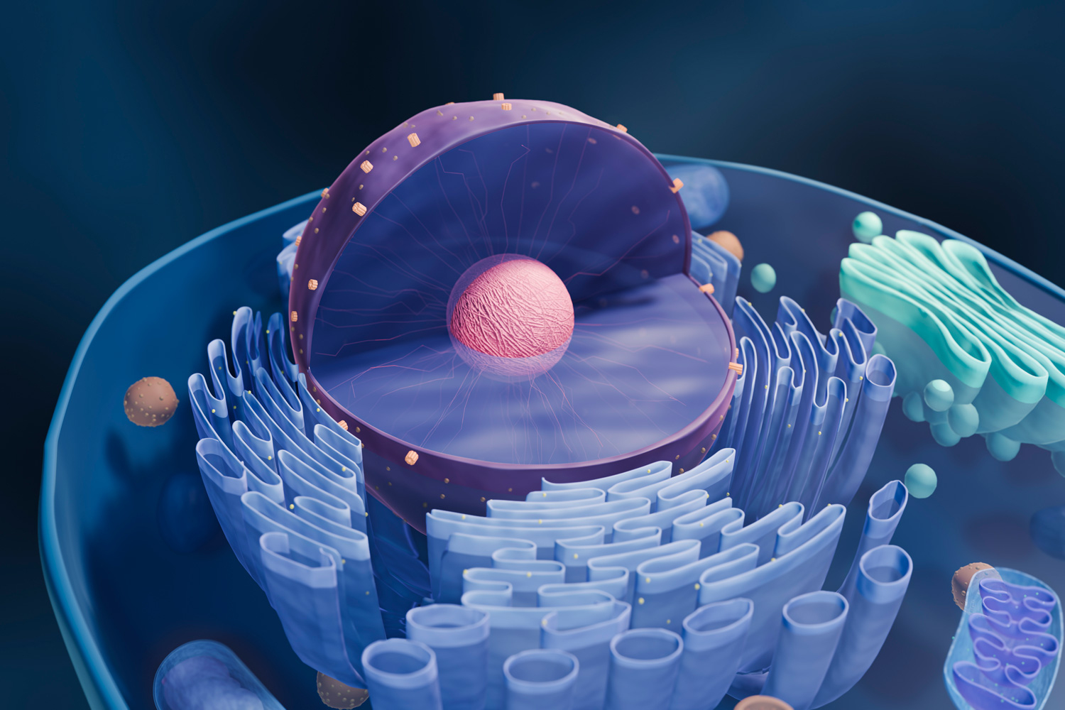
Seychelle Vos studies how DNA organization impacts gene expression at the atomic level, using cryogenic electron microscopy (cryo-EM), X-ray crystallography, biochemistry, and genetics. Human cells contain about 2 meters of DNA, which is packed so tightly that its entirety is contained within the nucleus, which is only a few microns across. Although DNA needs to be compacted, it also needs to be accessible to, and readable by, the cell’s molecular machinery.
Vos received a BS in genetics from the University of Georgia in 2008 and a PhD from University of California at Berkeley in 2013. During her postdoctoral research at the Max Planck Institute for Biophysical Chemistry in Germany, she determined how the molecular machine responsible for gene expression is regulated near gene promoters.
Vos joined MIT as an assistant professor of biology in fall 2019.
“I am very humbled and honored to have been named a HHMI Freeman Hrabowski Scholar,” Vos says. “It would not have been possible without the hard work of my lab and the help of my colleagues. It provides us with the support to achieve our ambitious research goals.”
Hernandez Moura Silva
Hernandez Moura Silva studies the role of immune cells in the maintenance and normal function of our bodies and tissues, beyond their role in battling infection. Specifically, he looks at a specific type of immune cell called a macrophage and its role in the proper function of white adipose tissue — our fat. White adipose tissue in a healthy state is highly populated by macrophages, including very abundant ones known as “vasculature-associated adipose tissue macrophages,” which are located around the blood vessels. When the activity of these adipose macrophages is disrupted, there are changes in the proper function of the white adipose tissue, which may ultimately link to disease. By understanding macrophage function in healthy tissues, Hernandez hopes to learn how to restore tissue homeostasis in disease.
Hernandez Moura Silva received a BS in biology in 2005 and an MSc in molecular biology in 2008 from the University of Brazil. He received his PhD in 2011 from the University of São Paulo Heart Institute. Silva pursued his postdoctoral work as the Bernard Levine Postdoctoral Fellow in immunology and immuno-metabolism at the New York University School of Medicine Skirball Institute of Biomolecular Medicine.
He joined MIT as an assistant professor of biology in 2022. He is also a core member of the Ragon Institute.
“For an immigrant coming from an underrepresented group, it’s a huge privilege to be granted this opportunity from HHMI that will empower me and my lab to shape the next generation of scientists and provide an environment where people can feel welcome and encouraged to do the science that they love and be successful,” Silva says. “It also aligns with MIT’s commitment to increase diversity and opportunity across the Institute and to become a place where all people can thrive.”
Billions of times a day, every day of our lives, cells receive signals to initiate the process of cell death. This strategic cell death, also called apoptosis, is one of the tools multicellular organisms use to maintain tissues and regulate immune responses: damaged, old, or superfluous cells are given the green light to, as it were, turn out the lights for the last time.
Programmed cell death is both extremely powerful and extremely regulated: for example, the careful culling of cells between our digits during embryonic development reveals fingers and toes. When programmed cell death goes awry, however, it can have serious consequences. Cells left unchecked can divide unstoppably and aggressively, leading to cancer. Dysregulated apoptotic pathways have also been implicated in neurodegenerative diseases like Alzheimer’s, where unrestrained cell death may play a part in the severity of the disease.
MIT Professor H. Robert Horvitz ‘68 shared a Nobel prize in 2002 for his foundational research on the genetics of programmed cell death and organ development in the nematode, a microscopic roundworm. Horvitz discovered that ced-9, a key gene in programmed cell death in nematodes, was similar in structure and function to the human gene bcl-2.
Targeting members of the BCL-2 protein family has already shown promise in the fight against cancer. For example, approved by the FDA in 2016, the oral drug Venetoclax is a BCL-2 inhibitor used to treat certain types of leukemia.
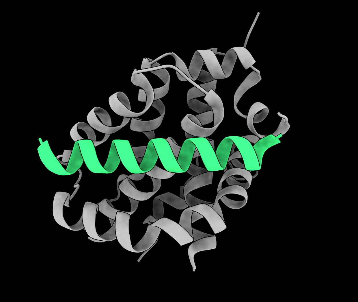
In a study published online Jan. 26 in Structure, Fiona Aguilar PhD ‘22 (Keating lab) and collaborators focused on a member of the BCL-2 protein family called BAK. When it is active, BAK promotes mitochondrial outer membrane disruption, leading to cell death, and is therefore referred to as a pro-apoptotic protein. But precisely how BAK becomes activated – or inhibited – is unknown.
“A greater understanding of BAK activation is interesting both from a fundamental biochemical and biophysical perspective as well as from the more translational one of BAK as a potential therapeutic target,” says lead author Fiona Aguilar.
BAK exists in two different forms: an inactive monomer and an active oligomer. A few activators of BAK (BIM, truncated BID, and PUMA) have already been identified and these proteins bind directly to BAK, leading to the model that binding of activators trigger changes in protein shape that allow BAK to transition from the inactive to active forms. To further explore this idea, Aguilar identified and characterized a number of other peptides that bind to and regulate BAK. To identify new peptide binders, the team used cell-surface display screening and computational protein design methods, including techniques developed by Keating lab alum Gevorg Grigoryan– dTERMen and TERMify – that use protein structural data to generate new protein sequences likely to bind a protein of interest.
In total, Aguilar et al. discovered 10 diverse new peptide binders of BAK that regulate its function.
Interestingly, some of the BAK-binding peptides inhibited activation rather than promoting it. Aguilar et al. found that inhibitors and activators of BAK shared many characteristics including structure as well as binding affinity and kinetics – the strength and rate that binders associate with and dissociate from BAK.
Newly identified activators had sequences both dissimilar from one another and from the previously known BAK activators BIM, truncated BID, and PUMA. The similarity of the sequence was not necessarily a good indicator of activation or inhibition. For example, an inhibitor and an activator differed by just two amino acids.
Aguilar and colleagues solved the crystal structures of two inhibitor-BAK complexes and one activator-BAK complex and found that the activator interacted with BAK with similar geometry as the two inhibitors. Also, the two inhibitors have only about 40% sequence identity, but bind very similarly to BAK.
Amy Keating, the senior author on the study, says “Fiona was tireless in identifying new peptides, testing their interactions with BAK, determining their functions, and solving structures to look for differences between activators and inhibitors. We were surprised that peptides with such different behaviors shared such common interaction properties.”
Although the puzzle is not yet solved, Aguilar believes the “transition state” between inactive and active forms of BAK is key.
“We think of activators as peptides that preferentially bind to the BAK transition state, whereas inhibitors are those that preferentially bind to the monomeric state,” Aguilar says. “Overall, we should be thinking more about the transition state, what steps are necessary to reach the transition state, and how to target the transition state.”
This study also added two sequences in the human proteome – BNIP5 and PXT1 – to the repertoire of known BAK binders. Not much is known about these sequences, Aguilar says, but the fact that they activate BAK could indicate that they may play a role in apoptotic pathways that have not yet been determined.
“The finding is something that people in the field are pretty excited about,” Aguilar says.
Ultimately, work remains to establish what characteristics of the binders determine their function, and how binding to BAK triggers the conformational changes that activate or inhibit this complex protein.
“It’s still unclear what it is about these sequences that trigger the allosteric network leading to BAK activation, but at least for now we can rule out the hypothesis that binding mode, affinity, and kinetics fully determine how this occurs,” Aguilar says.
Aguilar suggests that it will be interesting also to explore how these peptides interact with BAX, another pro-apoptotic protein in the BCL-2 family that is both structurally and functionally similar to BAK.
Fiona Aguilar is lead author and Amy Keating is senior author; Bob Grant and graduate students Sebastian Swanson, Dia Ghose, and Bonnie Su contributed. Collaborators Stacey Yu and Kristopher Sarosiek, from the Harvard T.H. Chan School of Public Health, helped with cell-based experiments. The research was funded by a National Institute of General Medical Sciences award, the MIT School of Science Fellowship in Cancer Research award, the John W. Jarve (1978) Seed Fund for Science Innovation (MIT) award, an award from the National Cancer Institute, a National Institute of Diabetes and Digestive and Kidney Diseases award, and Alex’s Lemonade Stand Foundation for Childhood Cancers award.
Many of our body’s most important functions occur without our conscious knowledge, such as digestion, heartbeat, and breathing. These vital functions depend on the signals generated by the “interoceptive nervous system,” which enables the brain to monitor our internal organs and trigger responses that sometimes save our lives. One second you are breathing normally as you eat your salad and the next, when a vinegar-soaked crouton enters your throat, you are coughing or swallowing to protect and clear your airway. We know our bodies are sensitive to cues like irritants, but we still have a lot to learn about how the interoceptive system works to meet our physiological needs, keep organs safe and healthy, and affect our behavior. We can also learn how chronic insults may lead to organ dysfunction and use what we learn to create therapeutic interventions.
Focusing on the airway, Sara Prescott, a new faculty member in the Department of Biology and Investigator in The Picower Institute for Learning and Memory, seeks to understand the ways our nervous systems detect and respond to stimuli in health and disease.
Q: You’re interested in interoceptive biology. What makes the nervous system of mice a good model for doing that?
A: Our flagship system is the mammalian airway. We use a mouse model and modern molecular neuroscience tools to manipulate various neural pathways and observe what the effects are on respiratory function and animal health.
Neuroscience and mouse work have a reputation for being a little challenging and intense, but I think this is also where we can ask really important questions that are useful for our everyday lives — and the only place where we can fully recapitulate the complexity of nervous system signaling all the way down to our organs, back to our brain, and back to our organs.
It’s a very fun place to do science with lots of open questions.
One of the core discoveries from my postdoctoral work was focusing on the vagus nerve as a major body-to-brain conduit, as it innervates our lungs, heart and gastrointestinal tract. We found that there were about 40 different subtypes of sensory neurons within this small nerve, which is really a remarkable amount of diversity and reflects the massive sensory space within the body. About a dozen of those vagal neurons project to the airways.
We identified a rare neuron type specifically responsible for triggering protective responses like coughing when water or acid entered the airway. We also discovered a separate population of neurons that make us feel and act sick when we get a flu infection. The field now knows what four to five vagal populations of neurons are actually sensing in the airways, but the remaining populations are still a mystery to us; we don’t know what those populations of sensory neurons are detecting, what their anatomy is, and what reflex effects those neurons are evoking.
Looking ahead, there are many exciting directions for the interoceptive biology field. For example, there’s been a lot of focus on characterizing the circuits underlying acute motor reflexes, like rapid responses to visceral stimuli on the timescale of minutes to hours. But we don’t have a lot of information about what happens when these circuits are activated over long periods of time. For example, respiratory tract infections often last for weeks or longer. We know that the airways undergo changes in composition when they’re exposed to different types of infection or stress to better accommodate future threats. One of the hypotheses we’re testing is that chronically activating neural circuits may drive changes in organ composition. We have this idea, which we’re calling reflexive remodeling: neurons may be communicating with stem cells and progenitor cells in the periphery to drive adaptive remodeling responses.
We have the genetic, molecular and circuit scale tools to explore this phenomenon in mice. In parallel, we’re also setting up some in vitro models of the mouse airway mucosa to expedite receptor screening and to explore basic mechanisms of neuron-epithelium crosstalk. We hope this will inform our understanding of how the airway surface senses and responds to different types of irritants or damage.
Q: Why is understanding the peripheral nervous system important, and what parts of your background are you drawing on for your current research?
A: The lab focuses on really trying to explore the body-brain connection.
People often think that our mind exists in a vacuum, but in reality, our nervous system is heavily integrated with the rest of the body, and those neural interfaces are important, both for taking information from our body or environment and turning it into an internal representation of the world, and, in reverse, being able to process that information and being able to enact changes throughout the body. That includes things like autonomic reflexes, basic functions of the body like breathing, blood-gas regulation, digestion, and heart rate.
I’ve integrated both my graduate training and postdoctoral training into thinking about biology across multiple scales.
Graduate school for me was quite focused on deep molecular mechanism questions, particularly gene regulation, so I feel like that has been very useful for me in my general approach to neuroscience because I take a very molecular angle to all of this.
It also showed me the power of in vitro models as reductionist tools to explore fundamental aspects of cell biology. During my postdoc, I focused on larger, emergent phenotypes. We were able to manipulate specific circuits and see very impressive behavioral responses in animals. You could stimulate about 100 neurons in a mouse and see that their breathing would just stop until you remove the stimulation, and then the breathing would return to normal.
Both of those experiences inform how we approach a problem in my research. We need to understand how these circuits work, not just their connectivity at the anatomical level but what is driving their changes in sensitivity over time, the receptor expression programs that affect how they sense and signal, how these circuits emerge during development, and their gene expression.
There are still so many foundational questions that haven’t been answered that there’s enough to do in the mouse for quite some time.
Q: This all sounds fascinating. Where does it lead?
A: Human health has been my north star for a long time and I’ve taken a long, wandering path to find particular areas where I can scratch whatever intellectual itch that I have.
I originally thought I would be a doctor and then realized that I felt like I could have a more lasting impact by discovering fundamental truths about how our bodies work. I think there are a number of chronic diseases in which autonomic imbalance is actually a huge clinical component of the disorder.
We have a lot of interest in some of these very common airway remodeling diseases, like chronic obstructive pulmonary disorder—COPD—asthma, and potentially lung cancer. We want to ask questions like how autonomic circuits are altered in disease contexts, and when neurons actually drive features of disease.
Perhaps this research will help us come up with better molecular, cellular or tissue engineering approaches to improve the outcomes for a variety of autonomic diseases.
It’s very easy for me to imagine how one day not too far from now we can turn these findings into something actionable for human health.
Inside cells, events can unfold quickly. Sub-cellular compartments constantly re-arrange while proteins move along structural fibers and membranes fuse and divide. By attaching fluorescent tags to sub-cellular structures, researchers can watch events unfold in real time using light microscopes. But to see the finest details of these processes, scientists need to shift from using light microscopy to using beams of electrons to generate even higher resolution images using a technique called electron microscopy. Using these techniques together is a powerful and rapidly growing strategy called correlative light electron microscopy (CLEM). In CLEM, light microscopy images are used to target regions of interest, and then the same sample is interrogated with electron microscopy to see the same areas at higher resolution.
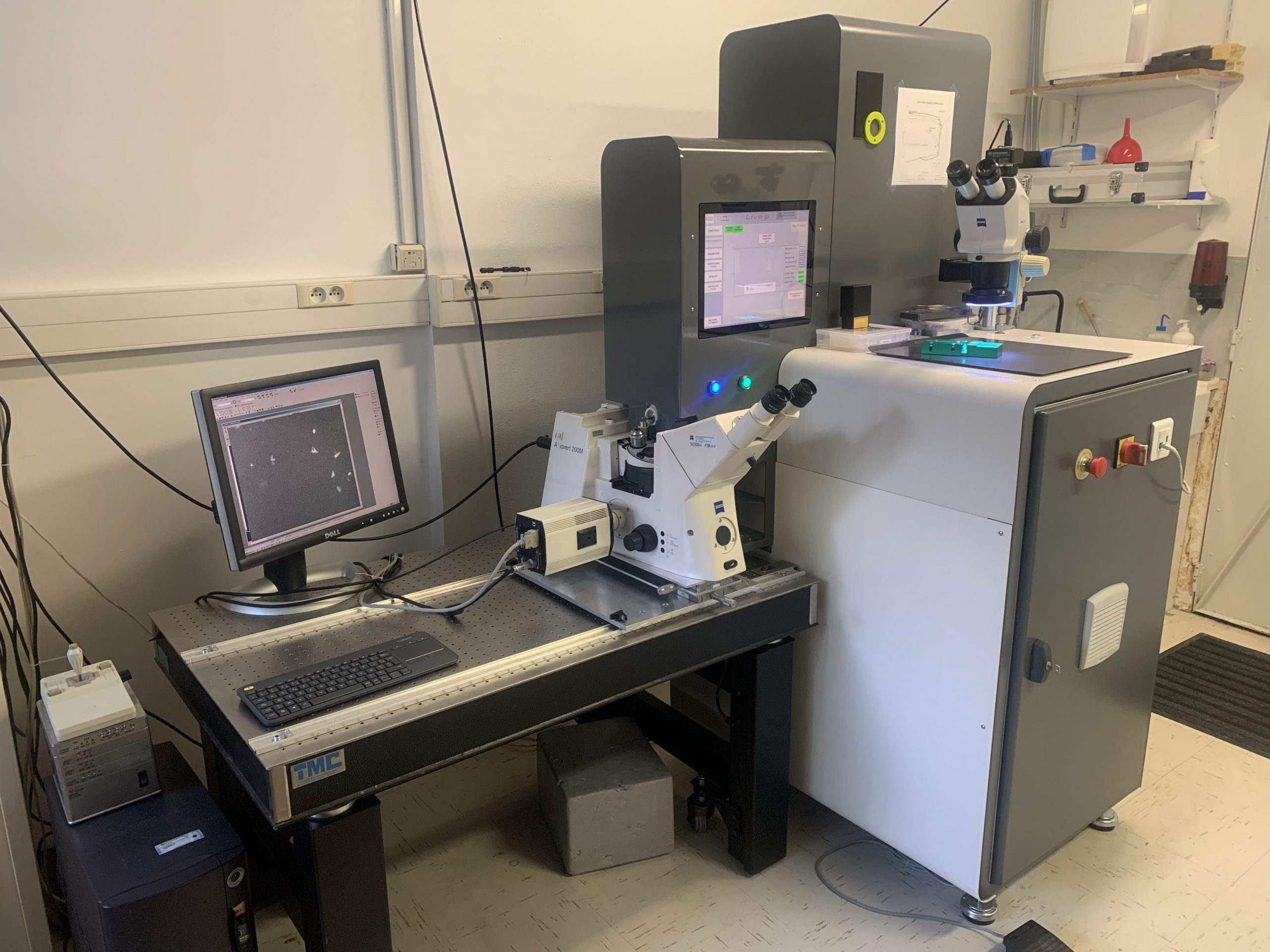
The Peterson (1957) Nanotechnology Materials Core Facility in the Robert A. Swanson (1969) Biotechnology Center at the Koch Institute for Integrative Cancer Research at MIT recently acquired a high pressure freezer called the Live μ that will let researchers do just that. This instrument allows scientists to image the same biological sample using fluorescent light microscopy and electron microscopy in close succession. These two techniques are usually performed on separate samples, but with the Live μ, researchers will be able to identify fleeting sub-cellular events using light microscopy, then preserve cells and observe the same events in high resolution using electron microscopy — a combination that was not previously available to researchers at MIT. In fact, the Live μ, which is sold by the Paris-based company CryoCapCell, will be the first instrument of its kind in the country.
Although high-pressure freezers like the Live μ have been around for decades, integration with a light microscope is what makes the Live μ special. The instrument itself is a washing machine-sized freezing instrument, equipped with an arm to hold a biological sample under a nearby light microscope. When researchers observe an interesting biological event using the light microscope, they can quickly retract the arm and insert the sample into the Live μ’s inner chamber, exposing it to low temperature and high pressure and freezing it in less than two seconds. Cells must be preserved before they can be observed using an electron microscope, and by freezing samples faster than ice crystals can form, the Live μ creates pristine samples that accurately represent the state of cells before preservation. Superimposing pictures taken using the light microscope on top of images from an electron microscope allows researchers to use the fluorescent signals like a “treasure map,” says Abigail Lytton-Jean, the director of the Peterson Facility.
Exocytosis is a vital sub-cellular event that could be studied using the Live μ. In this cellular process, cells use bubble-like vesicles to ferry proteins from the internal compartments where they’re made to the cell’s surface, where they can sense the external environment, attach cells to one another, or carry information to other cells. Exocytosis is important for many aspects of biology, and a variety of scientists, from ecologists to cancer researchers to microbiologists, would benefit from a greater understanding of this process. With the Live μ, researchers may be able to use light microscopy to catch the vesicles that mediate exocytosis when they dock with the cell’s surface, then use electron microscopy to understand the details of this association.
Researchers creating artificial materials to replace human tissues could also benefit from the Live μ, Lytton-Jean says. These materials are thick and contain a lot of water, but the Live μ is capable of freezing them without generating ice crystals that change their structure. Using this instrument, scientists can examine the internal structure of these synthetic materials and assess their similarities to live tissue.
“People who want to use the Live μ are coming from all sorts of labs,” Lytton-Jean says.
The world of biology and electron microscopy is wildly exciting right now, she adds, thanks in part to instruments like this. “People who have worked with electron microscopes for decades have told me that this is the most exciting time they’ve ever lived in.”
The Live μ recently took its place in the back of the Peterson Facility, under a picture of the Eiffel Tower that Lytton-Jean brought back from Paris when she first went to test the Live μ at CryoCapCell’s headquarters years ago. The Live μ is only the latest addition to a vast suite of instrumentation focused on cutting-edge cryo-electron microscopy and CLEM workflows, expanding the facility’s unusually large portfolio of workflows.
“There aren’t many places in the country that can do all of the different workflows we offer, and all in one place,” said Lytton-Jean. “High pressure freezing is the first step in the preservation process, so having this instrument in our lab will further enable many new workflows with our existing instrumentation. Although these workflows are challenging and sophisticated, our team of dedicated scientists are familiar with conducting this work.”
Different cells take on an astonishing variety of shapes, which are often critical to be able to perform specialized cell functions like absorbing nutrients or contracting muscles. We study how different cell shapes arise and how cells control the spatial distribution of their internal constituents. We take advantage of the tractability of fungal model systems, and address these questions using approaches from cell biology, genetics, and computational biology to understand molecular mechanisms.