BA, 2003, Computer Science, University of California, Berkeley
BS, 2003, Biological Engineering, University of California, Berkeley
Research Summary
The Davis lab is working to uncover how cells construct and degrade complex molecular machines rapidly and efficiently. We apply a variety of biochemical, biophysical, and structural approaches including quantitative mass spectrometry and single particle cryo-electron microscopy to understand the detailed molecular mechanisms of these processes. Ongoing projects in the lab are focused on autophagy, an essential eukaryotic protein and organelle degradation pathway, and assembly of the ribosome, which is essential in all cells.
Awards
Sloan Research Fellowship, Alfred P. Sloan Foundation, 2021
National Institute on Aging R00 Fellowship, 2017
National Institute on Aging K99 Fellowship, 2015
Education
PhD, 1990, University of California, Berkeley
BS, 1985, Integrated Science Program and Biochemistry, Molecular Biology and Cell Biology, Northwestern University
Research Summary
We focus on the events that occur at the starting points of chromosome duplication. These DNA sequences — called “origins of replication” — are found at multiple sites on each eukaryotic chromosome and direct the assembly of replisomes, which replicate the DNA on both sides of the origin. We study this assembly process to understand how chromosomes are replicated, and how these events are regulated during the cell cycle to ensure genome maintenance.
Awards
National Academy of Sciences, Member, 2017
National Academy of Sciences Award in Molecular Biology, 2009
Howard Hughes Medical Institute, HHMI Investigator, 2000
Whitehead Institute researchers detect the chemical mistakes of a common herbicide-resistance enzyme, then successfully re-engineer it for enhanced precision.
Nicole Davis | Whitehead Institute
November 29, 2017
A research team led by MIT’s Whitehead Institute for Biomedical Research has harnessed metabolomic technologies to unravel the molecular activities of a key protein that enables plants to withstand a common herbicide.
Their findings reveal how the protein — a kind of catalyst or enzyme first isolated in bacteria and introduced into plants such as corn and soybeans in the 1990s — can sometimes act imprecisely, and how it can be successfully re-engineered to be more precise. The new study, which appears online in the journal Nature Plants, raises the standards for bioengineering in the 21st century.
“Our work underscores a critical aspect of bioengineering that we are now becoming technically able to address,” says senior author Jing-Ke Weng, a member of the Whitehead Institute and an assistant professor of biology at MIT. “We know that enzymes can behave indiscriminately. Now, we have the scientific capabilities to detect their molecular side effects, and we can leverage those insights to design smarter enzymes with enhanced specificity.”
Plants provide an extraordinary model for scientists to study how metabolism changes over time. Because they cannot escape from predators or search for new food sources when supplies run low, plants must often grapple with an array of environmental insults using what is readily available — their own internal biochemistry.
“Although they appear to be stationary, plants have rapidly evolving metabolic systems,” Weng explains. “Now, we can gain an unprecedented view of these changes because of cutting-edge techniques like metabolomics, allowing us to analyze metabolites and other biochemicals on a broad scale.”
Key players in this evolutionary process, and a major focus of research in Weng’s laboratory, are enzymes. Traditionally, these naturally occurring catalysts have been viewed as mini-machines, taking the proper starting material (or substrate) and flawlessly converting it to the correct product. But Weng and other scientists now recognize that they make mistakes, often by latching on to an unintended substrate.
“This concept, known as enzyme promiscuity, has a variety of implications, both in enzyme evolution and more broadly, in human disease,” Weng says.
It also has implications for bioengineering, as Bastien Christ, a postdoctoral fellow in Weng’s laboratory, and his colleagues recently discovered.
Christ, then a graduate student in Stefan Hörtensteiner’s lab at the University of Zurich in Switzerland, was studying a particular strain of the flowering plant Arabidopsis thaliana as part of a separate project when he made a puzzling observation. He found that two biochemical compounds were present at unusually high levels in the plant’s leaves.
Strangely, these compounds (called acetyl-aminoadipate and acetyl-tryptophan) weren’t present in any of the normal, so-called wild type plants. As he and his colleagues searched for an explanation, they narrowed in on the source: an enzyme, called BAR, that was engineered into the plants as a kind of chemical beacon, enabling scientists to more readily study them.
But BAR is more than just a tool for scientists. It is also one of the most commonly deployed traits in genetically modified crops such as soybeans, corn, and cotton, enabling them to withstand a widely-used herbicide (known as phosphinothricin or glufosinate).
For decades, scientists have known that BAR, originally isolated from bacteria, can render the herbicide inactive by tacking on a short string of chemicals, made of two carbons and one oxygen (also called an acetyl group). As the researchers describe in their Nature Plants paper, BAR has a promiscuous side, and can work on other substrates, too, such as the amino acids tryptophan and aminoadipate (a lysine derivative).
That explains why they can detect the unintended products (acetyl-tryptophan and acetyl-aminoadipate) in crops genetically engineered to carry BAR, such as soybeans and canola.
Their research included detailed studies of the BAR protein, including crystal structures of the protein bound to its substrates. This provided them with a blueprint for how to strategically modify BAR to make it less promiscuous, and favor only the herbicide as a substrate and not the amino acids. Christ and his colleagues created several versions that lack the non-specific activity of the original BAR protein.
“These are natural catalysts, so when we borrow them from an organism and put them into another, they may not necessarily be perfect for our purposes,” Christ says. “Gathering this kind of fundamental knowledge about how enzymes work and how their structure influences function can teach us how to select the best tools for bioengineering.”
There are other important lessons, too. When the BAR trait was first evaluated by the U.S. Food and Drug Administration (FDA) in 1995 for use in canola, and in subsequent years for other crops, metabolomics was largely non-existent as a technology for biomedical research. Therefore, it could not be applied toward the characterization of genetically engineered plants and foods, as part of their regulatory review. Nevertheless, acetyl-aminoadipate and acetyl-tryptophan, which are normally present in humans, have been reviewed by the FDA and are safe for human and animal consumption.
Weng and his colleagues believe their study makes a strong case for considering metabolomics analyses as part of the review process for future genetically engineered crops.
“This is a cautionary tale,” Weng says.
The work was supported by the Swiss National Science Foundation, the EU-funded Plant Fellows program, the Pew Scholar Program in the Biomedical Sciences, and the Searle Scholars Program.
Whitehead Institute researchers have pinpointed distinct muscle subsets that orchestrate and pattern regrowth.
Nicole Davis | Whitehead Institute
November 22, 2017
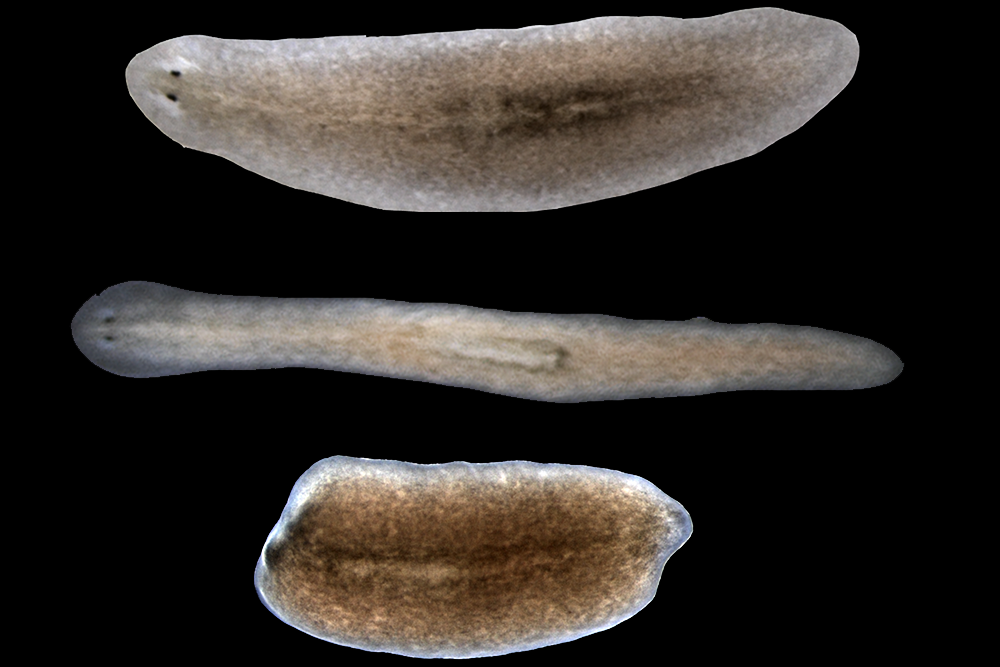
Researchers at the Whitehead Institute have illuminated an important role for different subtypes of muscle cells in orchestrating the process of tissue regeneration.
In a paper appearing online today in Nature, they reveal that a subtype of muscle fibers in flatworms is required for triggering the activity of genes that initiate the regeneration program. Notably, in the absence of these muscles, regeneration fails to proceed. Another type of muscle, they report, is required for giving regenerated tissue the proper pattern — for example, forming one head instead of two.
“One of the central mysteries in organ and tissue regeneration is: How do animals initiate all of the cellular and molecular steps that lead to regeneration?” says senior author Peter Reddien, a member of Whitehead Institute, professor of biology at MIT, and investigator with the Howard Hughes Medical Institute. “We’ve helped answer this question by revealing a surprising molecular program that operates within a subgroup of muscle cells that helps establish the molecular information required for proper tissue regeneration after injury.”
For more than a decade, Reddien and the researchers in his laboratory have studied the biological mechanisms that underlie regeneration in a tiny flatworm called planarians. These worms possess some impressive regenerative capabilities: When sliced in two, each piece of the worm can regrow the body parts needed to form two complete organisms. In previous studies, Reddien’s team identified a set of always-on genes, known as position control genes (PCGs), that provide cells with region-specific instructions, like a set of GPS coordinates, that tell cells where they are in the body, and thus what body part to regenerate. Interestingly, PGCs are active in planarian muscle cells, suggesting muscle may play a major role in the regeneration process.
“This discovery raised a lot of questions about how muscle participates in this process,” Reddien says.
In planarians, there are a handful of muscle cell types. For example, if you imagine the worms as simple cylindrical tubes, there are longitudinal muscle fibers, which run head-to-tail along the tubes’ long axis. There are also circular fibers, which are perpendicular to the longitudinal fibers and hug the tubes’ outer circumference.
To assess the roles of these different muscle cell types in regeneration, first author Lucila Scimone and her colleagues needed a method to selectively remove them. When myoD, a gene found specifically in the longitudinal fibers, is inhibited, those fibers fail to form. Similarly, the nkx1-1 gene marks the circular fibers, and when its function is reduced, they do not develop. Using these genes as molecular scalpels, Scimone and her co-authors could test the effects of ablating these distinct muscle groups on regeneration.
Surprisingly, when the longitudinal fibers were removed, the results were dramatic. The worms live quite normally, but when they are injured (the head removed, for example) they cannot regenerate the missing parts.
“This is an amazing result; it tells us that these longitudinal fibers are essential for orchestrating the regeneration program from the very beginning,” says Scimone, a scientist in Reddien’s lab.
As the researchers dug deeper into the finding, they learned that the functions of two critical genes are disrupted when longitudinal fibers are missing. These genes, called notum and follistatin, are known for their fundamental roles in regeneration, controlling head-versus-tail decisions and sustained cell proliferation, respectively, following tissue injury.
In addition to this essential role for longitudinal fibers, the research team also uncovered a key role for circular fibers. When these muscles are missing, planarians are able to regenerate missing body parts, but what regrows is abnormally patterned. For example, two heads may be regenerated within a single outgrowth, instead of one.
These results underscore an important and previously unappreciated role for muscle, widely known for its contractile properties, in instructing the tissue regeneration program. The Whitehead researchers will continue to probe the role of different muscle cell types in planarian regeneration and also explore whether other animals with regenerative capabilities possess a similar muscle-localized program for conferring positional information.
“It’s hard to understand what limits humans’ abilities to regenerate and repair wounds without first knowing what mechanisms are enabling some animals, like planarians, to do it so amazingly well,” Reddien says.
This work was supported by the National Institutes of Health, Howard Hughes Medical Institute, and the Eleanor Schwartz Charitable Foundation.
Education
PhD, 1991, Stanford University
BSc, 1982, Biology and Biochemistry, Hebrew University, Israel
Research Summary
The property of the brain that allows it to constantly adapt to change is termed plasticity, and is a prominent feature not only of learning and memory in the adult, but also of brain development. Connections between neurons (synapses) that are frequently used become stronger, while those that are unstimulated gradually dwindle away. The Nedivi lab works to identify the cellular mechanisms that underlie the addition and elimination of synaptic connections in response to activity using genetic and in vivo imaging approaches.
Awards
Elected Member at Large, AAAS, 2019-2023
Elected Member, Dana Alliance, 2019
BCS Award for Excellence in Undergraduate Teaching, 2018
American Association for the Advancement of Science (AAAS), Fellow, 2016
AFAR Julie Martin Mid-Career Award in Aging Research, 2007 – 2011
Edgerly Innovation Fund Award, 2006
Dean’s Education and Student Advising Award, 2003
NSF Powre Award, 1999
Alfred P . Sloan Research Fellowship, 1999 – 2001
Ellison Medical Foundation New Scholar Award, 1997 – 2002
Education
PhD, 1969, University of Illinois, Urbana-Champaign
BA, 1966, Chemistry and Math, Union College
Research Summary
We investigate small, non-coding RNAs called microRNAs (miRNAs), which regulate over half of the genes in mammalian cells at the stages of translation and mRNA stability. We are also interested in the processes underlying transcription from the anti-sense strand (so-called “divergent” transcription), as well as the relationship between elongation of transcription, RNA splicing, and chromatin modifications.
Awards
AACR Award for Lifetime Achievement in Cancer Research, 2020
AACR Distinguished Award for Extraordinary Scientific Innovation and Exceptional Leadership in Cancer Research and Biomedical Science, 2018
Royal Society of London, Foreign Fellow, 2011
National Science Foundation, National Medal of Science, 2004
The Nobel Foundation, Nobel Prize in Physiology or Medicine, 1993
National Academy of Medicine, Member, 1991
American Association for the Advancement of Science, Fellow, 1987
American Academy of Arts and Sciences, Fellow, 1987
National Academy of Sciences, Member, 1983
Education
PhD, 1966, Rockefeller University
BS, 1962, Chemistry and Mathematics, Kenyon College
Research Summary
Harvey Lodish has been a leader in molecular cell biology as well as a biotechnology entrepreneur for over five decades. Much of his early research focused on the regulation of messenger RNA translation and the biogenesis of plasma membrane glycoproteins. Beginning in the 1980s, his research focused on cloning and characterizing many proteins, microRNAs, and long noncoding RNAs important for red cell development and function. His laboratory was the first to clone and sequence mRNAs encoding many hormone receptors, mammalian glucose transport proteins, and proteins important for adipose cell formation and function. He went on to identify and characterize several genes and proteins involved in insulin resistance and stress responses in adipose cells. Over the years, he has mentored hundreds of undergraduates, PhD and MD/PhD students, and postdoctoral fellows, and continues to teach award-winning undergraduate and graduate classes on biotechnology.
Harvey Lodish closed his lab in 2020 and is no longer accepting students.
Awards
Wallace H. Coulter Award for Lifetime Achievement in Hematology, American Society of Hematology, 2021
Donald Metcalf Award, International Society for Experimental Hematology, 2020
American Society for Cell Biology WICB Sandra K. Masur Senior Leadership Award, 2017
Mentor Award in Basic Science, American Society of Hematology, 2010
President, American Society for Cell Biology, 2004
Associate Member, European Molecular Biology Organization (EMBO), 1996
National Academy of Sciences, Member, 1987
American Academy of Arts and Sciences, Fellow, 1986
John Simon Guggenheim Memorial Foundation, Guggenheim Fellowship, 1977
Education
PhD, 2002, University of California, Berkeley
BS, 1997, Biology, Duke University
Research Summary
Our lab is fascinated by the molecular machinery that directs core cellular processes, and in particular how these processes are modulated and rewired across different physiological contexts. Our work has focused on the proteins that direct chromosome segregation and cell division, including the macromolecular kinetochore structure that mediates chromosome-microtubule interactions. Although cell division is an essential cellular process, this machinery is remarkably flexible in its composition and properties, which can vary dramatically between species and are even modulated within the same organism — over the cell cycle, during development, and across diverse physiological situations. To define the basis by which the kinetochore and other core cellular structures are rewired to adapt to diverse situations and functional requirements, we are currently investigating diverse transcriptional, translational, and post-translational mechanisms that act to generate proteomic variability both within individual cells and across tissues, cell state, development, and disease.
Awards
Global Consortium for Reproductive Longevity and Equality (GCRLE) Scholar Award, 2020
MIT Undergraduate Research Opportunities Program (UROP) Outstanding Mentor – Faculty, 2019
American Society for Cell Biology (ASCB) Early Career Life Scientist Award, 2012
PhD, 1994, Baylor College of Medicine; MD, 1997, Baylor College of Medicine
BS, 1989, Biochemistry, Louisiana State University
Research Summary
Using Drosophila, we study how neurons form synaptic connections, as well as how synapses transmit information and change during learning and memory. We also investigate how alterations in neuronal signaling underlie several neurological diseases, including epilepsy, autism, and Huntington’s Disease. We hope to bridge the gap between the molecular components of the synapse and the physiological responses they mediate.
Whitehead researchers unravel fundamental molecular machinery that propels chromosome movement
November 16, 2017
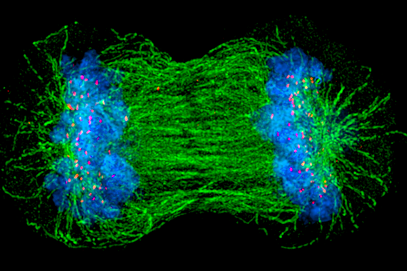
Each day, billions of cells in the human body undergo a vital ritual, wherein one cell divides to form two. This process, known as cell division, is as beautiful as it is essential, undergirding the body’s growth in times of both health and disease. Despite the fact that cell division (or “mitosis”) has been a basic topic in high school biology classes for the past 70 years, the mechanisms by which cells conduct this critical event remain poorly understood. In particular, there are lingering uncertainties about how chromosomes — large units of DNA that include our genes — get properly allocated so that both daughter cells receive intact, complete copies of their genetic blueprint.
“People have been watching chromosomes move, align, and segregate for more than a century — it’s such a fundamental aspect of biology,” says Iain Cheeseman, a member of the Whitehead Institute for Biomedical Research and an associate professor of biology at Massachusetts Institute of Technology. “It’s also much more elegant and complicated than we ever anticipated.”
Cheeseman and members of his Whitehead laboratory have discovered many of the molecular movers and shakers that ensure chromosomes get to the right place at the right time. These components assemble together — like the parts of an engine — to establish robust connections with chromosomes and ultimately power their movement within cells. “The most important unanswered question in the field is how do these components work together? That is, how do you build a machine that is more than the sum of its parts?” he says.
Now, in two recent papers in the journals eLife and Current Biology, Cheeseman and his colleagues help shed light on this central question.
Back to the drawing board
As recently as 15 years ago, scientists assumed that in dividing cells, chromosomes move the way many other cellular objects move — transported by tiny molecular motors. These mini-motors are specifically designed to travel along roads made from rod-like structures called microtubules. Like a car cruising on a highway, they can carry cargo over long distances. Although such microtubule-based vehicles seemed a logical suspect, when scientists inactivate them in human cells, chromosomes can still move and segregate just fine. So something else must contribute the necessary molecular muscle.
“We basically had to throw out the major hypothesis that was out there and go back to the drawing board,” says Cheeseman.
Over the last several years, he and other scientists in the field have helped develop a clearer view of how this process works and who the key players are that enable a very different type of movement. Consider a car sitting motionless on a highway. Rather than revving its own engine to generate motion, the highway itself moves, shrinking or growing while the car hangs on. “It is a radically different way of imagining this movement process,” says Cheeseman. “An important part of my lab’s mission has been to figure out how do you build a motor like that? What are the factors required and how do they act?”
Of course, the highway — or more precisely, the microtubule — must grow and shrink as needed. But even more important, there must also be an apparatus that can enable chromosomes to hold on to such a dynamic structure. As Cheeseman and his colleagues have uncovered, this coupling requires a suite of highly sophisticated molecular players.
Building an unusual machine
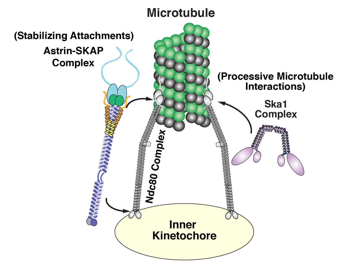
Cheeseman and his laboratory have focused on three key groups or complexes of proteins that play essential roles in chromosome segregation in human cells. These components assemble together to form a kind of molecular tether point on chromosomes (called the kinetochore) where microtubules attach.
Diagram of the kinetochore/microtube interface Courtesy: David Kern/Whitehead Institute
Among this trio of parts, the most critical is the Ndc80 complex. “It is the major connection between the kinetochore and the microtubule,” says Cheeseman. As a postdoc, he discovered the biochemical properties that enable this Ndc80 complex to grab on to microtubules, research that sparked his lab’s quest to study the various pieces of the kinetochore machinery and how they work.
While Ndc80 forms a critical linkage, it lacks some key capabilities, like processivity — the ability to keep ahold of something while it moves. In a series of papers, one published in 2009, another in 2012, and a new one in Current Biology, Cheeseman’s team revealed that Ska1 can perform this crucial function. That is, it has the biochemical capacity to enable chromosomes to hang onto microtubules while they grow and as they shrink, an activity that it can impart to the Ndc80 complex. “These are pretty powerful properties,” says Cheeseman.
Diving even deeper into Ska1’s bag of tricks, Julie Monda and Ian Whitney, lead authors of the Current Biology paper, went on to decipher the precise molecular features that enable the complex’s dynamic capabilities, uncovering multiple surfaces that associate with microtubules and enable Ska1 to undergo something akin to molecular somersaults. These somersaults are what allow it to maintain its association with microtubules.
The third complex, Astrin-SKAP, also plays a unique role. As Cheeseman’s team described in their recent eLife paper, led by first author David Kern, it serves as a master stabilizer — like a final drop of superglue to secure everything in place. “It’s the last thing that comes in and helps lock down these interactions, so you can stabilize and maintain them,” says Cheeseman.
Uncovering its role was no easy feat. Astrin-SKAP proved to be rather temperamental biochemically, complicating Kern’s efforts to purify and manipulate it in the laboratory. Also, as he and his colleagues discovered, a tiny piece of the structure had previously gone undetected; it works alongside the rest of the complex and is required for its normal function. Perhaps the most important revelation was that Astrin-SKAP doesn’t just work alone — it also coordinates with Ndc80. “This is an important finding for how we think about these components as a whole,” saysCheeseman.
Although questions remain about how all of these parts work together and how other pieces may come into play, Cheeseman believes these studies provide an exciting start. “The first human kinetochore component wasn’t identified until 1987, when many of the other key processes in the cell had already been intensively studied,” he says. “There are so many exciting questions that are accessible now that we have these tools and knowledge.”
Now, he and his colleagues will continue to meld approaches in cell biology and biochemistry to decode the inner workings of the kinetochore. That includes understanding how the various components operate not only in individual cells, but also in multicellular organisms.
“We are currently thinking a lot about the physiological context— that is, what matters to cells and to an organism,” says Cheeseman. “The work that our lab and others have conducted over the past two decades has given us a molecular handle on this problem. I’m excited to be able to apply these finding to understanding the ways that cell division is altered in development and in disease states.”
Written by Nicole Davis
* * *
Iain Cheeseman’s primary affiliation is with Whitehead Institute for Biomedical Research, where his laboratory is located and all his research is conducted. He is also an associate professor of biology at Massachusetts Institute of Technology.
* * *
Full citations:
“Astrin-SKAP complex reconstitution reveals its kinetochore interaction with microtubule-bound Ndc80”
eLife 2017;6:e26866 August 25, 2017. DOI: 10.7554/eLife.26866
David M Kern (1,2), Julie K Monda (1,2), Kuan-Chung Su (1), Elizabeth M Wilson-Kubalek (3), and Iain M Cheeseman (1,2).
1. Whitehead Institute for Biomedical Research, 455 Main Street, Cambridge, MA 02142, USA
2. Department of Biology, Massachusetts Institute of Technology, Cambridge, MA 02142, USA
3. Department of Cell Biology, The Scripps Research Institute, La Jolla, CA 92037, USA
“Microtubule tip tracking by the spindle and kinetochore protein Ska1 requires diverse tubulin-interacting surfaces”
Current Biology, online November 16, 2017. DOI: 10.1016/j.cub.2017.10.018
Julie K. Monda (1,2,6), Ian P. Whitney (1,6), Ekaterina V. Tarasovetc (3,4), Elizabeth Wilson-Kubalek (5), Ronald A. Milligan (5), Ekaterina L. Grishchuk (3), and Iain M. Cheeseman (1,2).
1. Whitehead Institute for Biomedical Research, 455 Main Street, Cambridge, MA 02142, USA
2. Department of Biology, Massachusetts Institute of Technology, Cambridge, MA 02142, USA
3. Department of Physiology, Perelman School of Medicine, University of Pennsylvania, Philadelphia, PA 19104, USA
4. Center for Theoretical Problems of Physicochemical Pharmacology, Russian Academy of Sciences, Moscow, Russia
5. Department of Cell Biology, The Scripps Research Institute, La Jolla, CA 92037, USA