Scientists develop a rapid gene-editing screen to find effects of cancer mutations

With the new technique, MIT researchers hope to identify mutations that could be targeted with new cancer therapies.

Anne Trafton | MIT News
March 12, 2024

Tumors can carry mutations in hundreds of different genes, and each of those genes may be mutated in different ways — some mutations simply replace one DNA nucleotide with another, while others insert or delete larger sections of DNA.

Until now, there has been no way to quickly and easily screen each of those mutations in their natural setting to see what role they may play in the development, progression, and treatment response of a tumor. Using a variant of CRISPR genome-editing known as prime editing, MIT researchers have now come up with a way to screen those mutations much more easily.

The researchers demonstrated their technique by screening cells with more than 1,000 different mutations of the tumor suppressor gene p53, all of which have been seen in cancer patients. This method, which is easier and faster than any existing approach, and edits the genome rather than introducing an artificial version of the mutant gene, revealed that some p53 mutations are more harmful than previously thought.

This technique could also be applied to many other cancer genes, the researchers say, and could eventually be used for precision medicine, to determine how an individual patient’s tumor will respond to a particular treatment.

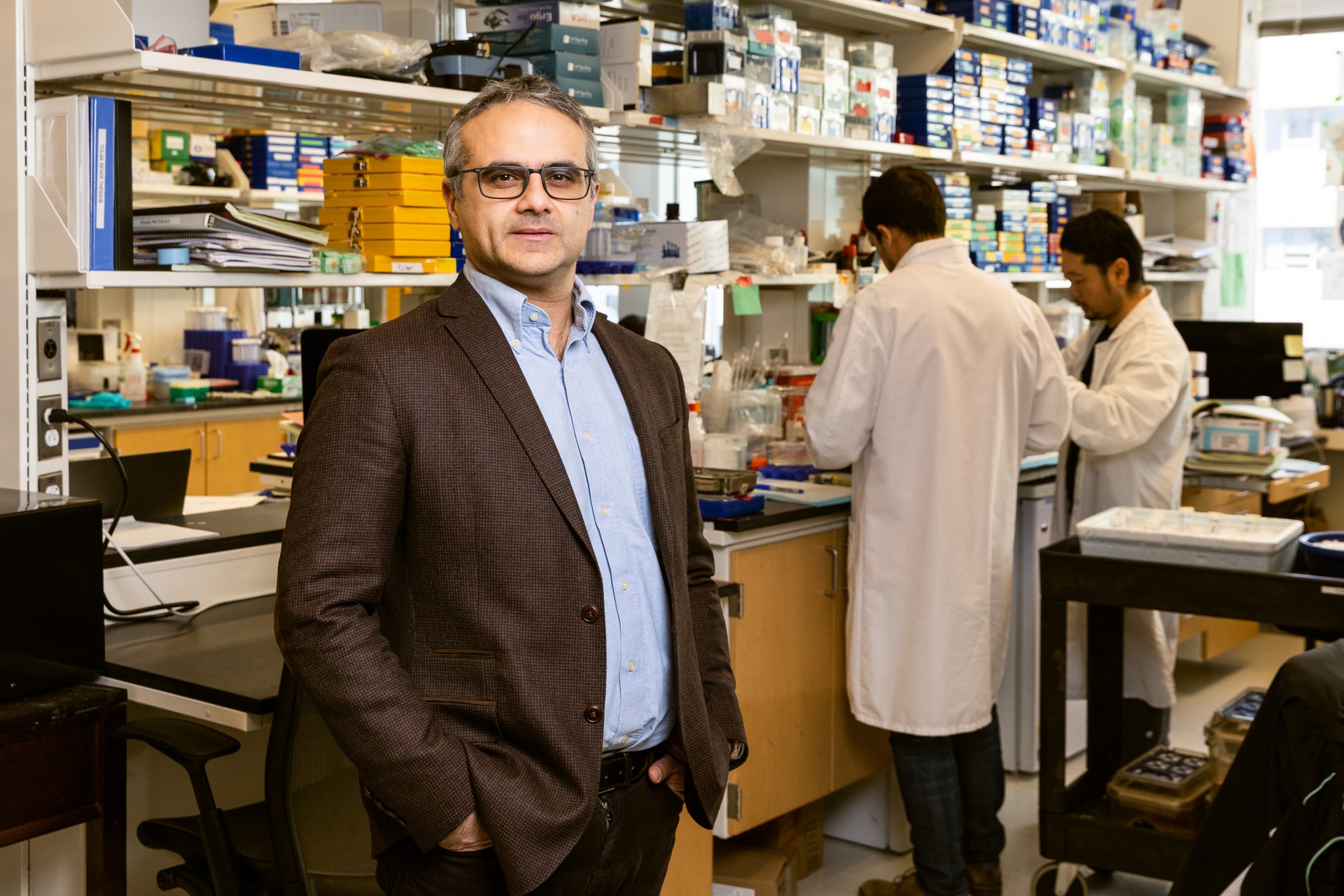
“In one experiment, you can generate thousands of genotypes that are seen in cancer patients, and immediately test whether one or more of those genotypes are sensitive or resistant to any type of therapy that you’re interested in using,” says Francisco Sanchez-Rivera, an MIT assistant professor of biology, a member of the Koch Institute for Integrative Cancer Research, and the senior author of the study.

MIT graduate student Samuel Gould is the lead author of the paper, which appears today in Nature Biotechnology.

Editing cells

The new technique builds on research that Sanchez-Rivera began 10 years ago as an MIT graduate student. At that time, working with Tyler Jacks, the David H. Koch Professor of Biology, and then-postdoc Thales Papagiannakopoulos, Sanchez-Rivera developed a way to use CRISPR genome-editing to introduce into mice genetic mutations linked to lung cancer.

In that study, the researchers showed that they could delete genes that are often lost in lung tumor cells, and the resulting tumors were similar to naturally arising tumors with those mutations. However, this technique did not allow for the creation of point mutations (substitutions of one nucleotide for another) or insertions.

“While some cancer patients have deletions in certain genes, the vast majority of mutations that cancer patients have in their tumors also include point mutations or small insertions,” Sanchez-Rivera says.

Since then, David Liu, a professor in the Harvard University Department of Chemistry and Chemical Biology and a core institute member of the Broad Institute, has developed new CRISPR-based genome editing technologies that can generate additional types of mutations more easily. With base editing, developed in 2016, researchers can engineer point mutations, but not all possible point mutations. In 2019, Liu, who is also an author of the Nature Biotechnology study, developed a technique called prime editing, which enables any kind of point mutation to be introduced, as well as insertions and deletions.

“Prime editing in theory solves one of the major challenges with earlier forms of CRISPR-based editing, which is that it allows you to engineer virtually any type of mutation,” Sanchez-Rivera says.

When they began working on this project, Sanchez-Rivera and Gould calculated that if performed successfully, prime editing could be used to generate more than 99 percent of all small mutations seen in cancer patients.

However, to achieve that, they needed to find a way to optimize the editing efficiency of the CRISPR-based system. The prime editing guide RNAs (pegRNAs) used to direct CRISPR enzymes to cut the genome in certain spots have varying levels of efficiency, which leads to “noise” in the data from pegRNAs that simply aren’t generating the correct target mutation. The MIT team devised a way to reduce that noise by using synthetic target sites to help them calculate how efficiently each guide RNA that they tested was working.

“We can design multiple prime-editing guide RNAs with different design properties, and then we get an empirical measurement of how efficient each of those pegRNAs is. It tells us what percentage of the time each pegRNA is actually introducing the correct edit,” Gould says.

Analyzing mutations

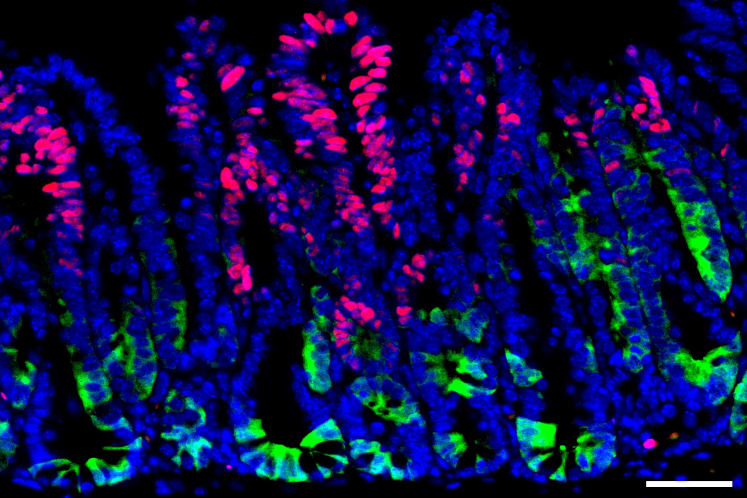
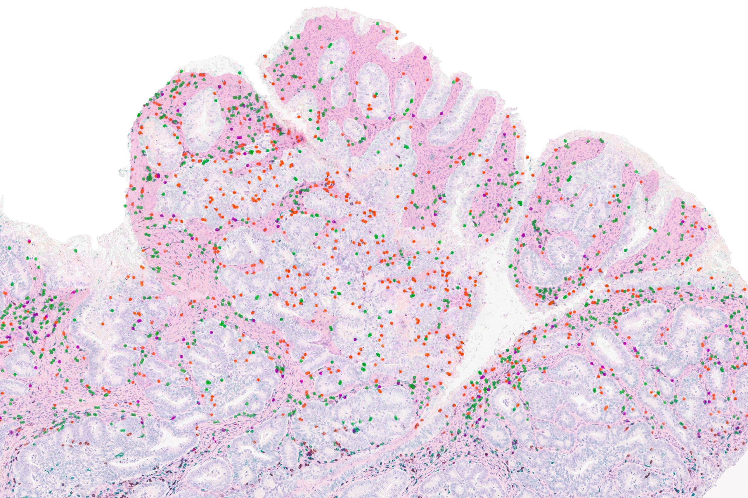
The researchers demonstrated their technique using p53, a gene that is mutated in more than half of all cancer patients. From a dataset that includes sequencing information from more than 40,000 patients, the researchers identified more than 1,000 different mutations that can occur in p53.

“We wanted to focus on p53 because it’s the most commonly mutated gene in human cancers, but only the most frequent variants in p53 have really been deeply studied. There are many variants in p53 that remain understudied,” Gould says.

Using their new method, the researchers introduced p53 mutations in human lung adenocarcinoma cells, then measured the survival rates of these cells, allowing them to determine each mutation’s effect on cell fitness.

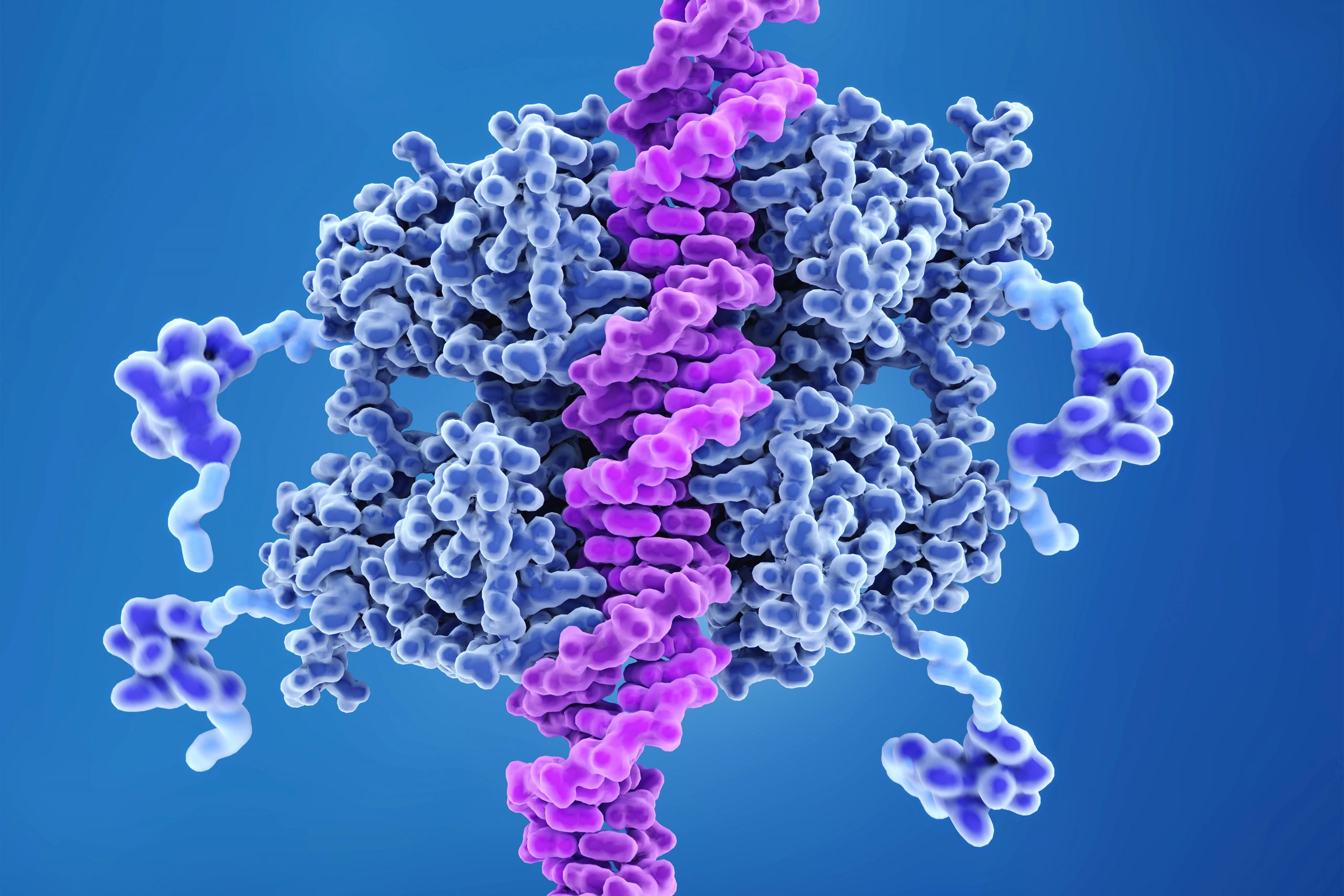
Among their findings, they showed that some p53 mutations promoted cell growth more than had been previously thought. These mutations, which prevent the p53 protein from forming a tetramer — an assembly of four p53 proteins — had been studied before, using a technique that involves inserting artificial copies of a mutated p53 gene into a cell.

Those studies found that these mutations did not confer any survival advantage to cancer cells. However, when the MIT team introduced those same mutations using the new prime editing technique, they found that the mutation prevented the tetramer from forming, allowing the cells to survive. Based on the studies done using overexpression of artificial p53 DNA, those mutations would have been classified as benign, while the new work shows that under more natural circumstances, they are not.

“This is a case where you could only observe these variant-induced phenotypes if you’re engineering the variants in their natural context and not with these more artificial systems,” Gould says. “This is just one example, but it speaks to a broader principle that we’re going to be able to access novel biology using these new genome-editing technologies.”

Because it is difficult to reactivate tumor suppressor genes, there are few drugs that target p53, but the researchers now plan to investigate mutations found in other cancer-linked genes, in hopes of discovering potential cancer therapies that could target those mutations. They also hope that the technique could one day enable personalized approaches to treating tumors.

“With the advent of sequencing technologies in the clinic, we’ll be able to use this genetic information to tailor therapies for patients suffering from tumors that have a defined genetic makeup,” Sanchez-Rivera says. “This approach based on prime editing has the potential to change everything.”

The research was funded, in part, by the National Institute of General Medical Sciences, an MIT School of Science Fellowship in Cancer Research, a Howard Hughes Medical Institute Hanna Gray Fellowship, the V Foundation for Cancer Research, a National Cancer Institute Cancer Center Support Grant, the Ludwig Center at MIT, a Koch Institute Frontier Award, the MIT Research Support Committee, and the Koch Institute Support (core) Grant from the National Cancer Institute.

How phase separation is revolutionizing biology

Postdocs from Whitehead Institute Member Richard A. Young's lab found that imaging and molecular manipulation reveal how biomolecular condensates form and offer clues to the role of phase separation in health and disease.

February 27, 2024
News brief: Calo Lab

How do cells respond to disruptions in splicing?

Lillian Eden | Department of Biology
March 4, 2024

New research from the Calo Lab in the Department of Biology has identified the protein Mdm2 generating a form that activates a cascade of cellular stress responses when splicing is disrupted.

To create proteins, DNA is transcribed into RNA, and that RNA is then “translated” into protein. Between the creation of the RNA and the translation to protein is often a step called splicing. During splicing, segments called introns are removed, and the remaining pieces, called exons, are joined together to form the blueprint for translation. By splicing together different exons, the cell can create different proteins from the same section of genetic code. When splicing goes awry, it can lead to diseases and cancers. 

New research recently published in Disease Models & Mechanisms from the Calo Lab in the Department of Biology at MIT has identified the mechanism for how cells respond to disruptions in splicing, which involves activating a cellular stress response. The stress response, once activated, causes widespread effects, including changes to cell metabolism. 

Researchers have discovered cellular stress responses for other core cellular processes, such as ribosome biogenesis. However, this is the first time researchers have identified how cells respond to perturbing the splicing process.

A particular protein acts as a kind of canary in a coal mine: Mdm2, which responds to a broad range of splicing disruptions. Mdm2 does not cause a stress response by itself. Rather, Mdm2 is itself spliced differently in response to splicing disruptions. Downstream, the alternative splicing of Mdm2 leads to the activation of a protein called p53, which is known to orchestrate a cascade of responses to stress.

Researchers have long wondered why some cell types seem more sensitive to splicing disruptions than others. For example, some disorders caused by mutations in proteins that perform RNA splicing, despite affecting the whole organism, induce more noticeable changes in tissues derived from the neural crest—a collection of stem cells that contributes to the formation of the face, jaw, retinas, limbs, and heart during development. Certain splicing inhibitors have also increased the effectiveness of some cancer treatments, but the mechanism is unknown. 

One of the p53-induced stress responses includes changing the metabolism of cells and how they use sugars, which may explain why some cells are more sensitive to splicing disruptions than others. Inhibiting glycolysis, the reactions that extract energy from glucose, can affect how cells divide and migrate. 

The way cells divide and migrate is critical during development; in experiments, zebrafish treated with glycolysis inhibitors exhibited similar changes to craniofacial features as those where splicing was disrupted. Cancerous cells, too, are known to require high levels of sugar metabolism and, therefore, may be especially sensitive to treatments that induce changes in the splicing pathway. 

The researchers knocked down genes to mimic milder splicing disruptions instead of knocking them out entirely. Splicing is so essential that knocking out the splicing machinery can lead to extreme responses like cell death. In organismal models like zebrafish, those severe phenotypes don’t accurately reflect how splicing disruptions present in human diseases.

First author Jade Varineau, a graduate student in the Calo lab, was drawn to the project because it allowed her to explore what was happening at the RNA and cellular level while also observing how splicing perturbations were affecting the whole organism. 

“I think this data can help us reframe the way we think about diseases and cancers that are impacted by splicing—that a treatment that works for one may work for another because all the symptoms may stem from the same cellular response,” Varineau says. 

Although the results indicate how cells broadly respond to splicing perturbations, the mechanism for how disruptions in splicing induce the alternate splicing of Mdm2 remains unclear. Senior author Eliezer Calo says the lab is also exploring how splicing mechanisms may be altered for things like cancer. Their work, he says, opens the door for further exploration of cell-type specificity of genetic disorders and improvements in cancer treatments using splicing inhibitors. 

 “We know that the sensor is encoded in the gene Mdm2—what are the molecules that allow Mdm2 to act as a sensor, and how does the sensor malfunction for things like cancer?” Calo says. “The next step is to find out how the sensor works.”  

How early-stage cancer cells hide from the immune system

A new study finds precancerous colon cells turn on a gene called SOX17, which helps them evade detection and develop into more advanced tumors.

Anne Trafton | MIT News
February 28, 2024

One of the immune system’s primary roles is to detect and kill cells that have acquired cancerous mutations. However, some early-stage cancer cells manage to evade this surveillance and develop into more advanced tumors.

A new study from MIT and Dana-Farber Cancer Institute has identified one strategy that helps these precancerous cells avoid immune detection. The researchers found that early in colon cancer development, cells that turn on a gene called SOX17 can become essentially invisible to the immune system.

If scientists could find a way to block SOX17 function or the pathway that it activates, this may offer a new way to treat early-stage cancers before they grow into larger tumors, the researchers say.

“Activation of the SOX17 program in the earliest innings of colorectal cancer formation is a critical step that shields precancerous cells from the immune system. If we can inhibit the SOX17 program, we might be better able to prevent colon cancer, particularly in patients that are prone to developing colon polyps,” says Omer Yilmaz, an MIT associate professor of biology, a member of MIT’s Koch Institute for Integrative Cancer Research, and one of the senior authors of the study.

Judith Agudo, a principal investigator at Dana-Farber Cancer Institute and an assistant professor at Harvard Medical School, is also a senior author of the study, which appears today in Nature. The paper’s lead author is MIT Research Scientist Norihiro Goto. Other collaborators include Tyler Jacks, a professor of biology and a member of MIT’s Koch Institute; Peter Westcott, a former Jacks lab postdoc who is now an assistant professor at Cold Spring Harbor Laboratory; and Saori Goto, an MIT postdoc in the Yilmaz lab.

Immune evasion

Colon cancer usually arises in long-lived cells called intestinal stem cells, whose job is to continually regenerate the lining of the intestines. Over their long lifetime, these cells can accumulate cancerous mutations that lead to the formation of polyps, a type of premalignant growth that can eventually become metastatic colon cancer.

To learn more about how these precancerous growths evade the immune system, the researchers used a technique they had previously developed for growing mini colon tumors in a lab dish and then implanting them into mice. In this case, the researchers engineered the tumors to express mutated versions of cancer-linked genes Kras, p53, and APC, which are often found in human colon cancers.

Once these tumors were implanted in mice, the researchers observed a dramatic increase in the tumors’ expression of SOX17. This gene encodes a transcription factor that is normally active only during embryonic development, when it helps to control development of the intestines and the formation of blood vessels.

The researchers’ experiments revealed that when SOX17 is turned on in cancer cells, it helps the cells to create an immunosuppressive environment. Among its effects, SOX17 prevents cells from synthesizing the receptor that normally detects interferon gamma, a molecule that is one of the immune system’s primary weapons against cancer cells.

Without those interferon gamma receptors, cancerous and precancerous cells can simply ignore messages from the immune system, which would normally direct them to undergo programmed cell death.

“One of SOX17’s main roles is to turn off the interferon gamma signaling pathway in colorectal cancer cells and in precancerous adenoma cells. By turning off interferon gamma receptor signaling in the tumor cells, the tumor cells become hidden from T cells and can grow in the presence of an immune system,” Yilmaz says.

Without interferon gamma signaling, cancer cells also minimize their production of molecules called MHC proteins, which are responsible for displaying cancerous antigens to the immune system. The cells’ insensitivity to interferon gamma also prevents them from producing immune molecules called chemokines, which normally recruit T cells that would help destroy the cancerous cells.

Targeting SOX17

When the researchers generated colon tumor organoids with SOX17 knocked out, and implanted those into mice, the immune system was able to attack those tumors much more effectively. This suggests that preventing cancer cells from turning off SOX17 could offer a way to treat colon cancer in its earliest stages.

“Just by turning off SOX17 in fairly complex tumors, we were able to essentially obliterate the ability of these tumor cells to persist,” Goto says.

As part of their study, the researchers also analyzed gene expression data from patients with colon cancer and found that SOX17 tended to be highly expressed in early-stage colon cancers but dropped off as the tumors became more invasive and metastatic.

“We think this makes a lot of sense because as colorectal cancers become more invasive and metastatic, there are other mechanisms that create an immunosuppressive environment,” Yilmaz says. “As the colon cancer becomes more aggressive and activates these other mechanisms, then there’s less importance for SOX17.”

Transcription factors such as SOX17 are considered difficult to target using drugs, in part because of their disorganized structure, so the researchers now plan to identify other proteins that SOX17 interacts with, in hopes that it might be easier to block some of those interactions.

The researchers also plan to investigate what triggers SOX17 to turn on in precancerous cells.

The research was funded by the MIT Stem Cell Initiative via Fondation MIT, the National Institutes of Health/National Cancer Institute, and a Koch Institute-Dana Farber Harvard Cancer Center Bridge Project grant.

Whitney Henry

Education

  • Graduate: PhD, 2016, Harvard University
  • Undergraduate: BS, 2010, Biology, Grambling State University

Research Summary

Ferroptosis is an iron-dependent form of cell death with profound implications in human health and disease. In the context of cancer, the use of ferroptosis inducers to target subpopulations of highly metastatic and therapy-resistant cancer cells has garnered much excitement over the last few years. However, to gain a comprehensive understanding of the full therapeutic potential of ferroptosis, our research focuses on (i) uncovering the molecular factors affecting ferroptosis susceptibility, (ii) studying its impact on the tumor microenvironment, and (iii) developing innovative ways to modulate ferroptosis resistance in vivo. We employ a multidisciplinary approach, combining functional genomics, metabolomics, bioengineering, and a range of in vitro and in vivo models to advance our understanding in this domain and to translate our findings into effective therapies.

Awards

  • The Margaret and Herman Sokol Postdoctoral Award, 2022
  • Ludwig Center at MIT Postdoctoral Fellowship, 2022
  • Jane Coffin Childs Memorial Fund Postdoctoral Fellowship, 2017
  • HHMI International Predoctoral Research Fellowship, 2013
Study explains why certain immunotherapies don’t always work as predicted

The findings could help doctors identify cancer patients who would benefit the most from drugs called checkpoint blockade inhibitors.

Anne Trafton | MIT News
September 14, 2023

Cancer drugs known as checkpoint blockade inhibitors have proven effective for some cancer patients. These drugs work by taking the brakes off the body’s T cell response, stimulating those immune cells to destroy tumors.

Some studies have shown that these drugs work better in patients whose tumors have a very large number of mutated proteins, which scientists believe is because those proteins offer plentiful targets for T cells to attack. However, for at least 50 percent of patients whose tumors show a high mutational burden, checkpoint blockade inhibitors don’t work at all.

A new study from MIT reveals a possible explanation for why that is. In a study of mice, the researchers found that measuring the diversity of mutations within a tumor generated much more accurate predictions of whether the treatment would succeed than measuring the overall number of mutations.

If validated in clinical trials, this information could help doctors to better determine which patients will benefit from checkpoint blockade inhibitors.

“While very powerful in the right settings, immune checkpoint therapies are not effective for all cancer patients. This work makes clear the role of genetic heterogeneity in cancer in determining the effectiveness of these treatments,” says Tyler Jacks, the David H. Koch Professor of Biology and a member of MIT’s Koch Institute for Cancer Research.

Jacks; Peter Westcott, a former MIT postdoc in the Jacks lab who is now an assistant professor at Cold Spring Harbor Laboratory; and Isidro Cortes-Ciriano, a research group leader at EMBL’s European Bioinformatics Institute (EMBL-EBI), are the senior authors of the paper, which appears today in Nature Genetics.

A diversity of mutations

Across all types of cancer, a small percentage of tumors have what is called a high tumor mutational burden (TMB), meaning they have a very large number of mutations in each cell. A subset of these tumors has defects related to DNA repair, most commonly in a repair system known as DNA mismatch repair.

Because these tumors have so many mutated proteins, they are believed to be good candidates for immunotherapy treatment, as they offer a plethora of potential targets for T cells to attack. Over the past few years, the FDA has approved a checkpoint blockade inhibitor called pembrolizumab, which activates T cells by blocking a protein called PD-1, to treat several types of tumors that have a high TMB.

However, subsequent studies of patients who received this drug found that more than half of them did not respond well or only showed short-lived responses, even though their tumors had a high mutational burden. The MIT team set out to explore why some patients respond better than others, by designing mouse models that closely mimic the progression of tumors with high TMB.

These mouse models carry mutations in genes that drive cancer development in the colon and lung, as well as a mutation that shuts down the DNA mismatch repair system in these tumors as they begin to develop. This causes the tumors to generate many additional mutations. When the researchers treated these mice with checkpoint blockade inhibitors, they were surprised to find that none of them responded well to the treatment.

“We verified that we were very efficiently inactivating the DNA repair pathway, resulting in lots of mutations. The tumors looked just like they look in human cancers, but they were not more infiltrated by T cells, and they were not responding to immunotherapy,” Westcott says.

The researchers discovered that this lack of response appears to be the result of a phenomenon known as intratumoral heterogeneity. This means that, while the tumors have many mutations, each cell in the tumor tends to have different mutations than most of the other cells. As a result, each individual cancer mutation is “subclonal,” meaning that it is expressed in a minority of cells. (A “clonal” mutation is one that is expressed in all of the cells.)

In further experiments, the researchers explored what happened as they changed the heterogeneity of lung tumors in mice. They found that in tumors with clonal mutations, checkpoint blockade inhibitors were very effective. However, as they increased the heterogeneity by mixing tumor cells with different mutations, they found that the treatment became less effective.

“That shows us that intratumoral heterogeneity is actually confounding the immune response, and you really only get the strong immune checkpoint blockade responses when you have a clonal tumor,” Westcott says.

Failure to activate

It appears that this weak T cell response occurs because the T cells simply don’t see enough of any particular cancerous protein, or antigen, to become activated, the researchers say. When the researchers implanted mice with tumors that contained subclonal levels of proteins that normally induce a strong immune response, the T cells failed to become powerful enough to attack the tumor.

“You can have these potently immunogenic tumor cells that otherwise should lead to a profound T cell response, but at this low clonal fraction, they completely go stealth, and the immune system fails to recognize them,” Westcott says. “There’s not enough of the antigen that the T cells recognize, so they’re insufficiently primed and don’t acquire the ability to kill tumor cells.”

To see if these findings might extend to human patients, the researchers analyzed data from two small clinical trials of people who had been treated with checkpoint blockade inhibitors for either colorectal or stomach cancer. After analyzing the sequences of the patients’ tumors, they found that patients’ whose tumors were more homogeneous responded better to the treatment.

“Our understanding of cancer is improving all the time, and this translates into better patient outcomes,” Cortes-Ciriano says. “Survival rates following a cancer diagnosis have significantly improved in the past 20 years, thanks to advanced research and clinical studies. We know that each patient’s cancer is different and will require a tailored approach. Personalized medicine must take into account new research that is helping us understand why cancer treatments work for some patients but not all.”

The findings also suggest that treating patients with drugs that block the DNA mismatch repair pathway, in hopes of generating more mutations that T cells could target, may not help and could be harmful, the researchers say. One such drug is now in clinical trials.

“If you try to mutate an existing cancer, where you already have many cancer cells at the primary site and others that may have disseminated throughout the body, you’re going to create a super heterogeneous collection of cancer genomes. And what we showed is that with this high intratumoral heterogeneity, the T cell response is confused and there is absolutely no response to immune checkpoint therapy,” Westcott says.

The research was funded by the Koch Institute Support (core) Grant from the U.S. National Cancer Institute, the Howard Hughes Medical Institute, and a Damon Runyon Fellowship Award.

Exploring the links between diet and cancer

Omer Yilmaz’s work on how diet influences intestinal stem cells could lead to new ways to treat or prevent gastrointestinal cancers.

Anne Trafton | MIT News Office
May 25, 2023

Every three to five days, all of the cells lining the human intestine are replaced. That constant replenishment of cells helps the intestinal lining withstand the damage caused by food passing through the digestive tract.

This rapid turnover of cells relies on intestinal stem cells, which give rise to all of the other types of cells found in the intestine. Recent research has shown that those stem cells are heavily influenced by diet, which can help keep them healthy or stimulate them to become cancerous.

“Low-calorie diets such as fasting and caloric restriction can have antiaging effects and antitumor effects, and we want to understand why that is. On the other hand, diets that lead to obesity can promote diseases of aging, such as cancer,” says Omer Yilmaz, the Eisen and Chang Career Development Associate Professor of Biology at MIT.

For the past decade, Yilmaz has been studying how different diets and environmental conditions affect intestinal stem cells, and how those factors can increase the risk of cancer and other diseases. This work could help researchers develop new ways to improve gastrointestinal health, either through dietary interventions or drugs that mimic the beneficial effects of certain diets, he says.

“Our findings have raised the possibility that fasting interventions, or small molecules that mimic the effects of fasting, might have a role in improving intestinal regeneration,” says Yilmaz, who is also a member of MIT’s Koch Institute for Integrative Cancer Research.

A clinical approach

Yilmaz’s interest in disease and medicine arose at an early age. His father practiced internal medicine, and Yilmaz spent a great deal of time at his father’s office after school, or tagging along at the hospital where his father saw patients.

“I was very interested in medicines and how medicines were used to treat diseases,” Yilmaz recalls. “He’d ask me questions, and many times I wouldn’t know the answer, but he would encourage me to figure out the answers to his questions. That really stimulated my interest in biology and in wanting to become a doctor.”

Knowing that he wanted to go into medicine, Yilmaz applied and was accepted to an eight-year, combined bachelor’s and MD program at the University of Michigan. As an undergraduate, this gave him the freedom to explore areas of interest without worrying about applying to medical school. While majoring in biochemistry and physics, he did undergraduate research in the field of protein folding.

During his first year of medical school, Yilmaz realized that he missed doing research, so he decided to apply to the MD/PhD program at the University of Michigan. For his PhD research, he studied blood-forming stem cells and identified new markers that allowed such cells to be more easily isolated from the bone marrow.

“This was important because there’s a lot of interest in understanding what makes a stem cell a stem cell, and how much of it is an internal program versus signals from the microenvironment,” Yilmaz says.

After finishing his PhD and MD, he thought about going straight into research and skipping a medical residency, but ended up doing a residency in pathology at Massachusetts General Hospital. During that time, he decided to switch his research focus from blood-forming stem cells to stem cells found in the gastrointestinal tract.

“The GI tract seemed very interesting because in contrast to the bone marrow, we knew very little about the identity of GI stem cells,” Yilmaz says. “I knew that once GI stem cells were identified, there’d be a lot of interesting questions about how they respond to diet and how they respond to other environmental stimuli.”

Dietary questions

To delve into those questions, Yilmaz did postdoctoral research at the Whitehead Institute, where he began investigating the connections between stem cells, metabolism, diet, and cancer.

Because intestinal stem cells are so long-lived, they are more likely to accumulate genetic mutations that make them susceptible to becoming cancerous. At the Whitehead Institute, Yilmaz began studying how different diets might influence this vulnerability to cancer, a topic that he carried into his lab at MIT when he joined the faculty in 2014.

One question his lab has been exploring is why low-calorie diets often have protective effects, including a boost in longevity — a phenomenon that has been seen in many studies in animals and humans.

In a 2018 study, his lab found that a 24-hour fast dramatically improves stem cells’ ability to regenerate. This effect was seen in both young and aged mice, suggesting that even in old age, fasting or drugs that mimic the effects of fasting could have a beneficial effect.

On the flip side, Yilmaz is also interested in why a high-fat diet appears to promote the development of cancer, especially colorectal cancer. In a 2016 study, he found that when mice consume a high-fat diet, it triggers a significant increase in the number of intestinal stem cells. Also, some non-stem-cell populations begin to resemble stem cells in their behavior. “The upshot of these changes is that both stem cells and non-stem-cells can give rise to tumors in a high-fat diet state,” Yilmaz says.

To help with these studies, Yilmaz’s lab has developed a way to use mouse or human intestinal stem cells to generate miniature intestines or colons in cell culture. These “organoids” can then be exposed to different nutrients in a very controlled setting, allowing researchers to analyze how different diets affect the system.

Recently, his lab adapted the system to allow them to expand their studies to include the role of immune cells, fibroblasts, and other supportive cells found in the microenvironment of stem cells. “It would be remiss of us to focus on just one cell type,” Yilmaz says. “We’re looking at how these different dietary interventions impact the entire stem cell neighborhood.”

While Yilmaz spends most of his time running his lab at MIT, he also devotes six to eight weeks per year to his work at MGH, where he is an associate pathologist focusing on gastrointestinal pathology.

“I enjoy my clinical work, and it always reminds me about the importance of the research we do,” he says. “Seeing colon cancer and other GI cancers under the microscope, and seeing their complexity, reminds me of the importance of our mission to figure out how we can prevent these cancers from forming.”

Gene-editing technique could speed up study of cancer mutations

With the new method, scientists can explore many cancer mutations whose roles are unknown, helping them develop new drugs that target those mutations.

Anne Trafton | MIT News Office
May 11, 2023

Genomic studies of cancer patients have revealed thousands of mutations linked to tumor development. However, for the vast majority of those mutations, researchers are unsure of how they contribute to cancer because there’s no easy way to study them in animal models.

In an advance that could help scientists make a dent in that long list of unexplored mutations, MIT researchers have developed a way to easily engineer specific cancer-linked mutations into mouse models.

Using this technique, which is based on CRISPR genome-editing technology, the researchers have created models of several different mutations of the cancer-causing gene Kras, in different organs. They believe this technique could also be used for nearly any other type of cancer mutation that has been identified.

Such models could help researchers identify and test new drugs that target these mutations.

“This is a remarkably powerful tool for examining the effects of essentially any mutation of interest in an intact animal, and in a fraction of the time required for earlier methods,” says Tyler Jacks, the David H. Koch Professor of Biology, a member of the Koch Institute for Integrative Cancer Research at MIT, and one of the senior authors of the new study.

Francisco Sánchez-Rivera, an assistant professor of biology at MIT and member of the Koch Institute, and David Liu, a professor in the Harvard University Department of Chemistry and Chemical Biology and a core institute member of the Broad Institute, are also senior authors of the study, which appears today in Nature Biotechnology.

Zack Ely PhD ’22, a former MIT graduate student who is now a visiting scientist at MIT, and MIT graduate student Nicolas Mathey-Andrews are the lead authors of the paper.

Faster editing

Testing cancer drugs in mouse models is an important step in determining whether they are safe and effective enough to go into human clinical trials. Over the past 20 years, researchers have used genetic engineering to create mouse models by deleting tumor suppressor genes or activating cancer-promoting genes. However, this approach is labor-intensive and requires several months or even years to produce and analyze mice with a single cancer-linked mutation.

“A graduate student can build a whole PhD around building a model for one mutation,” Ely says. “With traditional models, it would take the field decades to catch up to all of the mutations we’ve discovered with the Cancer Genome Atlas.”

In the mid-2010s, researchers began exploring the possibility of using the CRISPR genome-editing system to make cancerous mutations more easily. Some of this work occurred in Jacks’ lab, where Sánchez-Rivera (then an MIT graduate student) and his colleagues showed that they could use CRISPR to quickly and easily knock out genes that are often lost in tumors. However, while this approach makes it easy to knock out genes, it doesn’t lend itself to inserting new mutations into a gene because it relies on the cell’s DNA repair mechanisms, which tend to introduce errors.

Inspired by research from Liu’s lab at the Broad Institute, the MIT team wanted to come up with a way to perform more precise gene-editing that would allow them to make very targeted mutations to either oncogenes (genes that drive cancer) or tumor suppressors.

In 2019, Liu and colleagues reported a new version of CRISPR genome-editing called prime editing. Unlike the original version of CRISPR, which uses an enzyme called Cas9 to create double-stranded breaks in DNA, prime editing uses a modified enzyme called Cas9 nickase, which is fused to another enzyme called reverse transcriptase. This fusion enzyme cuts only one strand of the DNA helix, which avoids introducing double-stranded DNA breaks that can lead to errors when the cell repairs the DNA.

The MIT researchers designed their new mouse models by engineering the gene for the prime editor enzyme into the germline cells of the mice, which means that it will be present in every cell of the organism. The encoded prime editor enzyme allows cells to copy an RNA sequence into DNA that is incorporated into the genome. However, the prime editor gene remains silent until activated by the delivery of a specific protein called Cre recombinase.

Since the prime editing system is installed in the mouse genome, researchers can initiate tumor growth by injecting Cre recombinase into the tissue where they want a cancer mutation to be expressed, along with a guide RNA that directs Cas9 nickase to make a specific edit in the cells’ genome. The RNA guide can be designed to induce single DNA base substitutions, deletions, or additions in a specified gene, allowing the researchers to create any cancer mutation they wish.

Modeling mutations

To demonstrate the potential of this technique, the researchers engineered several different mutations into the Kras gene, which drives about 30 percent of all human cancers, including nearly all pancreatic adenocarcinomas. However, not all Kras mutations are identical. Many Kras mutations occur at a location known as G12, where the amino acid glycine is found, and depending on the mutation, this glycine can be converted into one of several different amino acids.

The researchers developed models of four different types of Kras mutations found in lung cancer: G12C, G12D, G12R, and G12A. To their surprise, they found that the tumors generated in each of these models had very different traits. For example, G12R mutations produced large, aggressive lung tumors, while G12A tumors were smaller and progressed more slowly.

Learning more about how these mutations affect tumor development differently could help researchers develop drugs that target each of the different mutations. Currently, there are only two FDA-approved drugs that target Kras mutations, and they are both specific to the G12C mutation, which accounts for about 30 percent of the Kras mutations seen in lung cancer.

The researchers also used their technique to create pancreatic organoids with several different types of mutations in the tumor suppressor gene p53, and they are now developing mouse models of these mutations. They are also working on generating models of additional Kras mutations, along with other mutations that help to confer resistance to Kras inhibitors.

“One thing that we’re excited about is looking at combinations of mutations including Kras mutations that drives tumorigenesis, along with resistance associated mutations,” Mathey-Andrews says. “We hope that will give us a handle on not just whether the mutation causes resistance, but what does a resistant tumor look like?”

The researchers have made mice with the prime editing system engineered into their genome available through a repository at the Jackson Laboratory, and they hope that other labs will begin to use this technique for their own studies of cancer mutations.

The research was funded by the Ludwig Center at MIT, the National Cancer Institute, a Howard Hughes Medical Institute Hanna Grey Fellowship, the V Foundation for Cancer Research, a Koch Institute Frontier Award, the MIT Research Support Committee, a Helen Hay Whitney Postdoctoral Fellowship, the David H. Koch Graduate Fellowship Fund, the National Institutes of Health, and the Lustgarten Foundation for Pancreatic Cancer Research.

Other authors of the paper include Santiago Naranjo, Samuel Gould, Kim Mercer, Gregory Newby, Christina Cabana, William Rideout, Grissel Cervantes Jaramillo, Jennifer Khirallah, Katie Holland, Peyton Randolph, William Freed-Pastor, Jessie Davis, Zachary Kulstad, Peter Westcott, Lin Lin, Andrew Anzalone, Brendan Horton, Nimisha Pattada, Sean-Luc Shanahan, Zhongfeng Ye, Stefani Spranger, and Qiaobing Xu.

Why lung cancer doesn’t respond well to immunotherapy

A new study reveals that lymph nodes near the lungs create an environment that weakens T-cell responses to tumors.

Anne Trafton | MIT News Office
February 2, 2023

Immunotherapy — drug treatment that stimulates the immune system to attack tumors — works well against some types of cancer, but it has shown mixed success against lung cancer.

A new study from MIT helps to shed light on why the immune system mounts such a lackluster response to lung cancer, even after treatment with immunotherapy drugs. In a study of mice, the researchers found that bacteria naturally found in the lungs help to create an environment that suppresses T-cell activation in the lymph nodes near the lungs.

The researchers did not find that kind of immune-suppressive environment in lymph nodes near tumors growing near the skin of mice. They hope that their findings could help lead to the development of new ways to rev up the immune response to lung tumors.

“There is a functional difference between the T-cell responses that are mounted in the different lymph nodes. We’re hoping to identify a way to counteract that suppressive response, so that we can reactivate the lung-tumor-targeting T cells,” says Stefani Spranger, the Howard S. and Linda B. Stern Career Development Assistant Professor of Biology, a member of MIT’s Koch Institute for Integrative Cancer Research, and the senior author of the new study.

MIT graduate student Maria Zagorulya is the lead author of the paper, which appears today in the journal Immunity.

Failure to attack

For many years, scientists have known that cancer cells can send out immunosuppressive signals, which leads to a phenomenon known as T-cell exhaustion. The goal of cancer immunotherapy is to rejuvenate those T cells so they can begin attacking tumors again.

One type of drug commonly used for immunotherapy involves checkpoint inhibitors, which remove the brakes on exhausted T cells and help reactivate them. This approach has worked well with cancers such as melanoma, but not as well with lung cancer.

Spranger’s recent work has offered one possible explanation for this: She found that some T cells stop working even before they reach a tumor, because of a failure to become activated early in their development. In a 2021 paper, she identified populations of dysfunctional T cells that can be distinguished from normal T cells by a pattern of gene expression that prevents them from attacking cancer cells when they enter a tumor.

“Despite the fact that these T cells are proliferating, and they’re infiltrating the tumor, they were never licensed to kill,” Spranger says.

In the new study, her team delved further into this activation failure, which occurs in the lymph nodes, which filter fluids that drain from nearby tissues. The lymph nodes are where “killer T cells” encounter dendritic cells, which present antigens (tumor proteins) and help to activate the T cells.

To explore why some killer T cells fail to be properly activated, Spranger’s team studied mice that had tumors implanted either in the lungs or in the flank. All of the tumors were genetically identical.

The researchers found that T cells in lymph nodes that drain from the lung tumors did encounter dendritic cells and recognize the tumor antigens displayed by those cells. However, these T cells failed to become fully activated, as a result of inhibition by another population of T cells called regulatory T cells.

These regulatory T cells became strongly activated in lymph nodes that drain from the lungs, but not in lymph nodes near tumors located in the flank, the researchers found. Regulatory T cells are normally responsible for making sure that the immune system doesn’t attack the body’s own cells. However, the researchers found that these T cells also interfere with dendritic cells’ ability to activate killer T cells that target lung tumors.

The researchers also discovered how these regulatory T cells suppress dendritic cells: by removing stimulatory proteins from the surface of dendritic cells, which prevents them from being able to turn on killer-T-cell activity.

Microbial influence

Further studies revealed that the activation of regulatory T cells is driven by high levels of interferon gamma in the lymph nodes that drain from the lungs. This signaling molecule is produced in response to the presence of commensal bacterial — bacteria that normally live in the lungs without causing infection.

The researchers have not yet identified the types of bacteria that induce this response or the cells that produce the interferon gamma, but they showed that when they treated mice with an antibody that blocks interferon gamma, they could restore killer T cells’ activity.

Interferon gamma has a variety of effects on immune signaling, and blocking it can dampen the overall immune response against a tumor, so using it to stimulate killer T cells would not be a good strategy to use in patients, Spranger says. Her lab is now exploring other ways to help stimulate the killer T cell response, such as inhibiting the regulatory T cells that suppress the killer-T-cell response or blocking the signals from the commensal bacteria, once the researchers identify them.

The research was funded by a Pew-Stewart Scholarship, the Koch Institute Frontier Research program, the Ludwig Center at the Koch Institute, and an MIT School of Science Fellowship in Cancer Research.

Enzyme “atlas” helps researchers decipher cellular pathways

Biologists have mapped out more than 300 protein kinases and their targets, which they hope could yield new leads for cancer drugs.

Anne Trafton | MIT News Office
January 11, 2023

One of the most important classes of human enzymes are protein kinases — signaling molecules that regulate nearly all cellular activities, including growth, cell division, and metabolism. Dysfunction in these cellular pathways can lead to a variety of diseases, particularly cancer.

Identifying the protein kinases involved in cellular dysfunction and cancer development could yield many new drug targets, but for the vast majority of these kinases, scientists don’t have a clear picture of which cellular pathways they are involved in, or what their substrates are.

“We have a lot of sequencing data for cancer genomes, but what we’re missing is the large-scale study of signaling pathway and protein kinase activation states in cancer. If we had that information, we would have a much better idea of how to drug particular tumors,” says Michael Yaffe, who is a David H. Koch Professor of Science at MIT, the director of the MIT Center for Precision Cancer Medicine, a member of MIT’s Koch Institute for Integrative Cancer Research, and one of the senior authors of the new study.

Yaffe and other researchers have now created a comprehensive atlas of more than 300 of the protein kinases found in human cells, and identified which proteins they likely target and control. This information could help scientists decipher many cellular signaling pathways, and help them to discover what happens to those pathways when cells become cancerous or are treated with specific drugs.

Lewis Cantley, a professor of cell biology at Harvard Medical School and Dana Farber Cancer Institute, and Benjamin Turk, an associate professor of pharmacology at Yale School of Medicine, are also senior authors of the paper, which appears today in Nature. The paper’s lead authors are Jared Johnson, an instructor in pharmacology at Weill Cornell Medical College, and Tomer Yaron, a graduate student at Weill Cornell Medical College.

“A Rosetta stone”

The human genome includes more than 500 protein kinases, which activate or deactivate other proteins by tagging them with a chemical modification known as a phosphate group. For most of these kinases, the proteins they target are unknown, although research into kinases such as MEK and RAF, which are both involved in cellular pathways that control growth, has led to new cancer drugs that inhibit those kinases.

To identify additional pathways that are dysregulated in cancer cells, researchers rely on phosphoproteomics using mass spectrometry — a technique that separates molecules based on their mass and charge — to discover proteins that are more highly phosphorylated in cancer cells or healthy cells. However, until now, there has been no easy way to interrogate the mass spectrometry data to determine which protein kinases are responsible for phosphorylating those proteins. Because of that, it has remained unknown how those proteins are regulated or misregulated in disease.

“For most of the phosphopeptides that are measured, we don’t know where they fit in a signaling pathway. We don’t have a Rosetta stone that you could use to look at these peptides and say, this is the pathway that the data is telling us about,” Yaffe says. “The reason for this is that for most protein kinases, we don’t know what their substrates are.”

Twenty-five years ago, while a postdoc in Cantley’s lab, Yaffe began studying the role of protein kinases in signaling pathways. Turk joined the lab shortly after, and the three have since spent decades studying these enzymes in their own research groups.

“This is a collaboration that began when Ben and I were in Lew’s lab 25 years ago, and now it’s all finally really coming together, driven in large part by what the lead authors, Jared and Tomer, did,” Yaffe says.

In this study, the researchers analyzed two classes of kinases — serine kinases and threonine kinases, which make up about 85 percent of the protein kinases in the human body — based on what type of structural motif they put phosphate groups onto.

Working with a library of peptides that Cantley and Turk had previously created to search for motifs that kinases interact with, the researchers measured how the peptides interacted with all 303 of the known serine and threonine kinases. Using a computational model to analyze the interactions they observed, the researchers were able to identify the kinases capable of phosphorylating every one of the 90,000 known phosphorylation sites that have been reported in human cells, for those two classes of kinases.

To their surprise, the researchers found that many kinases with very different amino acid sequences have evolved to bind and phosphorylate the same motifs on their substrates. They also showed that about half of the kinases they studied target one of three major classes of motifs, while the remaining half are specific to one of about a dozen smaller classes.

Decoding networks

This new kinase atlas can help researchers identify signaling pathways that differ between normal and cancerous cells, or between treated and untreated cancer cells, Yaffe says.

“This atlas of kinase motifs now lets us decode signaling networks,” he says. “We can look at all those phosphorylated peptides, and we can map them back onto a specific kinase.”

To demonstrate this approach, the researchers analyzed cells treated with an anticancer drug that inhibits a kinase called Plk1, which regulates cell division. When they analyzed the expression of phosphorylated proteins, they found that many of those affected were controlled by Plk1, as they expected. To their surprise, they also discovered that this treatment increased the activity of two kinases that are involved in the cellular response to DNA damage.

Yaffe’s lab is now interested in using this atlas to try to find other dysfunctional signaling pathways that drive cancer development, particularly in certain types of cancer for which no genetic drivers have been found.

“We can now use phosphoproteomics to say, maybe in this patient’s tumor, these pathways are upregulated or these pathways are downregulated,” he says. “It’s likely to identify signaling pathways that drive cancer in conditions where it isn’t obvious what the genetics that drives the cancer are.”

The research was funded by the Leukemia and Lymphoma Society, the National Institutes of Health, Cancer Research UK, the Brain Tumour Charity, the Charles and Marjorie Holloway foundation, the MIT Center for Precision Cancer Medicine, and the Koch Institute Support (core) grant from the National Cancer Institute.