PhD, 2011, University of São Paulo Heart Institute
MSc, Molecular Biology, 2008, University of Brasilia
BS, 2005, Biology, University of Brasilia
Research Summary
By utilizing an innovative and intersectional approach, our lab main goal is to reveal fundamental immune-related pathways that modulate organ and tissue physiology. Our work will help to develop new strategies to tune these molecular pathways in health and disease, leading to the development of much-needed therapeutic approaches for human diseases.
Awards
CAPES Thesis Award – Brazil, 2012
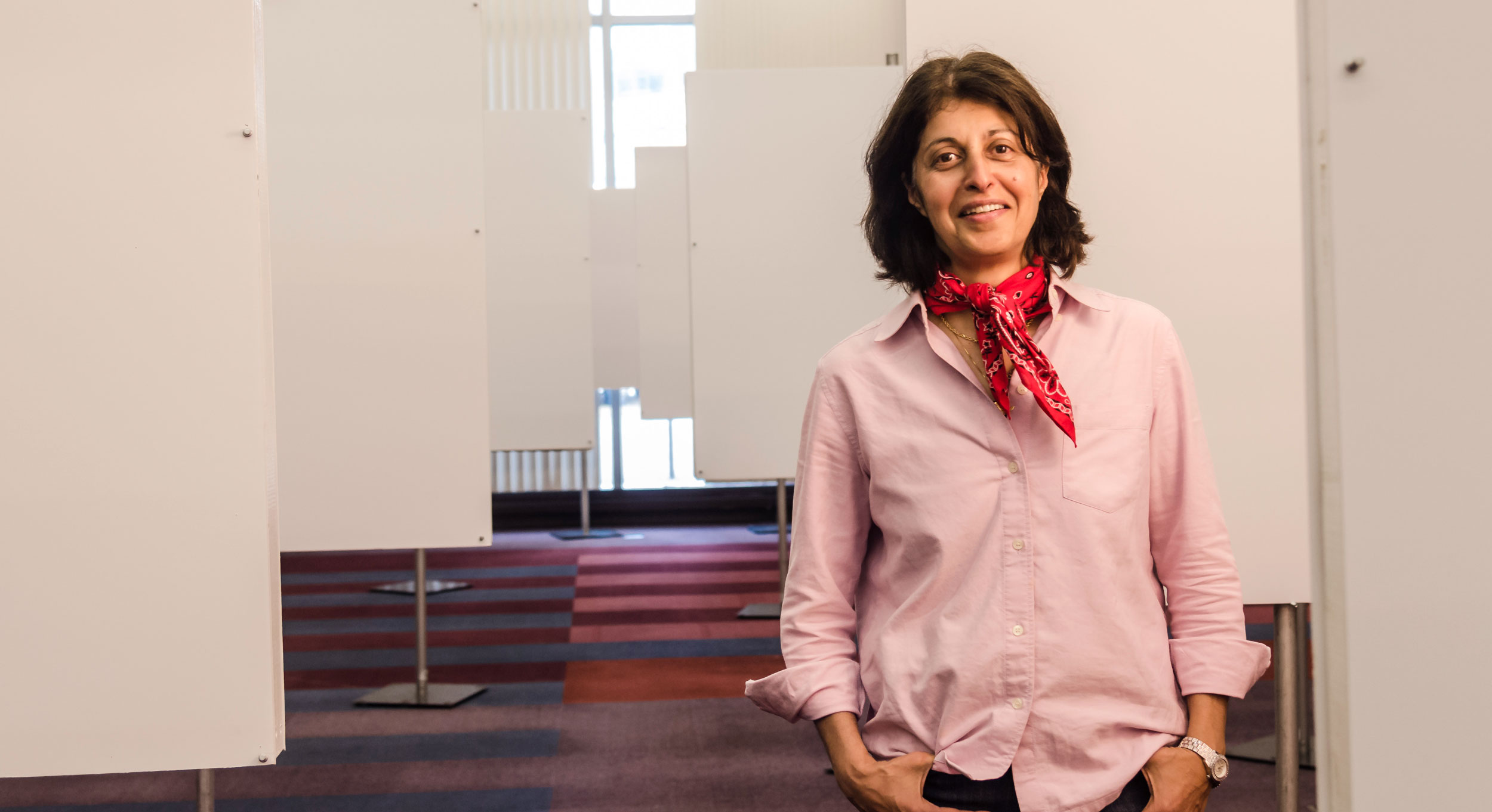
A lifelong interest in teaching brought Mandana Sassanfar to MIT, where she has established programs to engage diverse students and forged partnerships with institutes across the country.
Raleigh McElvery
May 25, 2021
Of all the offices in Building 68, Mandana Sassanfar’s is perhaps the most colorful. Her walls are lined with photos of students past and present, each of whom completed one or more of the six outreach programs she heads as the Department of Biology’s director of outreach. Over the last two decades, Sassanfar has forged partnerships with communities across the country, in an effort to engage historically underrepresented groups in science — and increase access to MIT’s on-site and online resources.
Although she was born in Switzerland, Sassanfar spent most of her childhood moving between France and Iran for her father’s job. No matter where her family lived, she always attended French-speaking schools. As early as fourth grade, she remembers analyzing her instructors’ teaching strategies, and practicing how she would explain the same concepts to make them clearer. While this interest in education continued to percolate, she also discovered that her favorite subjects were chemistry and math.
By 1983, she’d earned a master’s in biochemistry from Pierre and Marie Curie University in Paris, and moved to the US to start a PhD at Cornell University. Although she nearly switched tracks to study plant science, she ultimately stuck with biochemistry in the hopes of studying under well-known scientist Jeffery Roberts. Although Roberts was not taking new students at the time, Sassanfar convinced him to let her complete an eight-week rotation in his lab.
“I scheduled that rotation as my last, so I would have made every mistake before working with Jeff’s group,” she says. “At the end of the eight weeks, I literally told him, ‘If you don’t take me, I’m going back to France.’ And he took me in.”
While everyone else was probing various aspects of transcription antitermination, Sassanfar was an outlier investigating the role of DNA replication in the bacterial SOS repair pathway following DNA damage. She was among the first researchers to design a quantitative western blot assay to measure the level of LexA and RecA proteins in vivo. “Jeff’s lab was a wonderful place to work and I received a rigorous scientific training,” she recalls. “He was an excellent mentor.”
After graduating from Cornell in 1988, Sassanfar completed two postdocs: one with Leona Samson at the Harvard School of Public Health, and another with Jack Szostak at Massachusetts General Hospital (MGH). Szostak later went on to earn a Nobel Prize in Physiology or Medicine for discovering how chromosomes are protected by telomeres and telomerase enzymes. While Sassanfar was in his lab, she overlapped with many prominent scientists, including David Bartel, Jennifer Doudna, Rachel Green, and John Lorsch.
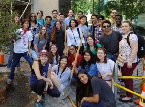
Sassanfar (back row, left) planting a tree with the 2017 MSRP-Bio cohort.
As Sassanfar’s time at MGH drew to a close, Szostak introduced her to Paul Schimmel, a long-time faculty member at the MIT Department of Biology, who was hiring research scientists for his new biotech startup, Cubist, which he had co-founded with chemistry professor Julius Rebek. The company intended to explore aminoacyl-tRNA synthetases as potential antibiotic targets. Sassanfar already knew Schimmel as the co-author of one of her favorite books, Biophysical Chemistry. But working with him for nearly four years taught her additional skills that she couldn’t have gleaned from a book.
“I came to understand a tremendous amount about the biotech culture while I was at Cubist,” she says. “Paul was a great mentor, and I learned a lot from him about writing papers, and watching the even-keeled way he interacts with people.”
When Schimmel eventually moved to The Scripps Research Institute, Sassanfar joined Harvard University’s Department of Molecular and Cellular Biology as a teaching fellow. There, professor Stephen Harrison, a Howard Hughes Medical Institute (HHIM) Investigator, offered her a chance to become involved in her first outreach program — a week-long workshop for high school teachers that she continues to run today from MIT. She was also charged with coordinating a summer program that placed non-Harvard undergraduates in campus labs each summer. But, in 2002, just a couple months before a student cohort was slated to arrive, the program was abruptly canceled and Sassanfar resigned.
“I had to transfer six undergraduates to other summer programs and find a space for the teacher’s summer workshop,” she remembers. “I just needed some lab space for two weeks.”
She called the MIT Department of Biology, and within a few days she not only had lab spaces for the teachers workshop, but a job offer as well. She accepted, and teamed up with professor Graham Walker. Together, they worked to expand the department’s pre-college and undergraduate outreach programs, creating a pipeline to graduate school in the process.
While many graduate institutions are quick to recruit students from Ivy League schools, Sassanfar saw an opportunity to widen the applicant pool. “If you decide that all the top students are from the Ivies — which is not true — then you’re missing out on many phenomenal applicants,” she says. “So I started reaching out to undergraduate institutions with limited research resources that serve diverse student bodies. Graham and I wanted to offer these students a comprehensive summer research experience, which would inspire them to apply to rigorous PhD programs like MIT Biology.”
MIT already offered some programs in this vein — such as the MIT Summer Research Program (now called “MSRP General”) — but none of them focused specifically on the life sciences. However, MSRP General was not specifically designed to be a recruiting tool for the Department of Biology. As a result, Walker and Sassanfar decided to establish the MIT Summer Research Program in Biology (MSRP-Bio), which would offer additional, biology-specific programming to help these trainees succeed and prepare them for the next stage of their careers.
Walker was the long-time program director of the HHMI Undergraduate Science Education Program at MIT, and was also named an HHMI professor the year Sassanfar arrived. He and Sassanfar used some of the accompanying funds to establish synergetic programs focused on education outreach and diversity. These included MSRP-Bio, the Quantitative Biology Workshop, the HHMI special seminar series, and a summer mini-sabbatical for faculty at institutions serving students from disadvantaged backgrounds and minority groups.
At first, Sassanfar says, she didn’t know much about the MIT Biology philosophy or the graduate program. “I spent a lot of time just talking to the grad students. And I realized that if we were going to use MSRP-Bio as a recruiting tool, then we had to set admission standards similar to those of the graduate program.”“When Mandana began at MIT, she realized that to compete for the most talented students we needed to strengthen the biology component of MIT’s summer research programs, by increasing our outreach efforts and developing an enriched summer experience,” Walker recalls. “Since then, her leadership, energy, enthusiasm, and humanity have helped MSRP-Bio develop into the strikingly successful, high-impact program that it is today.”
She began by tweaking the admissions process, raising the minimum GPA and requiring additional letters of recommendation. That first summer, Sassanfar and Walker had only a few months to prepare, so the inaugural 2003 cohort was just 11 students.
Today, the program is known as the Bernard S. and Sophie G. Gould MIT Summer Research Program in Biology (BSG-MSRP-Bio), and hosts up to 20 students. Participants perform full-time research for 10 weeks between June and August. They also attend academic seminars and weekly meetings with faculty. They visit biotech labs, take tours of Boston, learn about the grad school application process, practice their presentation skills, and share their research projects at the MSRP poster session and other conferences around the country.
In order to attract applicants from across the country, Sassanfar began traveling annually to schools with large populations of under-represented minority students, such as historically black colleges and universities; Hispanic-serving institutions; and large state schools in Texas, Florida, New York, Maryland, and Puerto Rico. She often relied on MSRP-Bio alumni to introduce her to science faculty during her campus visits.
At first it was difficult to connect with administrators and meet students. But Sassanfar slowly built sturdy relationships, and even started inviting faculty to join their students at MIT for seminars and summer sabbaticals. In 2004, the biotechnology program at the University Puerto Rico at Mayagüez honored Sassanfar with an award to celebrate her work.
“It’s really important to create opportunities that allow diverse students and faculty to benefit from MIT, rather than the other way around,” she says. “You have to show that you are doing this because you care, and not because you want something in return.”
Since 2003, over 400 students from 39 countries have participated in MSRP-Bio. Over 75% have gone on to graduate school (including 87 at MIT), 12 have become professors, and many others are leading successful careers in industry or medicine. One alumnus from the 2005 cohort, Eliezer Calo, is now a faculty member in the Department of Biology, and another from the 2007 cohort, Francisco Sánchez-Rivera, will start his own lab at the Koch Institute in 2022. Many of the MSRP-Bio alumni who complete their PhDs and postdocs at MIT stay actively engaged in outreach programs until they graduate, and help Sassanfar with many of the programs she coordinates.
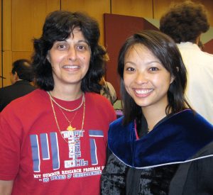
Sassanfar (left) and Lee (right) at Lee’s graduation from MIT in 2010.
Mary Lee, a member of MSRP-Bio’s inaugural cohort who later completed her PhD at MIT, says she applied to the program in hopes of experiencing cutting-edge biology research in a new city. “Mandana was an integral part of my experience in MSRP-Bio,” she explains. “From my first encounter with her to even now, 20 years later, it is clear how committed she is to connecting students like myself to MIT and the research community. It was a short summer but the experience unlocked opportunities for me that I would not have had otherwise.”
Sassanfar also serves as the director of diversity and science outreach for the Department of Brain and Cognitive Sciences, as well as the diversity coordinator for the Center for Brains, Minds and Machines. These additional roles have allowed her to expand MSRP-Bio and the Quantitative Biology Workshop, now known as the Quantitative Methods Workshop. In addition, she’s spearheaded programs for local high school students, including field trips and the LEAH Knox Scholars Program.
Beyond her outreach work, each winter during MIT’s Independent Activities Period she teaches a class for first-year MIT undergraduates to introduce them to biology lab techniques. “My favorite thing is seeing the looks on students’ faces when they have been working so hard to learn and apply techniques, and they finally can see and interpret the results of their experiments,” she says. “That’s what I love.”
Although Sassanfar has mentored hundreds of students over the past 20 years, she works hard to connect with each while they’re on campus, and has stayed in touch with many of them. She enjoys getting visits and emails from summer program alums who share their successes and thank her for the role she’s played.
“The fact that we have so many students who have finished their PhDs and gone on to become postdocs, faculty, doctors and important players in industry is, I think, truly where the success lies,” she says. “My hope is to build a strong network of alums who are excited to meet current students and create a community.”
Most recently, Sassanfar has teamed up with students, staff, and faculty from the Department of Biology to begin a new initiative, which provides research training opportunities to local community college students.
“What has really worked for me is that the Biology Department gives me free rein,” she says. “They provide their full support, and let me take it from there.”
Computer Science + Biology — and powerful insights from a Women's & Gender Studies minor
MIT School of Humanities, Arts, and Social Sciences
May 21, 2021
When graduating senior Natasha Joglekar ’21 faced some serious medical issues in the fall of 2018, she found comfort in one particular class that term: WGS.229 / Race, Culture, and Gender in the US and Beyond: A Psychological Perspective. “I think that class was sometimes the only time I talked to people all week,” she recalls.
Following a medical leave, Joglekar was able to return to MIT full-time in the fall of 2020, and soon took another class in Women’s and Gender Studies (WGS): WGS.250 HIV/AIDS in American Culture. “That’s the class that made me want to be a WGS minor,” she says. “It was nice to get a broader perspective on illness, one that was not rooted in medicine, treatment, and doctors.”
Insight into societal outcomes
A Computer Science + Biology major (Course 6-7), Joglekar found that her WGS coursework provided her with meaningful insight into the human factors that drive so many societal outcomes. “WGS studies helped give me a framework for understanding the world,” she says, “in the same way that my Physics and Math classes did.” She adds that WGS classes helped her understand myths about various minority groups, as well as the ways children are socialized to believe them.
Joglekar, who was named a Burchard Scholar in 2019 for excellence in her humanistic WGS classes, says she always knew she wanted to study the humanities, as well as the STEM fields, in college. But she didn’t choose MIT only because the Institute pairs extraordinary technical and scientific education with a world-class School of Humanities, Arts, and Social Sciences. She was also impressed by the gender parity she saw on a visit to campus.
Support for women in tech
While at high school in a Boston suburb, her techie classes were predominantly male; at MIT, she saw both men and women pursuing science, technology, and math. “You come here and see, omigod, here are all these girls doing all these cool things,” she says. “I knew I would go into a technical field, and I wanted to go to a place with a lot of women in tech and a support system for women in tech.”
One of the supportive networks Joglekar found at the Institute was the lab of Tyler Jacks, a leader in the field of cancer genetics, the David H. Koch Professor of Biology, and Director of the Koch Institute for Integrative Cancer Research. Working through MIT’s Undergraduate Research Opportunities Program (UROP), Joglekar conducted cancer research in the Jacks lab, investigating the combination therapy potential of a small molecule inhibitor on tumor heterogeneity.
“The lab was a wonderful place to learn,” she says. “They were the community I needed.”
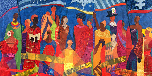
Detail, “The Ties That Bind,” artwork by Ekua Holmes; emblem for MIT Women’s & Gender Studies
Joglekar plans to work as a research assistant in a hospital, and says she expects her experience in Women and Gender Studies will help her understand patients better — and perhaps even address some of the social determinants of health.
Friendship and community
Community is of central important to Joglekar, whose family always emphasized the importance of friendship. That’s why she has spent much of her extracurricular time at MIT supporting community-building efforts. She is on the Executive Council of the Biology Undergraduate Student Association, which runs departmental study breaks and faculty dinners. She also serves on the Undergraduate Student Advisory Group for the Department of Electrical Engineering and Computer Science (EECS), which works to improve systemic issues, such as departmental communications.
The latter experience in particular gave Joglekar the chance to work directly with leaders in the EECS department. “That has been one of the highlights of my undergraduate experience,” she notes. “They’re all so good at listening and taking feedback, and they have influenced how I want to be one day if ever I’m in a leadership position.”
Leadership
In fact, Joglekar has served in several leadership roles already. In addition to her committee work, she serves as editor in chief of the MIT Undergraduate Research Journal, the Institute’s only peer-reviewed scientific journal serving the undergraduate population. And, like a good leader she is candid about her journey. “I don’t want people to think, ‘look at this person who’s flying through life.’ Far from it. I struggled at different times for different reasons,” she says. “But I’d still do it all over again!”
Joglekar is now planning to work as a research assistant in a hospital, and expects her experience in WGS will help her understand patients better — and perhaps even address some of the social determinants of health. “WGS gives you the tools to understand so many things, including underlying biases,” she says. “I think everybody should take a WGS class for this reason. it’s relevant regardless of what you do.”
People Magazine
May 20, 2021
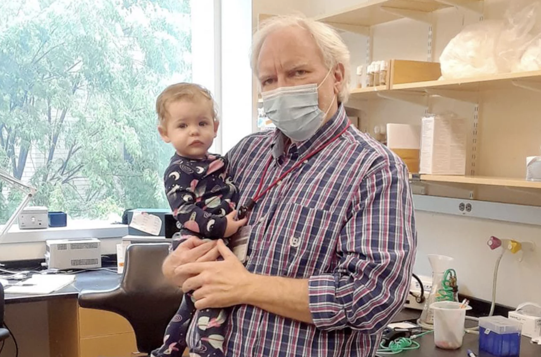
A Massachusetts professor is being praised for his kind actions after setting up a crib in his office to help a graduate student who has a 10-month-old child.
Dr. Troy Littleton, a professor of neuroscience at the Massachusetts Institute of Technology, recently went viral on Twitter after revealing that he had set up a travel crib for a grad student’s daughter.
Good Morning America identified the student as Karen Cunningham and her 10-month-old daughter, Katie.
“My favorite new equipment purchase for the lab – a travel crib to go in my office so my graduate student can bring her little girl to work when necessary and I get to play with her while her mom gets some work done. Win-win!!” Littleton tweeted on May 7 beside a photo of the crib.
That tweet has since captured the hearts of people around the world, who have commended the professor for his considerate gesture and raising awareness about the challenges working mothers face.
“This is what equity and support of women look like!” wrote one user. “This port-a-cot image has made my day. It is so NOT hard if you care to think of inclusive solutions.”
“This is beautiful!” added another person. “We need more workplaces like this that consider caretaking as a healthy & necessary part of an adult work life, not a distraction from it.”
“Usually in non-pandemic times we always have baby showers for expectant mothers and fathers where we give them gifts and we weren’t able to do that with Karen because of the pandemic, so this was sort of the lab gift for Karen, 10 months later,” he told the outlet. “It’s always a challenge [being a parent while in graduate school] so anything you can do in a lab to facilitate and help out, we try.”
Added Cunningham: “The first year of being a parent is hard and it’s helpful to have a lot of support. I think during the pandemic parents have been isolated from a lot of their support so [the crib] is definitely an add-on and a really wonderful one.”
Recently, Littleton decided to post the image of the crib after Cunningham brought her daughter to the lab for the first time, not knowing the massive response his tweet would receive, according to GMA.
“The tweet came from just being delighted to be able to see Katie for the first time and to have the opportunity on occasion, when Karen wants to bring her in, to be able to play with her a little bit,” Littleton explained to the outlet. “That was the genesis of the tweet, not from any idea it was going to create this large discussion about the challenges mothers face in the workplace.”
In response to his tweet, which has gained over 117,000 likes and 8,000 retweets, some users pointed out the difficulties that moms face with a work-life balance.
Though the response was completely unexpected, Littleton told GMA he was pleased with the productive conversations that were taking place on social media.
“I’m glad it had that effect because we need to be solving these issues, both in academia and on a broader level as well,” he shared with the outlet, noting that MIT does currently offer resources for working moms, including a daycare on its campus.
“It’s highlighted that this is a really important issue for our community,” he added.
According to Cunningham, her husband usually watches Katie each day and will continue to do so until she begins daycare in the fall. Still, she said, she appreciates having a backup option with MIT and Littleton.
“There’s the solid, focused six to eight hours of work that you wouldn’t want to bring a baby in for, but then there’s the lab errands that you do here and there and that’s when it’s really useful,” Cunningham explained to GMA. “I can put Katie down and just go do something quick and I can see her and talk to her and she can nap in there. It’s great.”
“One of the reasons I picked MIT was because I got a really positive response from the biology department when I brought up the fact that I was definitely going to want to have a baby during grad school,” she added. “I was thinking about that the whole time.”
While she’s grateful for the support from MIT and Littleton, Cunningham told GMA she hopes other schools, professors and employees will follow their lead.
“It’s really easy to look at the systemic challenges facing parents and moms in our country … and kind of throw up your hands and be like, ‘Well it’s huge. I can’t fix that,'” she explained. “But then these sort of local ways that people in positions of power can protect parents against the systemic things, like what Troy’s been doing in creating a really supportive and inclusive lab, I think that does make a really big difference and it’s great to have an example of that.”
As for Littleton, he said he doesn’t see his gesture as anything out of the ordinary and commended moms everywhere for their ability to balance both work and their personal life.
“I wish people were able to spot the real hero here,” he wrote on May 9 in response to his viral tweet. “It’s the graduate student mom, not me. She’s amazing to do all she has to with her daughter and still keep up her thesis project research. Happy Mother’s Day to all – they deserve it!”
Covid-19 class taps experts to help students and the public avoid misinformation as the crisis evolves.
Raleigh McElvery | Department of Biology
May 21, 2021
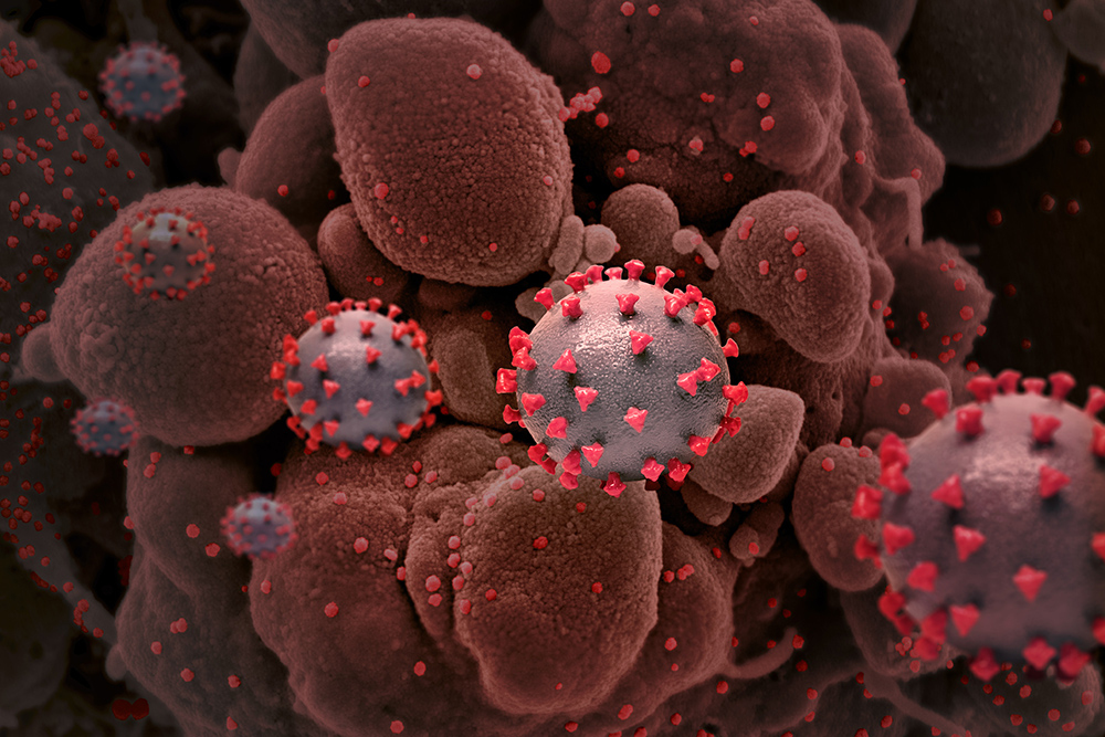
Just a few months after the Covid-19 pandemic took hold, Alan Grossman was already mulling over an idea for a new class to help people make sense of the virus. As head of MIT’s Department of Biology, he was aware of the key role fundamental research would play in the coming months. From RNA viruses and genomic sequencing to antibodies and vaccines, MIT students and the general public would need reliable scientific information to understand the evolving situation — and discern fact from fiction.
Not long after, the thoughts he’d feverishly scribbled on paper scraps scattered around his house began to take shape. With the support of the MIT School of Science, Accessibility Office, MIT Video Productions, and others around the institute, the Department of Biology added a new fall subject to the course catalog: 7.00 (Covid-19, SARS-CoV-2 and the Pandemic). Undergraduates could take the class for credit, as notable researchers stepped up to the virtual podium to share their expertise in front of a public livestream.
Grossman brought his nascent plans to associate department head and Whitehead Institute for Biomedical Research Member Peter Reddien, and together the two brainstormed individuals who might be willing to lead the class and queue speakers. They reached out to professor of biology and Whitehead Institute Member Richard Young, who served as an advisor to the World Health Organization and National Institutes of Health when a different virus of unknown origins was spreading — HIV. Young was also quick to mount a collaborative research campaign against SARS-CoV-2, the virus that causes Covid-19.
“I give Alan a lot of credit,” Young says. “He thought that it was the responsibility of the department to take the lead in filling the Covid-19 knowledge niche, and asked me if I would take this on and find a partner.”
Young contacted Ragon Institute Associate Director Facundo Batista, a world-class expert in immunology and infectious disease. Batista recalls being hesitant at first to co-lead the class; he couldn’t fathom condensing the global emergency into a single course. “But then I realized that the onslaught of information was the very reason we needed to organize this class — to help students and the public avoid misinformation,” he says. “We were filling a gap that the whole world was experiencing.”
Together, Batista and Young generated a list of 14 experts in an array of pandemic-related areas, including Anthony Fauci, director of the National Institute of Allergy and Infectious Diseases, David Baltimore of Caltech, and Kizzmekia Corbett of the National Institutes of Health. Each geared their lecture toward MIT undergraduates with a minimal biology background, and defined key terms and concepts so non-biologists watching the livestream could follow along as well.
Although Batista and Young agree that remote learning pales in comparison to in-person classes, the livestream format opened the talks up to thousands more viewers, and allowed the speakers to present their work without the need for travel. The recordings of each Tuesday lecture were posted on the Department of Biology’s website shortly thereafter, permitting asynchronous viewing for people around the world. The livestream audience regularly exceeded 1,000, and the YouTube views for each recording ranged from 4,000-97,000 and climbing. In many cases, the week’s topic fortuitously coincided with current events. For instance, Corbett spoke about vaccine development just days after the results of Pfizer-BioNTech’s first clinical trial were announced. As one of the NIH scientists who collaborated with Moderna to design another important mRNA-based vaccine, Corbett was able to discuss her reaction to the news and her expectations for Moderna’s imminent clinical trial results.
In addition to the livestream audience, each week roughly 300 MIT undergraduates would enter the Zoom room and get the opportunity to ask questions during the Q&A. Participation was unusually high, Young and Batista recall, thanks to the recitation sessions led by graduate student and teaching assistant Lena Afeyan. Afeyan would walk the students through the fundamentals of molecular biology, virology, and any other topics slated to feature heavily in the upcoming lecture. She also invited trainees and medical residents from various institutions to attend, in order to introduce students to the next generation of scientists and health-care professionals. The supplementary reading materials from these sessions are still available online, so biology teachers and other members of the public can access them.
“When I heard that this class was being put together, I hoped I could help make the content more accessible for the students and anyone else joining us,” Afeyan says. “The responses we got were overwhelming. It was incredible to hear from so many teachers, researchers, and alums across the world who watched the course every week.”
Even today, Afeyan, Young, and Batista continue to receive international kudos from scientists and non-scientists alike. At one point, Young was even interviewed by a radio station in Tasmania about the course.
“I learned a lot from 7.00 — not only about Covid-19, but about immunology and biology in general,” says Lucas Marden, a first-year undergraduate who enrolled in the class. “I particularly enjoyed the focus on the real-world response to the pandemic. We learned about everything from designing and developing different tests, treatments, and vaccines, to the scale-up of these technologies. The scientific community’s response to Covid-19 has been incredibly impressive, and I loved learning about it from the people at the forefront of their fields.”
Now, Grossman says, the department is planning to offer the class again this coming fall. “The initial idea stemmed from the need to share clear and reliable information about the pandemic as it began spreading,” he explains. “Although we’ve been living with Covid-19 for over a year now, that need is still present today — perhaps more so as we continue to learn what it will take to tame the virus.”
The next iteration of 7.00 will begin in September, and likely feature some of the same speakers and topics, along with new experts in areas that have recently emerged, such as the evolving viral variants. By arming the MIT community and the public with information from leading experts, Grossman, Batista, Young, and Afeyan hope to help the world navigate this pandemic — and prevent the next one.
Raleigh McElvery
May 19, 2021
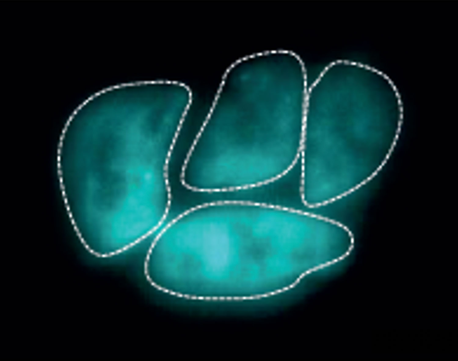
The DNA inside a single human cell is several meters long — yet it must be condensed to fit inside a space one-tenth the diameter of a hair. That’s like stretching a string from Philadelphia, Pennsylvania to Washington, D.C., and then trying to stuff it into a soccer ball. Imagine then organizing all of this information for each of the body’s 3 trillion cells! The DNA is condensed by proteins called histones that create a spool around which the DNA can wrap itself. How tightly the DNA is wound determines whether it is accessible enough for other proteins to bind to and copy into RNA, toggling gene expression levels up or down.
One specialized type of histone, H2A.Z, is ubiquitous and essential among multicellular organisms. But there have been conflicting reports about how it affects gene expression, especially during embryonic development.
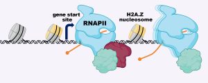
Several years ago, Laurie Boyer’s lab at MIT was the first to show that H2A.Z wraps the DNA located around the start sites of most genes, where the molecular machine RNA polymerase II (RNAPII) binds to copy the DNA into RNA. Boyer’s team demonstrated that removing H2A.Z prevented embryonic cells from turning on genes that are important for forming organs and tissues. But scientists still weren’t sure how H2A.Z exerted its effects.
Now, in a recent Nature Structural and Molecular Biology study, a team from the Boyer lab, led by former postdoc Constantine Mylonas, has revealed how H2A.Z regulates the ability of RNAPII to properly transcribe DNA into the messages that specify all cell types in the body. The researchers found that in embryonic stem cells, H2A.Z serves as a “yellow traffic light,” signaling RNAPII to slow the process of transcribing DNA into RNA. Although there are other proteins that also contribute to RNAPII pausing, H2A.Z establishes a second barrier to transcription that allows gene expression to be tuned in response to developmental signals.
“H2A.Z appears to regulate how fast RNAPII begins to transcribe DNA, and this allows the cell time to respond to important cues that ultimately direct a stem cell to become a brain or heart cell, for example,” says Boyer, a professor of biology and biological engineering. “This connection was a critical missing piece of the puzzle, and explains why H2A.Z is essential for development across all multicellular organisms.”
As RNAPII starts to transcribe a gene, it encounters a cluster of eight histones (a “nucleosome”) including H2A.z, which slows its progression — allowing for tuning of gene expression in response to developmental signals.
According to Boyer, H2A.Z’s role in gene expression has been difficult to pin down because previous approaches only provided static snapshots of how proteins interact with DNA days after loss of the histone. Boyer’s team overcame this shortcoming by leveraging a system that allowed for targeted degradation of H2A.Z within hours. They combined this technique with high-resolution genomic approaches and live cell imaging of RNAPII dynamics using super-resolution microscopy. With help from Ibrahim Cissé’s lab, they were able to visualize RNAPII dynamics in real time at the single molecule level in embryonic stem cells. Upon loss of H2A.Z, they found a remarkable increase in RNAPII movement in the cells, consistent with their genomic results showing a faster release of RNAPII and an increase in transcription in the absence of H2A.Z.
Next, the researchers plan to determine precisely how H2A.Z is targeted to the start sites of genes and how it forms a barrier to RNAPII passage.
Boyer says pinpointing the way histone variants like H2A.Z control gene expression is fundamental to understanding how developmental decisions are made, and will help researchers understand why misregulation of H2A.Z has been linked to diseases such as cancer.
“Emerging evidence indicates that DNA ‘packaging proteins’ like histones directly participate in how RNAPII can read and transcribe DNA,” she explains, “and that crucial connection wasn’t clear before.”
Image credits: courtesy of Laurie Boyer
Top image: Live cell super-resolution imaging showing RNAPII dynamics at a single molecule level in embryonic stem cells. The bright and colored clusters represent RNAPII molecules.
Citation: “A dual role for H2A. Z. 1 in modulating the dynamics of RNA Polymerase II initiation and elongation.” Nature Structural & Molecular Biology, online May 10, 2021, DOI: 10.1038/s41594-021-00589-3 Constantine Mylonas, Choongman Lee, Alexander L. Auld, Ibrahim I. Cisse, and Laurie A. Boyer.
MIT Biology junior Eduardo Canto tinkered with science long before he started studying Treacher Collins syndrome in the Calo lab.
Saima Sidik | Department of Biology
May 19, 2021
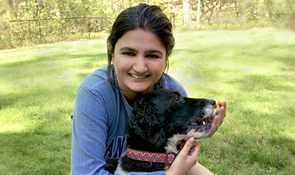
In seventh grade, Eduardo Canto wanted a dog. His mom said no, though. She didn’t want to spend her days vacuuming fur. They reached a compromise: Canto was allowed to have pet fish. Soon Canto’s disappointment with his new pets turned to curiosity. While he couldn’t train the fish to sit or roll over, he decided that breeding the fish could be a fun pastime.
An internet search told Canto that some aquarists use dried Indian almond leaves, a traditional Asian herbal remedy, to stimulate fish breeding, although no one is quite sure how the leaves do this. However, finding Indian almond leaves presented a problem for a kid without an Amazon account living far from the tree’s native habitat. On a whim, Canto picked up some similar-looking leaves in a park near his house in Puerto Rico. He knew they weren’t from an Indian almond tree, but he put them in the tank anyhow, just to see what would happen. A few days later, he noticed a collection of eggs attached to the bottom of a leaf!
Canto often took on little experiments like this, which caused his grandfather to predict early on that he would have a scientific career. Eight years after the breeding endeavor, Canto is fulfilling his grandfather’s prediction by studying Course 7 (Biology) at MIT, where he’s currently in his third year of a bachelor’s degree. Once again, fish have come into Canto’s life — he’s working in Eliezer Calo’s lab, where researchers use zebrafish to study a genetic disorder called Treacher Collins syndrome, which causes deformities in eyes, ears, cheekbones, and chins.
Throughout middle school and high school, Canto dipped his toes into many scientific disciplines. School science fairs motivated him to build a dry ice-powered trolley, a solar-powered water heater, and start a vegetable garden.
Sometimes, he admits, his motivation for joining science clubs wasn’t lofty. “I joined the math club because I got to miss a day of school every year for their annual competition,” he says with a laugh. But he also talks excitedly about his early experiments, particularly in biology. “I’ve always loved working with my hands,” he says.
Canto’s father, a medical doctor, encouraged his son’s interest by letting Canto shadow him at work. He also started a molecular biology summer program at Canto’s high school that taught students how to pipette and do simple experiments. By the time Canto applied to college, he was convinced he wanted to study biology, and MIT drew his attention because of its reputation as a top science school with excellent biology teachers. He knew it was the right choice for him when he attended Campus Preview Weekend, and found a large Puerto Rican community ready to welcome him. Even far from the island, he felt at home.
Canto has kept up with his roots since joining MIT by playing on a soccer team for Puerto Rican students. He’s also become part of a new community in a lab run by Eliezer Calo — who is a Puerto Rican himself. The lab is interested in ribosomes, the molecular machines that build proteins. Treacher Collins syndrome arises when cells can’t make ribosomes properly, and Canto wants to understand why that is.
Before Canto joined the Calo lab, the group had already started studying a protein called DDX21 that’s involved in making ribosomes in both humans and zebrafish. When genetic mutations in zebrafish cause DDX21 to go to the wrong part of the cell, the fish develop jaw deformations that mirror Treacher Collins syndrome. The Calo lab thinks cells with mislocalized DDX21 probably don’t produce ribosomes as well as normal cells, but they’re still testing this hypothesis.
Saima Sidik: Meet Eduardo Canto — a third-year undergraduate student in MIT Biology. When Canto isn’t studying, playing golf, or rooting for his favorite soccer team, Real Madrid, he works in Eliezer Calo’s lab where he’s studying a disorder called Treacher Collins syndrome. Keep listening to hear how Canto’s love of biology and a passion for helping people are shaping his career. What made you decide to apply to MIT?
Eduardo Canto: I just knew that it’s the best biology department, excellent teachers, there’s a lot of research being done here. But the reason I chose MIT, I saw that there were a lot of Puerto Ricans, and so I felt at home pretty easily. Feeling at home was definitely in the top part of my priorities.
Sidik: Are there any classes you’ve particularly enjoyed?
Canto: One of the classes that I really liked was human physiology because the teacher tried to explain how things worked, or why things worked, and that helped a lot. And also the TA helped with comical examples of the biology behind the concepts we were trying to learn.
Sidik: Cool! So what do you think you want to do after you finish your degree?
Canto: I’m hoping to go to medical school. I can learn more about the human body and use my hands to solve problems that are not easily solvable by everyone. Part of the Jesuit education I got in high school is that helping another people is one of the best things you can do.
Sidik: It sounds like you really like relieving people’s suffering
Canto: Yeah, because, I mean, I have a personal story. About a year ago, I ended up having to go to the hospital because I could barely swallow. It was very hard to talk. Eventually I got to the emergency room and the attending on the emergency room sees me, and even though I could barely talk correctly, he was trying to understand and being patient. I wanted to be that person, and to do the same thing for another person.
Sidik: If you’d like to learn more about Canto and other MIT Biology students, visit the department’s website at biology.mit.edu.
Produced by: Saima Sidik
Music:
“Heartland Flyer” — Blue Dot Sessions
“Liptis” — Blue Dot Sessions
Canto wants to probe the relationship between DDX21 and Treacher Collins syndrome further, but fish reproduce slowly, so they’re not ideal organisms for his research. Instead, he’s built a strain of Escherichia coli bacteria that carry DDX21 in place of the equivalent bacterial gene. DDX21 helps these bacteria survive the stress associated with cold temperatures, so without it, the bacteria will die in the cold. Canto hopes to take advantage of this trait by finding small molecules that stop the bacteria from growing at low temperatures — just like a DDX21 mutation would. Studying how these molecules bind DDX21 will help him understand which parts of this protein are important for its function.
The possibility that this work will one day reveal how Treacher Collins syndrome develops in patients is rewarding to Canto, and in fact he hopes helping patients will soon become his life’s focus. He wants to attend medical school, and eventually become a doctor. The human physiology class he took last semester was one of his favorites, even though it was over Zoom due to the COVID-19 pandemic. Becoming a doctor will let him help others while studying topics he finds fascinating. “Medicine is like biology on steroids!” he says.
And who knows — one day after he’s a doctor, maybe he’ll even get that pet he’s always wanted. But unlike Canto’s interest in biology, some of his interests have evolved over time. These days, he prefers cats over dogs.
Photo credit: Saima Sidik
Posted: 5.19.21
Education
PhD, 2017, MIT; MD, 2018, Harvard Medical School
Undergraduate: BS, 2010, Biology, Duke University
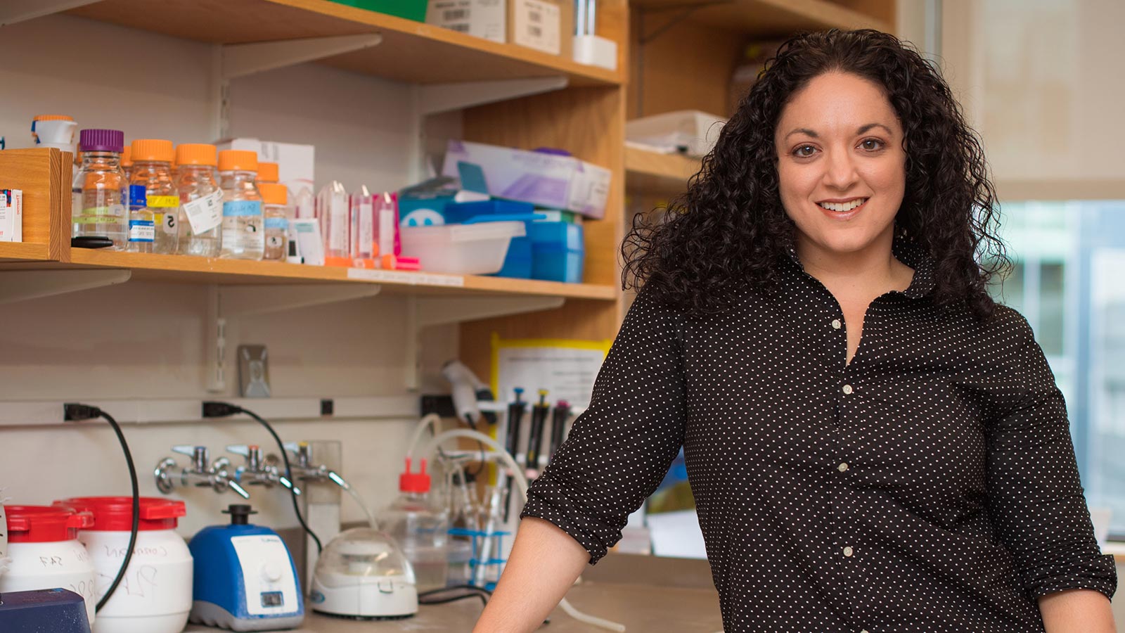
Research Summary
We aim to understand how tissues sense and respond to damage with the goal of developing novel treatments for diverse human diseases. We focus on the mammalian liver, which has the unique ability to completely regenerate itself, in order to identify the molecular requirements for effective organ repair. To this end, we innovate genetic, molecular, and cellular tools that allow us to investigate and modulate organ injury and regeneration directly within living organisms.
Awards
NIH Director’s Early Independence Award, 2018
Henry Asbury Christian Award, 2018
Education
PhD, 2015, Case Western Reserve University
BS, 2010, Biochemistry, Marquette University
Research Summary
Our lab studies genetic and epigenetic variation that contributes to human disease by disrupting gene expression programs. We utilize biological insights into the mechanisms of gene regulation in order to determine the impact of disease-associated variants on cellular function. We aim to identify actionable insights into disease pathogenesis by studying the confluence of genetic and epigenetic risk factors of human diseases, including multiple sclerosis and opioid use disorder.
Awards
NIH Director’s Pioneer Award Program Avenir Award, 2017
Merrill Meadow | Whitehead Institute
May 4, 2021
Whitehead Institute director Ruth Lehmann announced the appointment of two dynamic new Members: Olivia Corradin, currently a Whitehead Fellow, and Sinisa Hrvatin, currently an instructor and postdoctoral fellow at Harvard Medical School. Both will also become assistant professors of biology at Massachusetts Institute of Technology (MIT). Corradin’s joint appointments begin in July 2021, Hrvatin’s in January 2022.
“Both Olivia and Sinisa are creative, collaborative, and highly accomplished early-career scientists,” says Lehmann. “Each has impressed the Whitehead Institute and MIT faculties with their drive, intellect, and their scientific vision. We look forward to their contributions — as researchers, educators, and colleagues — for many years to come.”
Corradin investigates gene variants, small differences in DNA sequence, which can prompt disease-causing changes in gene regulation. During her nearly five years as a Whitehead Fellow, her lab defined the concept of “outside variants,” which helps to explain how genetic variants increase one’s likelihood of developing disease. She also developed a method to identify the cell type affected by a specific disease-linked variant; and then used it to single out oligodendrocytes as one type of brain cell involved in multiple sclerosis. Most recently, Corradin created an approach for defining epigenetic variation — which is caused by factors other than DNA sequence changes — in some individuals with opioid use disorder; this will help researchers’ identify genes associated with the disorder.
Before becoming the Scott Cook and Signe Ostby Fellow at Whitehead Institute in 2016, Corradin earned a PhD at Case Western Reserve University. There her research focused on genetic and epigenetic dysregulation in human disease, and she pioneered approaches to predict gene targets of regulatory DNA sequences associated with variants.
“I’m incredibly excited to be stepping into this new stage at Whitehead Institute and MIT Biology,” Corradin says. “I look forward to continued collaboration and to becoming a part of the rich history that shapes our Institute.”
Hrvatin investigates how organisms enter torpor and hibernation and how their cells adapt and survive in these states. As a postdoctoral research fellow in the lab of Harvard Medical School neurobiologist Michael Greenberg, Hrvatin established an experimental paradigm for studying a hibernation-like behavior in mice — and used this system to discover the neurons that control entry into this state. In addition, he pioneered the Paralleled Enhancer Single Cell Assay platform — a new method to generate cell-type-specific AAV vectors that can be used for targeted human gene therapy, as well as to control defined neuronal cell types across species, including in hibernating animals.
Hrvatin earned a PhD in stem cell and regenerative medicine from Harvard University, where he studied the process of directed differentiation from human embryonic stem cells to pancreatic beta cells. After his graduate work, he served as a postdoctoral associate at MIT in the lab of Daniel Anderson, where he investigated approaches for targeted siRNA delivery to pancreatic beta cells. Hrvatin also founded ReadCube, a startup dedicated to disseminating access to scientific literature and developing reference management tools for research scientists.
“I’ve always been inspired by the exceptional scientists, educators, pioneers, and visionaries at the Whitehead Institute and MIT Biology,” Hrvatin says. “I am absolutely thrilled for the opportunity to learn from and become a part of this extraordinary community.”