Placement: Promote to Homepage
September 16, 2021
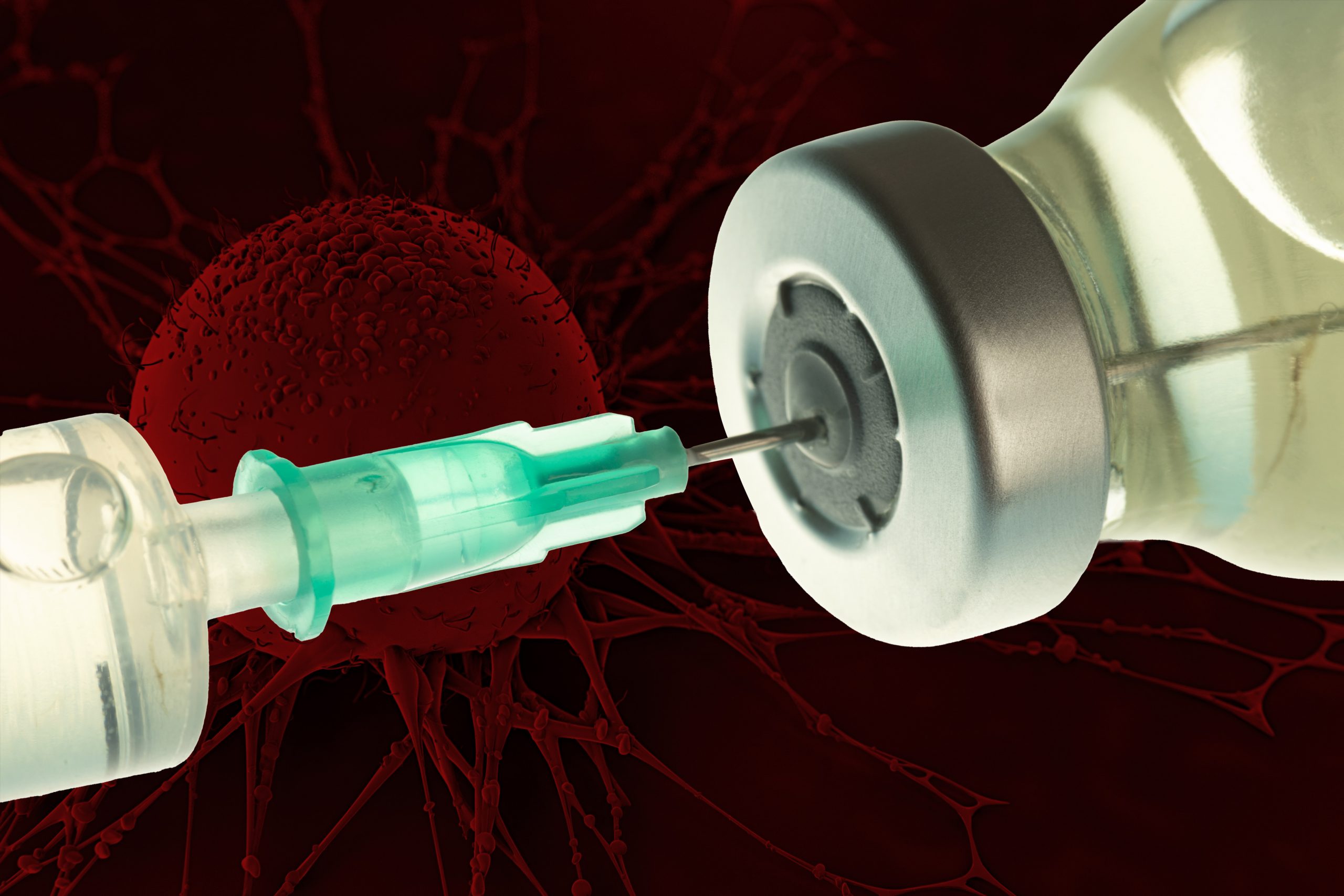
Vaccinating against certain proteins found on cancer cells could help to enhance the T cell response to tumors.
Anne Trafton | MIT News Office
September 16, 2021
Over the past decade, scientists have been exploring vaccination as a way to help fight cancer. These experimental cancer vaccines are designed to stimulate the body’s own immune system to destroy a tumor, by injecting fragments of cancer proteins found on the tumor.
So far, none of these vaccines have been approved by the FDA, but some have shown promise in clinical trials to treat melanoma and some types of lung cancer. In a new finding that may help researchers decide what proteins to include in cancer vaccines, MIT researchers have found that vaccinating against certain cancer proteins can boost the overall T cell response and help to shrink tumors in mice.
The research team found that vaccinating against the types of proteins they identified can help to reawaken dormant T cell populations that target those proteins, strengthening the overall immune response.
“This study highlights the importance of exploring the details of immune responses against cancer deeply. We can now see that not all anticancer immune responses are created equal, and that vaccination can unleash a potent response against a target that was otherwise effectively ignored,” says Tyler Jacks, the David H. Koch Professor of Biology, a member of the Koch Institute for Integrative Cancer Research, and the senior author of the study.
MIT postdoc Megan Burger is the lead author of the new study, which appears today in Cell.
T cell competition
When cells begin to turn cancerous, they start producing mutated proteins not seen in healthy cells. These cancerous proteins, also called neoantigens, can alert the body’s immune system that something has gone wrong, and T cells that recognize those neoantigens start destroying the cancerous cells.
Eventually, these T cells experience a phenomenon known as “T cell exhaustion,” which occurs when the tumor creates an immunosuppressive environment that disables the T cells, allowing the tumor to grow unchecked.
Scientists hope that cancer vaccines could help to rejuvenate those T cells and help them to attack tumors. In recent years, they have worked to develop methods for identifying neoantigens in patient tumors to incorporate into personalized cancer vaccines. Some of these vaccines have shown promise in clinical trials to treat melanoma and non-small cell lung cancer.
“These therapies work amazingly in a subset of patients, but the vast majority still don’t respond very well,” Burger says. “A lot of the research in our lab is aimed at trying to understand why that is and what we can do therapeutically to get more of those patients responding.”
Previous studies have shown that of the hundreds of neoantigens found in most tumors, only a small number generate a T cell response.
The new MIT study helps to shed light on why that is. In studies of mice with lung tumors, the researchers found that as tumor-targeting T cells arise, subsets of T cells that target different cancerous proteins compete with each other, eventually leading to the emergence of one dominant population of T cells. After these T cells become exhausted, they still remain in the environment and suppress any competing T cell populations that target different proteins found on the tumor.
However, Burger found that if she vaccinated these mice with one of the neoantigens targeted by the suppressed T cells, she could rejuvenate those T cell populations.
“If you vaccinate against antigens that have suppressed responses, you can unleash those T cell responses,” she says. “Trying to identify these suppressed responses and specifically targeting them might improve patient responses to vaccine therapies.”
Shrinking tumors
In this study, the researchers found that they had the most success when vaccinating with neoantigens that bind weakly to immune cells that are responsible for presenting the antigen to T cells. When they used one of those neoantigens to vaccinate mice with lung tumors, they found the tumors shrank by an average of 27 percent.
“The T cells proliferate more, they target the tumors better, and we see an overall decrease in lung tumor burden in our mouse model as a result of the therapy,” Burger says.
After vaccination, the T cell population included a type of cells that have the potential to continuously refuel the response, which could allow for long-term control of a tumor.
In future work, the researchers hope to test therapeutic approaches that would combine this vaccination strategy with cancer drugs called checkpoint inhibitors, which can take the brakes off exhausted T cells, stimulating them to attack tumors. Supporting that approach, the results published today also indicate that vaccination boosts the number of a specific type of T cells that have been shown to respond well to checkpoint therapies.
The research was funded by the Howard Hughes Medical Institute, the Ludwig Center at Harvard University, the National Institutes of Health, the Koch Institute Support (core) Grant from the National Cancer Institute, the Bridge Project of the Koch Institute and Dana-Farber/Harvard Cancer Center, and fellowship awards from the Jane Coffin Childs Memorial Fund for Medical Research and the Ludwig Center for Molecular Oncology at MIT.
This year’s projects address mobile evaporative vegetable preservation, portable water filtration, and dairy waste reduction.
Susanna Maize | Abdul Latif Jameel Water and Food Systems Lab
August 29, 2021
Today, the Abdul Latif Jameel Water and Food Systems Lab (J-WAFS) at the Massachusetts Institute of Technology announced the 2021 J-WAFS Solutions grant recipients. The J-WAFS Solutions program aims to propel MIT water and food-related research toward commercialization. Grant recipients receive one year of financial support, as well as mentorship, networking, and guidance from industry experts, to begin their journey into the commercial world — whether that be in the form of bringing innovative products to market or launching cutting-edge startup companies.
This year, three projects will receive funding across water, food, and agriculture spaces. The winning projects will advance nascent technologies for off-grid refrigeration, portable water filtration, and dairy waste recycling. Each provides an efficient, accessible solution to the respective challenge being addressing.
Since the start of the Solutions program in 2015, the grants have provided instrumental support in creating a number of key MIT startups that focus on major water and food challenges. A 2015-2016 Solutions grant helped the team behind Via Separations develop their business plan to massively decarbonize industrial separations processes. Other successful Solutions alumni include researchers who created a low-cost water filter made from tree branches and the team that launched the startup Xibus Systems, which is developing a handheld food safety sensor.
“New technological advances are being made at MIT every day, and J-WAFS Solutions grants provide critical resources and support for these technologies to make it to market so that they can transform our local and global water and food systems,” says J-WAFS executive director, Renee Robins. “This year’s grant recipients offer innovative tools that will provide more accessible food storage for smallholder farmers in places like Africa, safer drinking water, and a new approach to recycling food waste,” Robins notes. She adds, “J-WAFS is excited to work with these teams, and we look forward to seeing their impact on the water and food sectors.”
The J-WAFS Solutions program is implemented in collaboration with Community Jameel, the global philanthropic organization founded by MIT alumnus Mohammed Jameel, and is supported by the MIT Venture Mentoring Service and the iCorps New England Regional Innovation Node at MIT.
Read more about the 2021 J-WAFS Solutions grantee projects below.
Mobile evaporative cooling rooms for vegetable preservation
Food waste is a persistent problem across food systems supply chains, as 30-50% of food produced is lost before it reaches the table. The problem is compounded in areas without access to the refrigeration necessary to store food after it is harvested. Hot and dry climates in particular struggle to preserve food before it reaches consumers. A team led by Daniel Frey, faculty director for research at MIT D-Lab and professor of mechanical engineering, has pioneered a new approach to enable farmers to better preserve their produce and improve access to nutritious food in the community. The team includes Leon Glicksman, professor of building technology and mechanical engineering, and Eric Verploegen, a research engineer in MIT D-Lab.
Instead of relying on traditional refrigeration with high energy and cost requirements, the team is utilizing forced-air evaporative cooling chambers. Their design, based on retrofitting shipping containers, will provide a lower-cost, better performing solution enabling farmers to chill their produce without access to power. The research team was previously funded by J-WAFS through two different grants in 2019 to develop the off-grid technology in collaboration with researchers at the University of Nairobi and the Collectives for Integrated Livelihood Initiatives (CInI), Jamshedpur. Now, the cooling rooms are ready for pilot testing, which the MIT team will conduct with rural farmers in Kenya and India. The MIT team will deploy and test the storage chambers through collaborations with two Kenyan social enterprises and an NGO in Gujarat, India.
Off-grid portable ion concentration polarization desalination unit
Shrinking aquifers, polluted rivers, and increased drought is making fresh drinking water increasingly scarce, driving the need for improved desalination technologies. The water purifiers market, which was $45.0B in 2019, is expected to grow to $90.1B in 2025. However, current products on the market are limited in scope, in that they are designed to treat water that is already relatively low in salinity, and do not account for lead contamination or other technical challenges. A better solution is required to ensure access to clean and safe drinking water in the face of water shortages.
A team led by Jongyoon Han, professor of biological engineering and electrical engineering at MIT, has developed a portable desalination unit that utilizes an ion concentration polarization process. The compact and lightweight unit has the ability to remove dissolved and suspended solids from brackish water at a rate of one liter per hour, both in installed and remote field settings. The unit was featured in an award-winning video in the 2021 J-WAFS World Water Day Video Competition: MIT Research for a Water Secure Future. The team plans to develop the next-generation prototype of the desalination unit alongside a mass-production strategy and business model.
Converting dairy industry waste into food and feed ingredients
One of the trendiest foods in the last decade, Greek yogurt, has a hidden dark side: acid whey. This low-pH, liquid by-product of yogurt production has been a growing problem for producers as untreated disposal of the whey can pose environmental risks due to its high-organic content and acidic odor. With an estimated three million tons of acid whey generated in the U.S. each year, MIT researchers saw an opportunity to turn waste into a valuable resource for our food systems. Led by the Willard Henry Dow Professor in Chemical Engineering, Gregory Stephanopoulos, and Anthony J. Sinskey, professor of microbiology, the researchers are utilizing metabolic engineering to turn acid whey into carotenoids, the yellow and orange organic pigments found naturally in carrots, autumn leaves, and salmon. The team is hoping that these carotenoids can be utilized as food supplements or feed additives to make the most of what otherwise would have been wasted.
Schimmel Family Program for Life Sciences will benefit graduate students and research.
School of Science
August 30, 2021
Professor Emeritus Paul Schimmel PhD ’66 and his family recently committed $50 million to support the life sciences at MIT. They provided an initial gift of $25 million to establish the Schimmel Family Program for Life Sciences. This gift matches $25 million secured from other sources in support of the Department of Biology. The remaining $25 million from the Schimmel family will go to support the Schimmel Family Program in the form of matching funds as other gifts are secured over the next five years. Schimmel, who is the John D. and Catherine T. MacArthur Professor of Biochemistry and Biophysics Emeritus, is a lifelong supporter of the Institute in teaching, research, and philanthropy.
“I am tremendously grateful to Paul and his family for their generosity and support, and for their advocacy for our department and the life sciences,” says department head Alan D. Grossman, the Praecis Professor of Biology.
This most recent gift is one among many that Schimmel and his family have provided to MIT during their more than 50-year affiliation with the Institute, which includes Paul’s doctorate and his 30 years of teaching and research in the department. While at MIT, Paul and Cleo, Paul’s wife and philanthropic partner, provided an anonymous donation for the construction of Building 68, the most recent home for the Department of Biology.
“We cannot overstate our gratitude for our MIT experience. It was MIT that provided a ‘frontier of knowledge, which has no bounds’ and introduced us to some of the finest minds and people in the world,” Schimmel says.
“They educated and uplifted us, and convinced us of MIT’s singular role in making this a better world for all peoples,” says Cleo Schimmel, who was a past chair of the MIT Women’s League and, in her own right, contributed to the endowment of the league and other efforts to support women at MIT.
Currently, Paul Schimmel is the Ernst and Jean Hahn Professor at the Skaggs Institute for Chemical Biology at the Scripps Research Institute. Schimmel formally left MIT in 1997 to join Scripps Research, but he has remained actively involved in supporting the Institute’s research enterprise, specifically MIT graduate students.
Graduate funding for the future
Shortly after Paul left MIT, the Schimmels endowed four graduate fellowships for outstanding women in life sciences. “Since 2000, the Cleo and Paul Schimmel Scholars fellowships have helped the biology department recruit and retain the best talent,” says Grossman. Kristin Knouse PhD ’17 is a former Schimmel Scholar who rejoined the department this past July as an assistant professor.
“The MIT Department of Biology encompasses a remarkable breath of biology within a very close-knit community that places a strong emphasis on graduate training,” says Knouse. “Once in the lab, the resources and collaborations available through MIT provide unparalleled opportunities to accelerate and advance your research.”
Schimmel, who sits on the department’s Visiting Committee, continued to champion graduate student support by helping to endow the Teresa Keng Graduate Teaching Prize to support excellence in graduate student teaching in the department. In 2013, the Schimmel family donated the proceeds from the sale of their La Jolla, California, home for the purpose of training the next generation of MIT graduates in the life sciences. What formally became the department’s Graduate Training Initiative (GTI) was supported by others, including biology alumni Eric Schmidt PhD ’96 and Tracy Smith PhD ’96.
The GTI supports departmental efforts to enhance the graduate student experience in the form of both direct student support, including tuition and stipend, and indirect support, including programmatic activities such as seed funds for student-directed projects, shared computing facilities, and forums related to post-graduation employment.
This new gift to establish the Schimmel Family Program for Life Sciences will support not only the GTI in the Department of Biology, but also graduate students across MIT.
“The life sciences educational enterprise spreads across a dozen departments at MIT,” says Schimmel. “What makes the biology department and the life sciences at MIT so extraordinary is the singular ability to transfer knowledge and inventions to society for its benefit. That is much of why Kendall Square and Boston are what they are.”
To that end, Schimmel has also been an active player in shaping the MIT-Kendall Square innovation ecosystem, including the founding of companies such as Alnylam Pharmaceuticals in 2002. Alnylam — founded by Schimmel along with Institute Professor Phillip Sharp, MIT Professor David Bartel, MIT postdocs Thomas Tuschl and Phillip Zamore, and investors — has been a major player in the biopharma scene. Most recently, Alnylam partnered with Vir Biotechnology to develop therapeutics for coronavirus infections, including Covid-19.
Having a longstanding interest in the applications of basic biomedical research to human health, Schimmel holds numerous patents and is a co-founder or founding director of several biotechnology companies in addition to Alnylam, including aTyr Pharma, Alkermes, Cubist Pharmaceuticals, Metabolon, Repligen, and Sirtris Pharmaceuticals.
“I’ve been talking to the people that I’ve started companies with, reminding them that none of the extensive commercial and residential real estate development, restaurants, hotels, and the founding and locating of major biopharmaceutical enterprises would have happened without the MIT life sciences enterprise,” says Schimmel. “MIT’s Kendall Square is to biopharma what Silicon Valley is to technology. None of the robust economic impact would have occurred if it hadn’t been for MIT’s life sciences.”
The $50 million commitment was a capstone gift to MIT’s Campaign for a Better World, supporting important campaign priorities of human health and discovery science. In addition, Schimmel has future plans to continue supporting the life sciences at MIT through his estate plan with the Institute.
“We are extraordinarily grateful to Paul, Cleo, and the entire family,” says Nergis Mavalvala PhD ’97, the Curtis and Kathleen Marble Professor of Astrophysics and the dean of the MIT School of Science. “Not only do the Schimmels understand, from a firsthand perspective, the need to support graduate students, but they also understand that these young researchers are the future of our life sciences endeavors outside of MIT, in fundamental research, biopharma industries, and beyond.”
Schimmel graduated from Ohio Wesleyan University, earned a doctorate from MIT, and completed postdoc research at Stanford University. His many accomplishments include the publication of more than 500 scientific papers, numerous awards and honorary degrees, and elected membership to the American Academy of Arts and Sciences, the National Academy of Sciences, the American Philosophical Society, the Institute of Medicine (National Academy of Medicine), and National Academy of Inventors.
Education
- PhD, 2016, Stanford University School of Medicine
- BA, 2008, Molecular Biology, Princeton University
Research Summary
Our bodies are tuned to detect and respond to cues from the outside world and from within through exquisite collaborations between cells. For example, the cells lining our airways communicate with sensory neurons in response to chemical and mechanical signals, and evoke key reflexes such as coughing. This cellular collaboration protects our airways from damage and stabilizes breathing, but can become dysregulated in disease. Despite their vital importance to human health, fundamental questions about how sensory transduction is accomplished at these sites remain unsolved. We use the mammalian airways as a model system to investigate how physiological insults are detected, encoded, and addressed at essential barrier tissues — with the ultimate goal of providing new ways to treat autonomic dysfunction.
Awards
- Warren Alpert Distinguished Scholars Award, 2021
- Life Sciences Research Foundation Fellowship, 2018
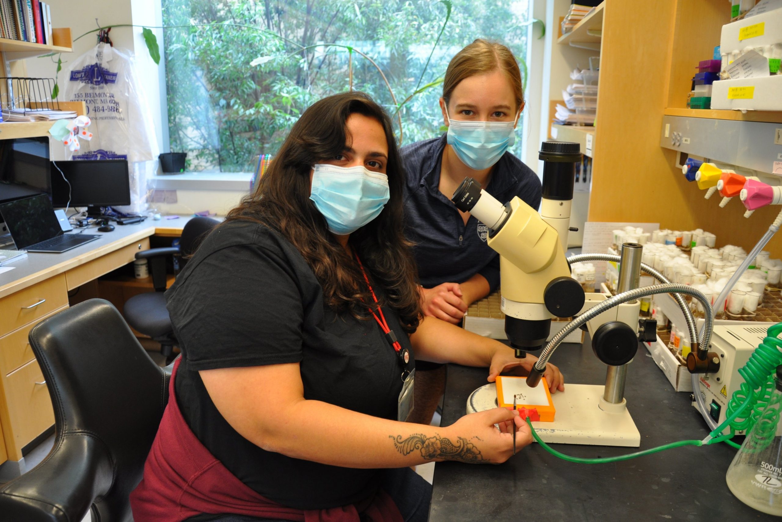
Through a summer research program at MIT, Patricia Pujols explored the neuromuscular junction, and a future in science.
Alison Gold | School of Science
August 26, 2021
Patricia Pujols grew up in the city of Ponce, Puerto Rico, fascinated by documentaries she had seen about human behavior and psychology. She wanted to learn the molecular roots of things like memory, love, hate, happiness, and anger. Despite her early curiosity, becoming a scientist and studying these phenomena didn’t seem like a possibility.
“Where I grew up, people didn’t really encourage me to study science,” she says. Instead, she initially pursued a career in accounting. “Later on, after the death of my father, I realized life is short. I prefer to do the thing that I love and am passionate about. And for me, that is teaching and learning science.”
With a strong network of mentors to inspire and push her, Pujols is now well on her way to becoming a scientist. She has a semester left in her undergraduate degree at Universidad Central de Bayamón in Puerto Rico, where she is pursuing a major in neuroscience and a minor in psychology. After she graduates, she plans to earn a PhD. This summer, she was part of the MIT Summer Research Program in Biology (MSRP-Bio), which invites non-MIT undergraduate science majors to the Institute for 10 weeks of summer research.
“MSRP-Bio is designed for students like Patricia, who are driven and passionate about science, with limited access to research at their own institution and ready for a challenging and rigorous research experience at MIT that will prepare them for graduate school and open a lot of doors,” says Mandana Sassanfar, the Department of Biology’s director of outreach. “In addition, the program greatly facilitates access to MIT faculty and graduate students and provides a strong community-building component to give students a sense of belonging.”
Pujols arrived at MIT through the guidance of one of her undergraduate professors, molecular neuroscientist Ramon Jorquera. Jorquera worked with Pujols back in Puerto Rico, and is now at the Universidad Andrés Bello in Santiago, Chile.
“He was the first person to invite me to a research lab,” Pujols says. “He has helped me a lot with everything, with gaining confidence, with my English language skills, and with seeing that I can really do this.”
Years ago, Jorquera worked as a fellow in the lab of Troy Littleton, the Menicon Professor of Biology at MIT and the Picower Institute for Learning and Memory. It was Jorquera who encouraged Pujols to apply to a research program at the University of North Carolina at Charlotte several summers ago, and then to apply to MSRP-Bio. Now, just like her mentor, Pujols is working in the Littleton lab to answer crucial questions about human behavior.
Every summer, the Littleton lab welcomes MSRP students.
“This year, while pairing candidates, Patricia was sort of an obvious match for us in terms of her prior research and interests,” Littleton says. “The major interest of my lab is to really understand how neurons talk to each other within the nervous system. The ability of neurons to rapidly communicate drives our behavior, ability to learn, and to remember. That biology all occurs at specific sites known as synapses, where neurons connect with each other.”
Problems in synapse formation or function contribute to the progression of brain disorders and diseases including Alzheimer’s, Parkinson’s, schizophrenia, and many others.
At each of the billions of synapses in the human nervous system, one neuron sends a chemical message and the next receives it –– just like two friends texting. The sender is known as the presynaptic neuron, and the receiver is called the postsynaptic neuron. To allow for seamless, rapid transit of information, the sites where the chemicals are released from on the presynaptic neuron must perfectly align with the receptors on the postsynaptic neuron.
“All of our work is built around genetics,” Littleton says. “We do manipulations where you take out a gene or alter its coding a bit and see how things change. This allows us to piece together how the individual proteins at synapses work to allow neurons to effectively talk to each other.”
To conduct their work, the Littleton lab uses Drosophila melanogaster, the common fruit fly whose genome is well-characterized and is widely used as a genetic model system. After removing a piece of genetic code, they can image the fly’s synapses to see if there was a change in the alignment of the synaptic chemical receptors. They also test if the synapses’ ability to actually transmit and receive chemical messages has changed.
This summer, Pujols is studying the neuromuscular junction, a particular type of synapse where a motor neuron communicates with a muscle cell. This communication enables movement.
In mammals, the motor neuron (the sender, in this case), secretes a protein called agrin that helps to align the key components of the synapse. Agrin is important for organizing acetylcholine receptors in the synapse. Acetylcholine is a neurotransmitter released from motor neurons that is essential for movement. Mutations in agrin in humans can therefore cause muscular dystrophies and various autoimmune disorders.
In Drosophila, it is a neurotransmitter called glutamate, not acetylcholine, that operates at the neuromuscular junction. Researchers want to know if the way that agrin organizes acetylcholine receptors in the mammalian neuromuscular junction is similar to the way that a protein called perlecan organizes the neuromuscular junctions in Drosophila.
To address this question, Pujols has spent her summer removing perlecan from either the sending motor neuron or the receiving muscle cell in Drosophila, and examining how synapse formation and clustering of glutamate receptors is altered. Pujols is working closely with PhD candidate Ellen Guss in a partnership she calls “the best experience ever.”
Both Littleton and Pujols stress the importance of mentorship in the journey to becoming a scientist. When he was an undergraduate at Louisiana State University, Littleton spent a summer at the University of Florida, working with a scientist whose guidance shaped him. That summer was one of his most influential experiences as a scientist, he says.
At MIT, Pujols says, “I stepped out of my comfort zone and strengthened my skills. MSRP gave me all the tools I needed to have an enriching experience in science, as well as the opportunity to meet colleagues that I will remember for the rest of my life.”
To other students thinking of pursuing a career as a scientist, Pujols says, “don’t be afraid.”
“You will get a lot of opinions about what to do, that it’s too difficult, or you don’t have the potential, or some other negative thing,” Pujols says. “I think the most important thing is that you do what you love, even though maybe you are going against the current. You don’t want to have regrets.”
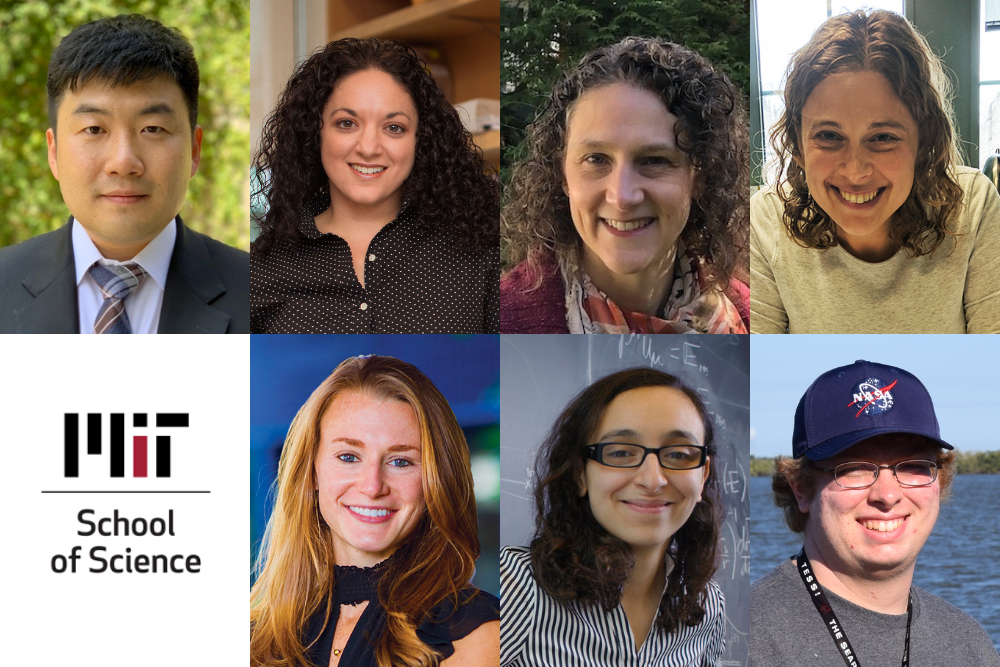
Seven professors begin in the departments of Biology; Chemistry; Earth, Atmospheric and Planetary Sciences; and Physics.
School of Science
August 25, 2021
This fall, MIT welcomes new faculty members — five assistant professors and two tenured professors — to the departments of Biology; Chemistry; Earth, Atmospheric and Planetary Sciences; and Physics.
A physicist, Soonwon Choi is interested in dynamical phenomena that occur in strongly interacting quantum many-body systems far from equilibrium and designing their applications for quantum information science. He takes a variety of interdisciplinary approaches from analytic theory and numerical computations to collaborations on experiments with controlled quantum degrees of freedom. Recently, Choi’s research has encompassed studying the phenomenon of a phase transition in the dynamics of quantum entanglement and information, drawing on machine learning to introduce a quantum convolutional neural network that can recognize quantum states associated with a one-dimensional symmetry-protected topological phase, and exploring a range of quantum applications of the nitrogen-vacancy color center of diamond.
After completing his undergraduate study in physics at Caltech in 2012, Choi received his PhD degree in physics from Harvard University in 2018. He then worked as a Miller Postdoctoral Fellow at the University of California at Berkeley before joining the Department of Physics and the Center for Theoretical Physics as an assistant professor in July 2021.
Olivia Corradin investigates how genetic variants contribute to disease. She focuses on non-coding DNA variants — changes in DNA sequence that can alter the regulation of gene expression — to gain insight into pathogenesis. With her novel outside-variant approach, Corradin’s lab singled out a type of brain cell involved in multiple sclerosis, increasing total heritability identified by three- to five-fold. A recipient of the Avenir Award through the NIH Director’s Pioneer Award Program, Corradin also scrutinizes how genetic and epigenetic variation influence susceptibility to substance abuse disorders. These critical insights into multiple sclerosis, opioid use disorder, and other diseases have the potential to improve risk assessment, diagnosis, treatment, and preventative care for patients.
Corradin completed a bachelor’s degree in biochemistry from Marquette University in 2010 and a PhD in genetics from Case Western Reserve University in 2016. A Whitehead Institute Fellow since 2016, she also became an institute member in July 2021. The Department of Biology welcomes Corradin as an assistant professor.
Arlene Fiore seeks to understand processes that control two-way interactions between air pollutants and the climate system, as well as the sensitivity of atmospheric chemistry to different chemical, physical, and biological sources and sinks at scales ranging from urban to global and daily to decadal. Combining chemistry-climate models and observations from ground, airborne, and satellite platforms, Fiore has identified global dimensions to ground-level ozone smog and particulate haze that arise from linkages with the climate system, global atmospheric composition, and the terrestrial biosphere. She also investigates regional meteorology and climate feedbacks due to aerosols versus greenhouse gases, future air pollution responses to climate change, and drivers of atmospheric oxidizing capacity. A new research direction involves using chemistry-climate model ensemble simulations to identify imprints of climate variability on observational records of trace gases in the troposphere.
After earning a bachelor’s degree and PhD from Harvard University, Fiore held a research scientist position at the Geophysical Fluid Dynamics Laboratory and was appointed as an associate professor with tenure at Columbia University in 2011. Over the last decade, she has worked with air and health management partners to develop applications of satellite and other Earth science datasets to address their emerging needs. Fiore’s honors include the American Geophysical Union (AGU) James R. Holton Junior Scientist Award, Presidential Early Career Award for Scientists and Engineers (the highest honor bestowed by the United States government on outstanding scientists and engineers in the early stages of their independent research careers), and AGU’s James B. Macelwane Medal. The Department of Earth, Atmospheric and Planetary Sciences welcomes Fiore as the first Peter H. Stone and Paola Malanotte Stone Professor.
With a background in magnetism, Danna Freedman leverages inorganic chemistry to solve problems in physics. Within this paradigm, she is creating the next generation of materials for quantum information by designing spin-based quantum bits, or qubits, based in molecules. These molecular qubits can be precisely controlled, opening the door for advances in quantum computation, sensing, and more. She also harnesses high pressure to synthesize new emergent materials, exploring the possibilities of intermetallic compounds and solid-state bonding. Among other innovations, Freedman has realized millisecond coherence times in molecular qubits, created a molecular analogue of an NV center featuring optical read-out of spin, and discovered the first iron-bismuth binary compound.
Freedman received her bachelor’s degree from Harvard University and her PhD from the University of California at Berkeley, then conducted postdoctoral research at MIT before joining the faculty at Northwestern University as an assistant professor in 2012, earning an NSF CAREER Award, the Presidential Early Career Award for Scientists and Engineers, the ACS Award in Pure Chemistry, and more. She was promoted to associate professor in 2018 and full professor with tenure in 2020. Freedman returns to MIT as the Frederick George Keyes Professor of Chemistry.
Kristin Knouse PhD ’17 aims to understand how tissues sense and respond to damage, with the goal of developing new approaches for regenerative medicine. She focuses on the mammalian liver — which has the unique ability to completely regenerate itself — to ask how organisms react to organ injury, how certain cells retain the ability to grow and divide while others do not, and what genes regulate this process. Knouse creates innovative tools, such as a genome-wide CRISPR screening within a living mouse, to examine liver regeneration from the level of a single-cell to the whole organism.
Knouse received a bachelor’s degree in biology from Duke University in 2010 and then enrolled in the Harvard and MIT MD-PhD Program, where she earned a PhD through the MIT Department of Biology in 2016 and an MD through the Harvard-MIT Program in Health Sciences and Technology in 2018. In 2018, she established her independent laboratory at the Whitehead Institute for Biomedical Research and was honored with the NIH Director’s Early Independence Award. Knouse joins the Department of Biology and the Koch Institute for Integrative Cancer Research as an assistant professor.
Lina Necib PhD ’17 is an astroparticle physicist exploring the origin of dark matter through a combination of simulations and observational data that correlate the dynamics of dark matter with that of the stars in the Milky Way. She has investigated the local dynamic structures in the solar neighborhood using the Gaia satellite, contributed to building a catalog of local accreted stars using machine learning techniques, and discovered a new stream called Nyx, after the Greek goddess of the night. Necib is interested in employing Gaia in conjunction with other spectroscopic surveys to understand the dark matter profile in the local solar neighborhood, the center of the galaxy, and in dwarf galaxies.
After obtaining a bachelor’s degree in mathematics and physics from Boston University in 2012 and a PhD in theoretical physics from MIT in 2017, Necib was a Sherman Fairchild Fellow at Caltech, a Presidential Fellow at the University of California at Irvine, and a fellow in theoretical astrophysics at Carnegie Observatories. She returns to MIT as an assistant professor in the Department of Physics and a member of the MIT Kavli Institute for Astrophysics and Space Research.
Andrew Vanderburg studies exoplanets, or planets that orbit stars other than the sun. Conducting astronomical observations from Earth as well as space, he develops cutting-edge methods to learn about planets outside of our solar system. Recently, he has leveraged machine learning to optimize searches and identify planets that were missed by previous techniques. With collaborators, he discovered the eighth planet in the Kepler-90 solar system, a Jupiter-like planet with unexpectedly close orbiting planets, and rocky bodies disintegrating near a white dwarf, providing confirmation of a theory that such stars may accumulate debris from their planetary systems.
Vanderburg received a bachelor’s degree in physics and astrophysics from the University of California at Berkeley in 2013 and a PhD in Astronomy from Harvard University in 2017. Afterward, Vanderburg moved to the University of Texas at Austin as a NASA Sagan Postdoctoral Fellow, then to the University of Wisconsin at Madison as a faculty member. He joins MIT as an assistant professor in the Department of Physics and a member of the Kavli Institute for Astrophysics and Space Research.
Eva Frederick | Whitehead Institute
August 23, 2021
More than 10 percent of our genome is made up of repetitive, seemingly nonsensical stretches of genetic material called satellite DNA that do not code for any proteins. In the past, some scientists have referred to this DNA as “genomic junk.”
Over a series of papers spanning several years, however, Whitehead Institute Member Yukiko Yamashita and colleagues have made the case that satellite DNA is not junk, but instead has an essential role in the cell: it works with cellular proteins to keep all of a cell’s individual chromosomes together in a single nucleus.
Now, in the latest installment of their work, published online July 24 in the journal Molecular Biology and Evolution, Yamashita and former postdoctoral fellow Madhav Jagannathan, currently an assistant professor at ETH Zurich, Switzerland, take these studies a step further, proposing that the system of chromosomal organization made possible by satellite DNA is one reason that organisms from different species cannot produce viable offspring.
“Seven or eight years ago when we decided we wanted to study satellite DNA, we had zero plans to study evolution,” said Yamashita, who is also a professor of biology at the Massachusetts Institute of Technology and an investigator with the Howard Hughes Medical Institute. “This is one very fun part of doing science: when you don’t have a preconceived idea, and you just follow the lead until you bump into something completely unexpected.”
The origin of species: DNA edition
Researchers have known for years that satellite DNA is highly variable between species. “If you look at the chimpanzee genome and the human genome, the protein coding regions are, like, 98 percent, 99 percent identical,” she says. “But the junk DNA part is very, very different.”
“These are about the most rapidly evolving sequences in the genome, but the prior perspective has been, ‘Well, these are junk sequences, who cares if your junk is different from mine?’” said Jagannathan.
But as they were investigating the importance of satellite DNA for fertility and survival in pure species, Yamashita and Jagannathan had their first hint that these repetitive sequences might play a role in speciation.
When the researchers deleted a protein called Prod that binds to a specific satellite DNA sequence in the fruit fly Drosophila melanogaster, the flies’ chromosomes scattered outside of the nucleus into tiny globs of cellular material called micronuclei, and the flies died. “But we realized at this point that this [piece of] satellite DNA that was bound by the Prod protein was completely missing in the nearest relatives of Drosophila melanogaster,” Jagannathan said. “It completely doesn’t exist. So that’s an interesting little problem.”
If that piece of satellite DNA was essential for survival in one species but missing from another, it could imply that the two species of flies had evolved different satellite DNA sequences for the same role over time. And since satellite DNA played a role in keeping all the chromosomes together, Yamashita and Jagannathan wondered whether these evolved differences could be one reason different species are reproductively incompatible.
“After we realized the function [of satellite DNA in the cell], the fact that satellite DNA is quite different between species really hit like lightning,” Yamashita said. “All of a sudden, it became a completely different investigation.”
A tale of two fruit fly species
To study how satellite DNA differences might underlie reproductive incompatibility, the researchers decided to focus on two branches of the fruit fly family tree: the classic lab model Drosophila melanogaster, and its closest relative, Drosophila simulans. These two species diverged from each other two to three million years ago.
Researchers can breed a Drosophila melanogaster female to a Drosophila simulans male, “but [the cross] generates very unhappy offspring,” Yamashita said. “Either they’re sterile or they die.”
Yamashita and Jagannathan bred the flies together, then studied the tissues of the offspring to see what was leading these “unhappy” hybrids to drop like flies. Right away they noticed something interesting: “When we looked at those hybrid tissues, it was very clear that their phenotype was exactly the same as if you had disrupted the satellite DNA [-mediated chromosomal organization] of a pure species,” Yamashita said. “The chromosomes were scattered, and not encapsulated in a single nucleus.”
Furthermore, the researchers could create a healthy hybrid fly by mutating certain genes in the parent flies called “hybrid incompatibility genes,” which have been shown to localize to satellite DNA in the cells of pure species. Via these experiments, the researchers were able to demonstrate how these genes affect chromosomal packaging in hybrids, and pinpoint the cellular phenotypes associated with them for the first time. “I think for me, that is probably the most critical part of this paper,” Jagannathan said.
Taken together, these findings suggest that because satellite DNA mutates relatively frequently, the proteins that bind the satellite DNA and keep chromosomes together must evolve to keep up, leading each species to develop their own “strategy” for working with the satellite DNA. When two organisms with different strategies interbreed, a clash occurs, leading the chromosomes to scatter outside of the nucleus.
In future studies, Yamashita and Jagannathan hope to put their model to the ultimate test: if they can design a protein that can bind the satellite DNA of two different species and hold the chromosomes together, they could theoretically ‘rescue’ a doomed hybrid, allowing it to survive and produce viable offspring.
This feat of bioengineering is likely years off. “Right now it’s just a pure conceptual thing,” Yamashita said. “In doing this tinkering, there’s probably a lot of specifics that will have to be solved.”
For now, the researchers plan to continue investigating the roles of satellite DNA in the cell, armed with their new knowledge of the part it plays in speciation. “To me, the surprising part of this paper is that our hypothesis was correct,” Jagannathan said. “I mean, in retrospect, there are so many ways things could have been inconsistent with what we hypothesized, so it’s kind of amazing that we’ve sort of been able to chart a clear path from start to finish.”
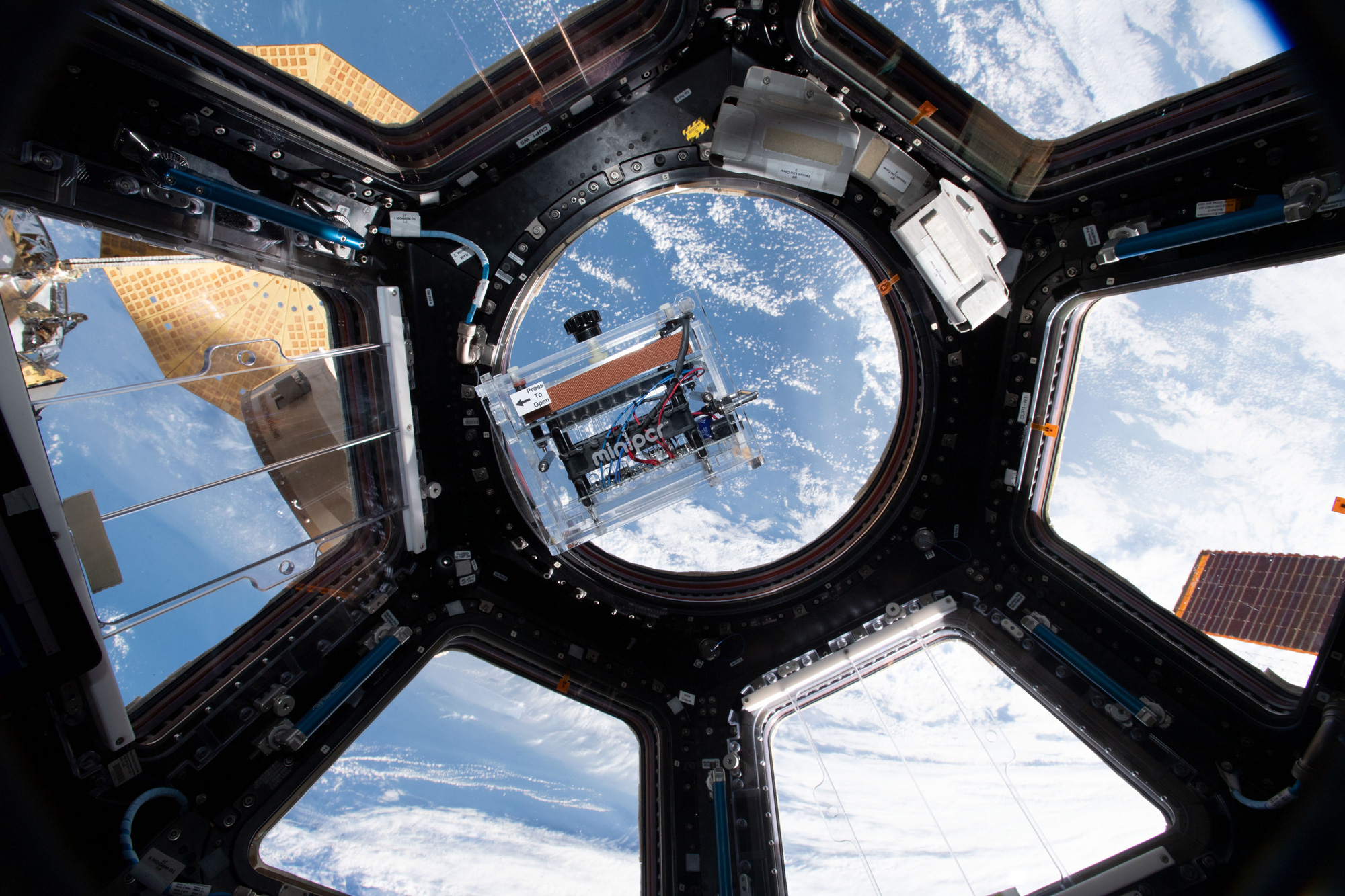
MiniPCR bio has sold thousands of its inexpensive polymerase chain reaction machines to researchers and schools around the world.
Zach Winn | MIT News Office
August 20, 2021
If you gave students around the world the power to study and manipulate genes in a test tube, what would they do with it?
MiniPCR bio first began selling its portable, inexpensive polymerase chain reaction (PCR) machines in 2013. The machines allow users to multiply specific strands of DNA in minutes, following along with experiments through a phone app.
Since then, the founders have been amazed at the amount of learning and research that has come from the devices.
Researchers have taken the machines into the Amazon rainforest, the deep oceans, and onto remote islands to do things like classify the DNA of the Ebola virus, sequence genes in endangered animals, and monitor for disease. Hundreds of thousands of students have used the machines for hands-on classroom experiments. The machines have even gone to the International Space Station as part of miniPCR bio’s Genes in Space initiative.
The space experiments are designed by middle and high school students as one of miniPCR bio’s projects in education, its main focus. To date, miniPCR bio has sold more than 20,000 of its machines to schools in 80 countries across the globe.
“I still find it shocking,” miniPCR bio’s co-founder Ezequiel Alvarez Saavedra PhD ’08 says of the company’s impact. “We get emails from teachers every week thanking us and telling us how much learning improved in the classroom because of our machine. I never would have thought this would happen.”
Making PCR mainstream
Alvarez Saavedra conducted thousands of experiments with PCR machines, which help researchers replicate specific pieces of DNA and RNA, as part of his PhD work at MIT studying the C. elegans worm. After completing his PhD in 2008, he wasn’t sure how to continue his research career, but he’d worked at MIT’s Hobby Shop in his free time and knew he liked building things, so he began working with a small engineering firm to design a simpler machine.
“I wasn’t thinking of starting a company at all,” Alvarez Saavedra says. “I just liked engineering and I was hoping to learn more about it.”
PCR machines work through a series of temperature changes. First, DNA is heated up inside the machine’s sample tubes. The heat breaks the DNA’s two strands apart. Then, during a cool down phase, molecules specifying the start and end point of the DNA that scientists want to replicate latch onto their targets. As the PCR machine heats the sample back up, an enzyme fills in the target section of DNA, matching the A nucleotides with Ts and the C nucleotides with Gs. The heat-cool-heat cycle is repeated over and over until millions of copies of the target section have been generated.
“PCR is really the workhorse of molecular biology,” Alvarez Saavedra says. “PCR lets you zoom into your region of interest — the starting material could be an entire genome or a small piece of DNA — and then do something with it. You can sequence it, for example, or you could remove a piece of it.”
Traditional PCR machines cost thousands of dollars and typically use thermoelectric cooling to change temperatures. MiniPCR’s machines, the most popular of which costs $650, use a fan and a thin-film heater, simplifying their design and making their operation far less energy-intensive.
Those changes make the machines cheap. They’re also far easier to use than their lab-based counterparts. A simple app lets users select what kind of experiment they want to run, and a temperature graph with animated depictions lets students and researchers follow along at every stage.
In 2013, Alvarez Saavedra partnered with Sebastian Kraves, a fellow Argentinian who’d earned his PhD at Harvard Medical School, to consider the best use case for the new invention. The co-founders decided to try expanding access to PCR machines for middle and high school students around the globe.
To show educators the machines for the first time, the founders attended a professional development training session for teachers at MIT.
“We showed it for 10 minutes and a teacher at the back of the room immediately said, ‘I want 10 of those,’” Alvarez Saavedra remembers. “We though okay, there’s something here.”
The founders ended up building the first 20 machines themselves, storing growing numbers of them in Ezequiel’s living room and basement until his wife suggested they find an office.
Fortunately, miniPCR bio was quickly gaining traction in the education space. Many schools buy batches of miniPCR machines for groups of students to work with directly.
“U.S. schools have been teaching PCR for years, but pretty much no one at the time had PCR machines,” Alvarez Saavedra says. “If a school did have a PCR machine, it would sit at the back of the classroom. When you’re teaching you want small groups of students doing experiments that allows each one to be more hands-on.”
As miniPCR bio’s impact on education scaled, it also gained a loyal following among researchers who appreciate the device’s low price point, efficiency, and suitability to travel to remote regions.
Researchers have run the machines off batteries charged with solar panels and done experiments without leaving the field. When one researcher was trying to sequence the Ebola virus in a makeshift lab in Sierra Leone, the miniPCR machines he’d brought to train lab technicians proved more effective than the traditional — far more expensive — PCR machines he’d brought for his work.
“It’s very nice to get reminded what you’re doing has an impact,” Alvarez Saavedra says.
PCR and beyond
Early on, the founders had the idea for students to design experiments for astronauts to run in space. The idea grew into a national competition held in partnership with Boeing that invites middle and high school students to propose pioneering DNA experiments that address challenges in space exploration. Finalist teams receive miniPCR machines for their schools, and winners get to see their experiments carried out in the International Space Station.
“Kids find space and molecular biology very exciting,” Alvarez Saavedra says.
MiniPCR has done eight missions so far. The program is just one example of the miniPCR team’s ability to keep innovating. The company also offers inexpensive systems for visualizing DNA and enzymes. It’s also developed projects for running classroom experiments using gene editing and synthetic biology. The latter project, called Biobits, was codeveloped in the lab of Jim Collins, the Termeer Professor of Medical Engineering and Science at MIT.
Biobits gives students a hands-on introduction to synthetic biology by letting them create molecular factories that churn out brightly colored proteins, functional enzymes, and more. Ally Huang, a grad student in Collins’ lab who helped develop Biobits, joined the miniPCR team to help launch the first Biobits labs and has helped scale the program to classrooms across the country.
“We try to go where the exciting science is,” Alvarez Saavedra says. “With all these programs, it’s been crazy. You put it out and you start hearing from people in all these crazy places. In the beginning, this wasn’t even supposed to be a company. But it’s incredibly simple. I guess that’s the beauty of it.”