Placement: Promote to Homepage
Researchers glean a more complete picture of a complex structure called the nuclear pore complex by studying it directly inside cells.
Raleigh McElvery | Department of Biology
October 13, 2021
Context matters. It’s true for many facets of life, including the tiny molecular machines that perform vital functions inside our cells.
Scientists often purify cellular components, such as proteins or organelles, in order to examine them individually. However, a new study published today in the journal Nature suggests that this practice can drastically alter the components in question.
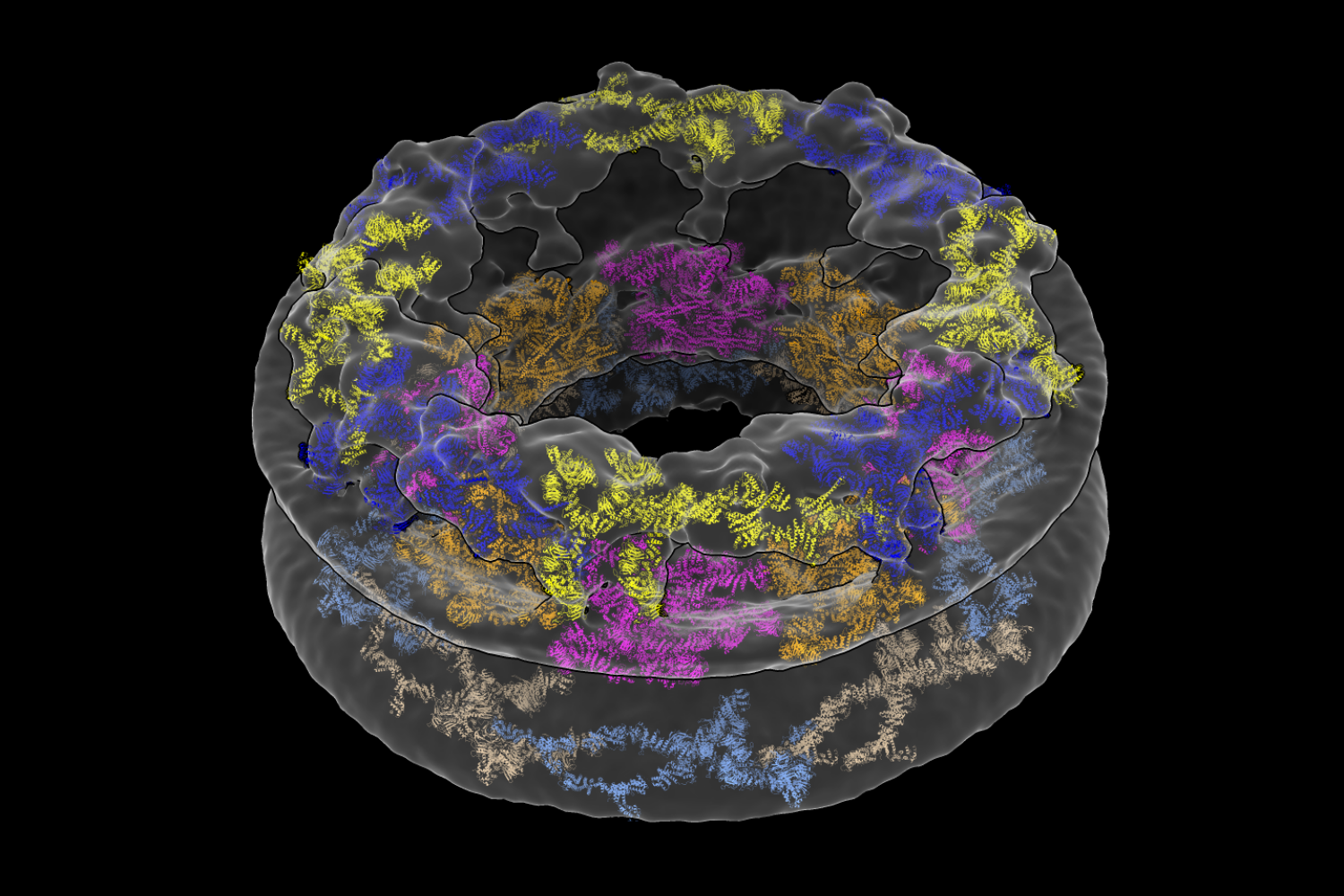
The researchers devised a method to study a large, donut-shaped structure called the nuclear pore complex (NPC) directly inside cells. Their results revealed that the pore had larger dimensions than previously thought, emphasizing the importance of analyzing complex molecules in their native environments.
“We’ve shown that the cellular environment has a significant impact on large structures like the NPC, which was something we weren’t expecting when we started,” says Thomas Schwartz, the Boris Magasanik Professor of Biology at MIT and the study’s co-senior author. “Scientists have generally thought that large molecules are stable enough to maintain their fundamental properties both inside and outside a cell, but our findings turn that assumption on its head.”
In eukaryotes like humans and animals, most of a cell’s DNA is stored in a rounded structure called the nucleus. This organelle is shielded by the nuclear envelope, a protective barrier that separates the genetic material in the nucleus from the thick fluid filling the rest of the cell. But molecules still need a way to come in and out of the nucleus in order to facilitate important processes, including gene expression. That’s where the NPC comes in. Hundreds — sometimes thousands — of these pores are embedded in the nuclear envelope, creating gateways that allow certain molecules to pass.
The study’s first author, former MIT postdoc Anthony Schuller, compares NPCs to gates at a sports stadium. “If you want to access the game inside, you have to show your ticket and go through one of these gates,” he explains.
The NPC may be tiny by human standards, but it’s one of the largest structures in the cell. It’s comprised of roughly 500 proteins, which has made its structure challenging to parse. Traditionally, scientists have broken it up into individual components to study it piecemeal using a method called X-ray crystallography. According to Schwartz, the technology required to analyze the NPC in a more natural environment has only recently become available.
Together with researchers from the University of Zurich, Schuller and Schwartz employed two cutting-edge approaches to solve the pore’s structure: cryo-focused ion beam (cryo-FIB) milling and cryo-electron tomography (cryo-ET).
An entire cell is too thick to look at under an electron microscope. But the researchers sliced frozen colon cells into thin layers using the cryo-FIB equipment housed at MIT.nano’s Center for Automated Cryogenic Electron Microscopy and the Koch Institute for Integrative Cancer Research’s Peterson (1957) Nanotechnology Materials Core Facility. In doing so, the team captured cross-sections of the cells that included NPCs, rather than simply looking at the NPCs in isolation.
“The amazing thing about this approach is that we’ve barely manipulated the cell at all,” Schwartz says. “We haven’t perturbed the cell’s internal structure. That’s the revolution.”
What the researchers saw when they looked at their microscopy images was quite different from existing descriptions of the NPC. They were surprised to find that the innermost ring structure, which forms the pore’s central channel, is much wider than previously thought. When it’s left in its natural environment, the pore opens up to 57 nanometers — resulting in a 75 percent increase in volume compared to previous estimates. The team was also able to take a closer look at how the NPC’s various components work together to define the pore’s dimensions and overall architecture.
“We’ve shown that the cellular environment impacts NPC structure, but now we have to figure out how and why,” Schuller says. Not all proteins can be purified, he adds, so the combination of cryo-ET and cryo-FIB will also be useful for examining a variety of other cellular components. “This dual approach unlocks everything.”
“The paper nicely illustrates how technical advances, in this case cryo-electron tomography on cryo-focused ion beam milled human cells, provide a fresh picture of cellular structures,” says Wolfram Antonin, a professor of biochemistry at RWTH Aachen University in Germany who was not involved in the study. The fact that the diameter of the NPC’s central transport channel is larger than previously thought hints that the pore could have impressive structural flexibility. “That may be important for the cell to adapt to increased transport demands,” Antonin explains.
Next, Schuller and Schwartz hope to understand how the size of the pore affects which molecules can pass through. For instance, scientists only recently determined that the pore was big enough to allow intact viruses like HIV into the nucleus. The same principle applies to medical treatments: only appropriately-sized drugs with specific properties will be able to access the cell’s DNA.
Schwartz is especially curious to know whether all NPCs are created equal, or if their structure differs between species or cell types.
“We’ve always manipulated cells and taken the individual components out of their native context,” he says. “Now we know this method may have much bigger consequences than we thought.”
October 8, 2021
Awards support high-risk, high-reward biomedical and behavioral research.
School of Science
October 8, 2021
On Oct. 5, the National Institutes of Health announced the names of 106 scientists who have been awarded grants through the High-Risk, High-Reward Research program to advance highly innovative biomedical and behavioral research. Seven of the recipients are MIT faculty members.
The High-Risk, High-Reward Research program catalyzes scientific discovery by supporting research proposals that, due to their inherent risk, may struggle in the traditional peer-review process despite their transformative potential. Program applicants are encouraged to pursue trailblazing ideas in any area of research relevant to the NIH’s mission to advance knowledge and enhance health.
“The science put forward by this cohort is exceptionally novel and creative and is sure to push at the boundaries of what is known,” says NIH Director Francis S. Collins. “These visionary investigators come from a wide breadth of career stages and show that groundbreaking science can happen at any career level given the right opportunity.”
New innovators
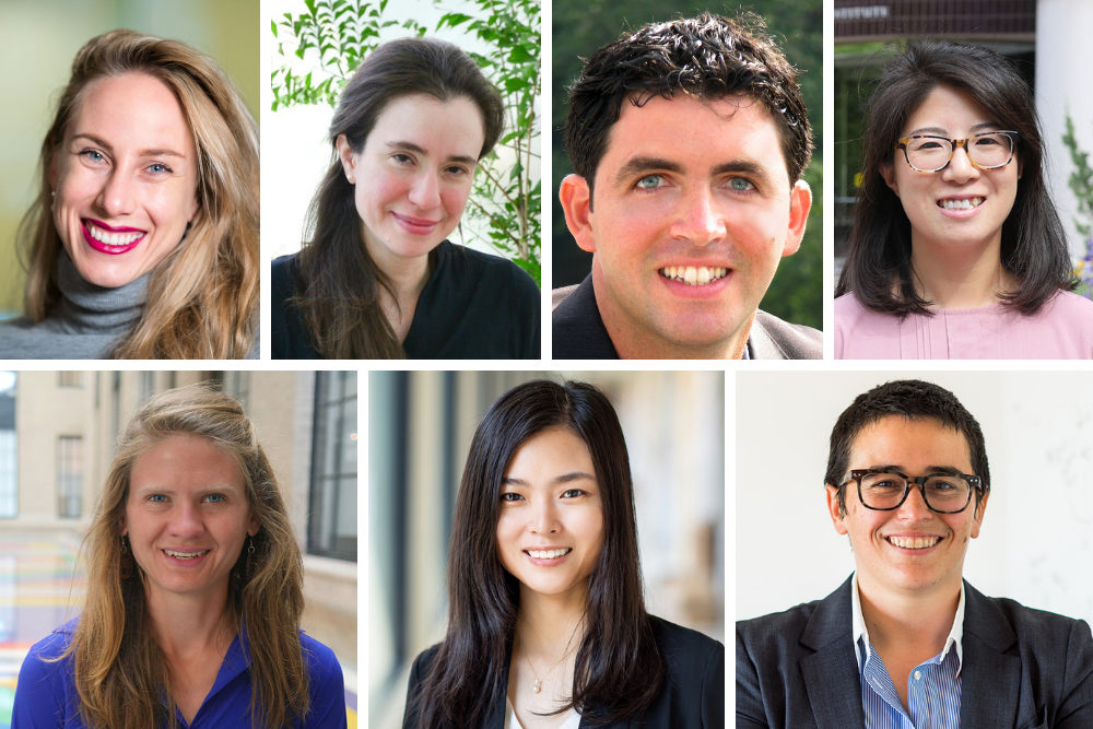
Four MIT researchers received New Innovator Awards, which recognize “unusually innovative research from early career investigators.” They are:
- Pulin Li is a member at the Whitehead Institute for Biomedical Research and an assistant professor in the Department of Biology. Li combines approaches from synthetic biology, developmental biology, biophysics and systems biology to quantitatively understand the genetic circuits underlying cell-cell communication that creates multicellular behaviors.
- Seychelle Vos, the Robert A. Swanson (1969) Career Development Professor of Life Sciences in the Department of Biology, studies the interplay of gene expression and genome organization. Her work focuses on understanding how large molecular machineries involved in genome organization and gene transcription regulate each others’ function to ultimately determine cell fate and identity.
- Xiao Wang, the Thomas D. and Virginia Cabot Assistant Professor of Chemistry and a member of the Broad Institute of MIT and Harvard, aims to develop high-resolution and highly-multiplexed molecular imaging methods across multiple scales toward understanding the physical and chemical basis of brain wiring and function.
- Alison Wendlandt is a Cecil and Ida Green Career Development Assistant Professor of Chemistry. Wendlandt focuses on the development of selective, catalytic reactions using the tools of organic and organometallic synthesis and physical organic chemistry. Mechanistic study plays a central role in the development of these new transformations.
Transformative researchers
Two MIT researchers have received Transformative Research Awards, which “promote cross-cutting, interdisciplinary approaches that could potentially create or challenge existing paradigms.” The recipients are:
- Manolis Kellis is a professor of computer science at MIT in the area of computational biology, an associate member of the Broad Institute, and a principal investigator with MIT’s Computer Science and Artificial Intelligence Laboratory. He aims to further our understanding of the human genome by computational integration of large-scale functional and comparative genomics datasets.
- Myriam Heiman is the Latham Family Career Development Associate Professor of Neuroscience in the Department of Brain and Cognitive Sciences and an investigator in the Picower Institute for Learning and Memory. Heiman studies the selective vulnerability and pathophysiology seen in two neurodegenerative diseases of the basal ganglia, Huntington’s disease, and Parkinson’s disease.
Together, Heiman, Kellis and colleagues will launch a five-year investigation to pinpoint what may be going wrong in specific brain cells and to help identify new treatment approaches for amyotrophic lateral sclerosis (ALS) and frontotemporal lobar degeneration with motor neuron disease (FTLD/MND). The project will bring together four labs, including Heiman and Kellis’ labs at MIT, to apply innovative techniques ranging from computational, genomic, and epigenomic analyses of cells from a rich sample of central nervous system tissue, to precision genetic engineering of stem cells and animal models.
Pioneering researchers
- Polina Anikeeva received a Pioneer Award, which “challenges investigators at all career levels to pursue new research directions and develop groundbreaking, high-impact approaches to a broad area of biomedical, behavioral, or social science.” Anikeeva is an MIT professor of materials science and engineering, a professor of brain and cognitive sciences, and a McGovern Institute for Brain Research associate investigator. She has established a research program that uniquely combines materials synthesis, device fabrication, neurophysiology, and animal models of behavior. Her group carries out projects that understand, invent, and design materials from the level of atoms to functional devices with applications in fundamental neuroscience.
The program is supported by the NIH Common Fund, which oversees programs that pursue major opportunities and gaps throughout the research enterprise that are of great importance to NIH and require collaboration across the agency to succeed. It issues four awards each year: the Pioneer Award, the New Innovator Award, the Transformative Research Award, and the Early Independence Award.
This year, NIH issued 10 Pioneer awards, 64 New Innovator awards, 19 Transformative Research awards (10 general, four ALS-related, and five Covid-19-related), and 13 Early Independence awards for 2021. Funding for the awards comes from the NIH Common Fund, the National Institute of General Medical Sciences, the National Institute of Mental Health, and the National Institute of Neurological Disorders and Stroke.
PhD students discuss their participation in The Poetry of Science project and the importance of bringing the arts into science communication.
Grace van Deelen | Department of Biology
October 5, 2021
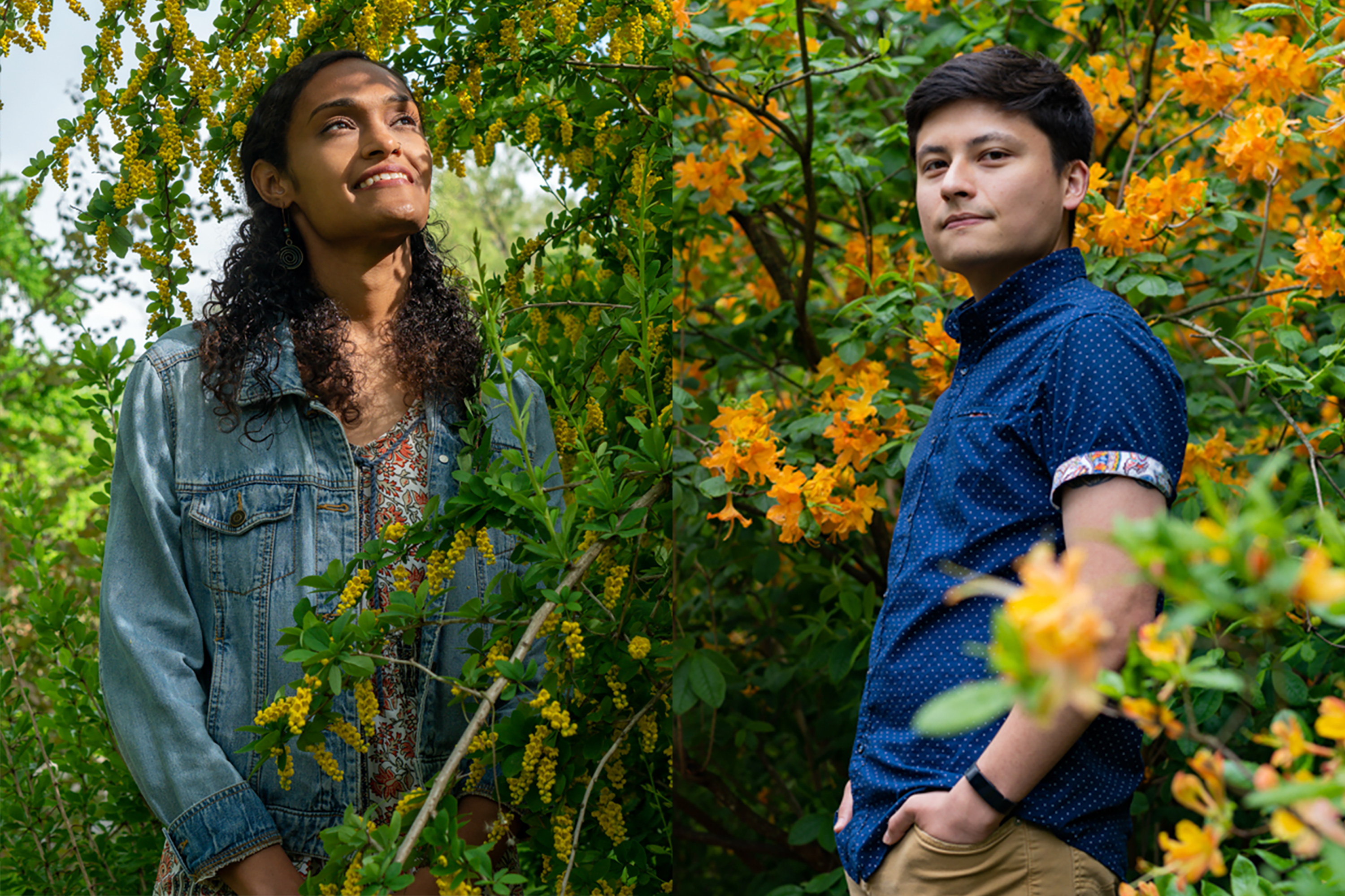
Christian Loyo of the Grossman lab and Sheena Vasquez of the Drennan lab, both graduate students in the Department of Biology, were recently selected to participate in The Poetry of Science. The project, founded by Joshua Sariñana PhD ’11, aims to advance racial justice at the intersection of science and art by bringing together Cambridge, Massachusetts-affiliated poets and scientists of color to create poems about scientific research. These poems will be on public display, along with the scientists’ portraits, at the Massachusetts General Hospital main lobby from Nov. 13 through Nov. 30 and the Rotch Library at MIT during Independent Activities Period (IAP) in January 2022.
For Loyo and Vasquez, The Poetry of Science was the ideal opportunity to create something of impact by combining their personal passions for poetry, science communication, and racial justice. They worked with two poets (Danielle Legros Georges and Luisa Fernanda Apolaya Torres, respectively) to create poems about their research. Loyo and Vasquez sat down to discuss the project.
Q: Both of you are accomplished scientists, but you also spend time working within your communities on various outreach programs. How have these experiences influenced the way you approach your scientific research or your roles as scientists?
Vasquez: I’ve conducted outreach from K-12 to the undergraduate level during my time here at MIT. My most recent outreach is targeted to local community colleges around MIT, including Bunker Hill Community College and Roxbury Community College. Outreach experiences really make me take a step back and think about how to make my science more accessible to the general public. Overall, experiences like these allow me to enhance my mentoring skills. Working with students from different backgrounds shows me how fortunate I am to conduct research at one of the top institutions in the world. If I can make it at an institute like MIT, I feel like anyone else can.
Loyo: For me, it was not easy to get into science. It took a lot of people who became my mentors to teach me what I now know about being a scientist and navigating academia. As an undergrad, I was looking for a research lab and I emailed probably 50 professors; none of them had room. I was about to give up when I finally found one professor who wanted to meet and take a chance on me. This meant a lot to me and is actually in Luisa’s poem. I ended up having a great experience and exploring research questions at a pretty high level for an undergraduate. This opportunity made me realize it’s really important to pay it forward. There are a lot of people who are having a tougher time than I ever did getting into science. Helping them ensures that the future of science is more inclusive.
Q: Science and art are often seen as two distinct disciplines, but The Poetry of Science project is all about bridging that gap. How do you think combining science and the arts can further the goal of advancing racial justice?
Loyo: I think science is about understanding the universe that we live in, and art is about understanding what it means to be human. Because we are human, we all have biases. One of those biases can be racial bias. If you look at who has historically been doing science, it’s mostly white men. That’s not because those people were the best at science; that’s because everyone else was not traditionally allowed to do science. Art gives us an opportunity to share our experiences as people from these historically excluded groups, and to highlight how we became scientists — even if, growing up, we didn’t often see scientists who looked like us.
Vasquez: The Poetry of Science exhibition also offers a chance to create new and positive representations of people of color. More examples of people of color in science helps us break down stereotypes and learn more about the individuals themselves. It allows for more stories to be told in different ways, which creates room for different perspectives. For example — and Danielle included this in her poem — something I really wish people would know is that I’m human, and, just like all scientists, I make mistakes. I am still learning and growing.
Q: What was it like to communicate your research through poetry, and how do you think the arts contribute to scientific literacy?
Vasquez: It was interesting to see what Danielle latched onto when I was explaining my research to her. For example, there’s part of the poem where she writes about how the proteins spiral, and she compares them to a girl’s curly hair. That was the alpha helix I was showing her, and it does spiral like a girl’s hair — like both of our hair. It was neat to see how she made connections between my science and general life.
The arts bring science to life, which helps improve scientific literacy. That’s important because it puts us all on the same page about what’s true and what’s not true. If we didn’t have certain scientific understandings about viruses, for example, we would not have made it this far in combating the pandemic.
Loyo: When you write a poem about science, it becomes much less about the nitty-gritty details, and instead captures the love behind the research. There’s this wonder and awe that we have for the natural world, and when we can discover something about the natural world that we didn’t know before, that feels so good. People really connect to your work when they can feel that same sort of excitement and emotion.
People also take pride in art. For example, I’m of Mexican descent, and I am a big fan of Frida Kahlo and Diego Rivera, who are Mexican painters. Using art can connect people to science even if they don’t really know what the science is about. If they can see that the person doing the experiments, for example, also grew up where they grew up, that can really be beneficial.
MIT alumnus and one other honored for their discoveries in how the nervous system senses temperature and touch.
Anne Trafton | MIT News Office
October 4, 2021
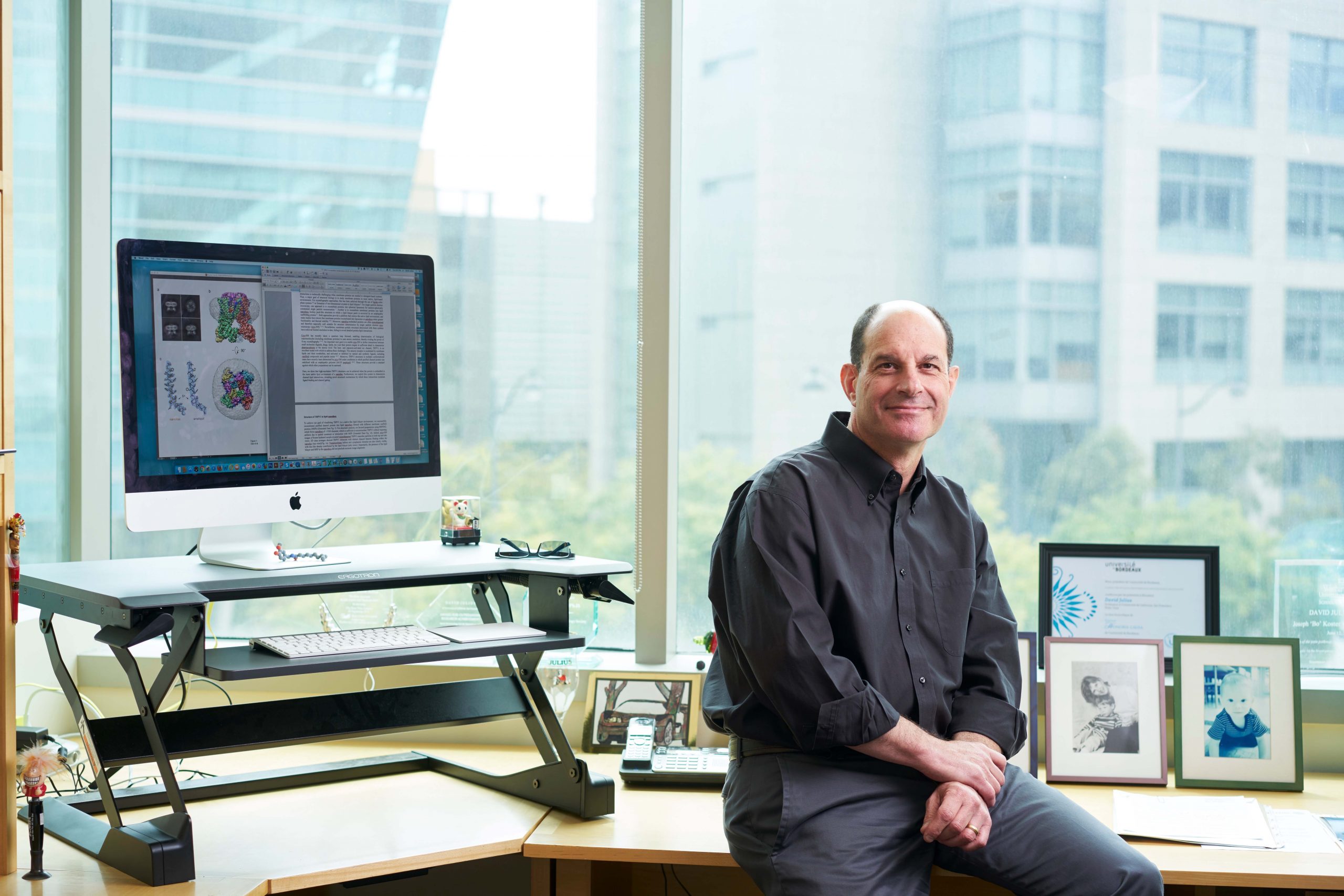
David Julius, a 1977 graduate of MIT, will share the 2021 Nobel Prize in physiology or medicine, the Royal Swedish Academy of Sciences announced this morning in Stockholm.
Julius, a professor at the University of California at San Francisco, shares the prize with Ardem Patapoutian, a professor at the Scripps Research Institute, for their discoveries in how the body senses touch and temperature.
Both scientists helped to answer a fundamental question regarding how the nervous system interprets our environment: How are temperature and mechanical stimuli converted into electrical impulses in the nervous system?
Using capsaicin, a compound that gives chili peppers their distinctive burning sensation, Julius was able to identify a receptor in the nerve endings of skin that responds to heat. His experiments revealed that this receptor, which he called TRPV1, is an ion channel that is activated by painful heat.
“David Julius’ discovery of TRPV1 was the breakthrough that allowed us to understand how differences in temperature can induce electrical signals in the nervous system,” according to today’s announcement by the Nobel committee.
Later, Julius and Patapoutian independently discovered a receptor called TRPM8, which responds to cold. Patapoutian was also honored for his discovery of receptors that respond to mechanical force in the skin and other organs. Their work on how the body senses temperature and mechanical stimuli is now being harnessed to develop treatments for a variety of diseases, including chronic pain.
Julius, who was born in New York, earned his bachelor’s degree in biology from MIT in 1977. He received a PhD in 1984 from University of California at Berkeley and was a postdoc at Columbia University before joining the faculty of the University of California at San Francisco in 1989.
He is the 39th MIT graduate to win a Nobel Prize.
October 1, 2021
How brain circuits integrate many sources of context to flexibly guide behavior
Picower Institute
September 29, 2021
Mating is instinctual for a mouse but sometimes, for instance when his potential partner smells sick, a male mouse will keep away. When Mark Hyman Jr. Career Development Associate Professor Gloria Choi and colleagues published a study in Nature in April revealing how this primal form of social distancing occurs, they provided an exquisite (and timely) example of how brain circuits factor context into behaviors, making them adaptive and appropriate even when they are innate, or “hardwired.”
When the odor of illness enters the mouse’s nose, that stimulates neurons in its vomeronasal organ to send an electrical signal through a nerve to the brain’s olfactory bulb. Cells there, Choi’s team discovered, relay the signal on to neurons in a region called the cortical amygdala that govern the mating instinct. Finally, completing the health-preserving circuit that will inhibit the mating instinct, those neurons pass on the message to brethren in the neighboring medial amygdalar nucleus. In so doing, this sequence feeds a sensory context, the female’s ill odor, into a circuit to override the default context of an internal state, the instinct to mate. The researchers even showed that by artificially stimulating cortical amygdala neurons they could prevent a mouse from mating with a healthy partner and by artificially silencing those same cells they could make a mouse mate with an ill-smelling one.
As you can learn below, the brain has much greater flexibility in how it operates than the electrical circuits that power your house or even the chips that drive your cell phone. But fundamentally it is the routing of electrical signals from neuron to neuron that forms the basis not only for how we behave, but also how we match behavior appropriately to the circumstances we encounter, Choi said.
“The closest component to behaviors and internal states, and changes in those, are still believed to be neurons and circuits,” she said.
Understanding how brain circuits produce behavior is an exciting area of neuroscience research, including in many Picower Institute labs. Their studies are helping to elucidate how the brain’s anatomy is arranged to process information, and how the many dimensions of flexibility that the central nervous system overlays upon that infrastructure can integrate context to guide appropriate behavior. Context, after all, comes from many sources in many forms—from the senses, like scents and sounds and sights; from internal states, like mating drive or hunger or sleepiness; and even from time and place and from what we’ve learned and remember.
So what were you thinking when you did “this” instead of “that”? You were thinking about the context and relying on your brain’s ability to account for it.
Chemical control
The popular “circuit” metaphor makes it easy to think of neurons as merely switches and wires that pass electrical transmissions from one point to another. And indeed they do that, although instead of being screwed and soldered to metal contacts, they use molecules called neurotransmitters to send signals across tiny junctions called synapses. But if that were all that was going on, the brain would be pretty static and it is anything but. Many members of the Institute’s faculty study how learning occurs and memories are formed when the brain changes its synapses to create or edit circuit connections, but none of that is strictly necessary for existing circuits to flexibly control behaviors that we’ve already learned or that are innate. The brain has other ways to flexibly change how it operates. Choi’s team, for instance, found that the behavioral change of inhibiting mating could not occur without the cortical amygdala neurons also sending a chemical, thyrotrophin releasing hormone (TRH), to the medial amygdalar nucleus neurons.
In the lab of Lister Brothers Associate Professor Steven Flavell, researchers study how internal states and behaviors emerge and change using a worm so simple that its complete, invariant “wiring diagram” has been completely mapped out for decades. Yet even in C. elegans, with its exact total of 302 neurons, scientists are still discovering how the animal adapts its actions to survive and thrive in a world of ever-changing contexts.
“Since 1986, that wiring diagram has been staring at researchers,” Flavell quipped. “Many of the small circuits embedded in the wiring diagram have been closely studied, while others haven’t. But a key question that we are trying to answer is how does the whole system work. How are these circuits coupled together to give rise to so-called ‘brain states’?”
In several studies Flavell has shown how a small number of neurons encode contexts and then signal that those circumstances are afoot by releasing chemicals called “neuromodulators” to many other neurons, giving rise to a brain state. Just as TRH may be doing in the circuit Choi uncovered, neuromodulators such as serotonin and dopamine, which are also ubiquitous in humans, add an extra dimension of tuning that can change, or “modulate,” how hardwired circuits process information and output behaviors, Flavell said. Neuromodulators can make neurons more or less electrically excitable given the same degree of input, Flavell explained. They can also make transmission at individual synapses more or less effective.
“The physical connections are like a roadmap, but the way that traffic is actually flowing on the road, the way that neurons are coupled to each other, is dynamic and changes with the animal’s context,” Flavell said. Neuromodulators are one way to make that happen.
For instance, in a 2019 paper in Cell, Flavell’s lab showed how a hungry worm knows to slow down and savor a patch of yummy bacteria when it finds one. A single neuron called NSM extends a little tendril called a neurite into the worm’s pharynx. Equipped with bacterial sensors (that turn out to also be present in the human intestine), the neurite detects when the worm has started to ingest and mash up its food. NSM releases serotonin, which finds its way to many of the neurons in worm’s brain that control locomotion. Upon sensing the serotonin, they hit the brakes.
In a more recent study in bioRxiv, the lab takes their investigation of neuromodulators even further. The study characterizes exactly how serotonin release from NSM modulates that activity of specific neurons in the C. elegans brain. In addition, Flavell’s group found that a neuron called AIA integrates information from sensory neurons about the smell of food. NSM can help determine what it does with that information, depending on whether it detects that the worm is eating or not. If it is, the smell of food (detected by AIA) reinforces that it should stick around to continue dining, a state maintained with serotonin. If the worm isn’t eating, the food smells signal that the animal should go exploring to find the source of that enticing odor. AIA, in that case, can instead trigger neurons that produce a different neuromodulator, called PDF, that cause the worm to start roaming (toward the food odor). Even in the simple circuitry of C. elegans, context changes how neurons interact, giving the animal flexibility to process sensory information.
That neurons capable of emitting neuromodulators can exert far-flung influence over behavior is illustrated by research in Newton Professor Mriganka Sur’s lab, too. There Sur’s team has a focus on a deeply situated, tiny brain region called the locus coeruleus (LC) that happens to supply most of the brain’s norepinephrine. Classically, neuroscientists have regarded norepinephrine from the LC as increasing the brain’s internal state of general arousal, but recent research in the Sur lab suggests it has profound, context-dependent effects on learning and behavior.
For instance, members of the lab have trained mice to expect a reward if they push a lever after hearing a high-pitched tone; the mice also receive an unexpected and irritating puff of air if they mistakenly press the lever after a low-pitched tone. By varying the loudness of the tones, the researchers can also vary the certainty the mice have about what tone they heard. Sur’s lab has found that the louder a high-pitched tone, the more norepinephrine a mouse will send to the motor cortex, which plans movement, before pushing the lever – as if greater certainty prompts it more strongly to push the lever.
Once the lever has been pushed and the mouse gets its feedback of reward or air puff, LC neurons producing norepinephrine then act to fine-tune learning by calling attention to any surprising feedback, Sur’s team has seen. For instance, if the tone was high pitched and faint, but the mouse took the risk to push the lever, the neurons will send a burst of norepinephrine to the prefrontal cortex to note that pleasant surprise. The biggest post-push surge of the neuromodulator, however, occurs when the mouse guesses wrong: that norepinephrine release to the prefrontal cortex appears to signal that the adverse result must be noted. Sure enough, Sur said, the team has seen that the mouse’s performance typically improves after making an error. The LC’s neuromodulatory actions may contribute to that behavioral improvement, though more research is needed to prove it.
Sur’s is not the only research in The Picower Institute showing that the LC communicates with the prefrontal cortex to improve task performance, though. Last November in the Proceedings of the National Academy of Sciences, Picower Professor Susumu Tonegawa’s lab showed that LC norepinephrine neurons connect via distinct circuits to two different parts of the prefrontal cortex to endow mice with both the ability to curb impulses (i.e. to not “jump the gun” when waiting to perform tasks) and to ignore distractions, such as false cues.
Rhythms among regions
Much as the Sur and Tonegawa labs have been investigating the LC, Fairchild Professor Matt Wilson’s lab studies how a different region appears to be a key hub for integrating contexts such as location, motion and memories of reward into behaviors such as navigation: the lateral septum (LS). As rats learn to find and return to the location of a reward in a maze, the lab’s extensive measurements of electrical activity among neurons in the LS shows that those cells are taking in and processing crucial contextual input from many other regions. The LS then appears to package that context to help direct the rat’s navigational plans and actions.
Over the past two years, Wilson and former graduate student Hannah Wirtshafter have published papers in Current Biology and in eLife showing that populations of LS neurons distinctively encode place information coming from the hippocampus, reward information coming from the ventral tegmental area and speed and acceleration information coming from the brainstem. The encoding is apparent in changes in the timing and rate at which the neurons “fire,” or electrically activate, in these different contexts. Some LS neurons, for example, become especially active specifically when the rat nears the reward location. In a new article published in Neuroscience and Biobehavioral Reviews in July, Wilson and Wirtshafter combined their observations with those of other labs to propose that the lateral septum packages all this contextual information into an “integrated movement value signal.”
“The lateral septum has a ton of different inputs,” Wirtshafter said. “What could the animal be doing with place-related firing that’s reward modulated and then velocity and acceleration? The answer, we think, based on where the LS outputs to, is that it is sending a signal about the context and whatever reward is part of that context. It includes what movement needs to be done and whether that movement is worth it in that context.”
While there are ample signs in the research that neuromodulators such as dopamine help the LS communicate about contexts like the feeling of reward, the studies also highlight the key role of another mechanism of flexibility: brain rhythms. Also known as brain waves or oscillations, these rhythms arise from the coordinated fluctuation of electrical activity among neurons that are working in concert. They allow neurons in brain regions to broadcast information and neurons in other regions to tune into those broadcasts, so that they can work together to perform a function, Wilson said.
“These brain dynamics ensure that whoever is sending the information and whoever is receiving the information are doing it at the same time,” Wilson said.
In fact, Picower Professor Earl Miller, who has published numerous studies on how brain rhythms guide the flow of information across the many regions of the brain’s cortex, uses much the same kind of traffic analogy in talking about the function of rhythms that Flavell uses when talking about neuromodulators. Much as those chemicals can, oscillations also flexibly direct the flow of information on the network of “roads” that physical circuit connections create. The traffic metaphor perhaps combines well with the broadcasting one: Just like drivers who tune into a radio traffic report can decide to take an alternate route when they hear about an accident ahead, neurons in a brain region may act differently when they tune into new contextual information coming in from another brain region.
Wilson and Wirtshafter’s research, for example, demonstrates that lateral septum neurons tune into the hippocampus’s broadcast of location information via a specific “theta” frequency of brain waves. In particular, movement through a place is represented by the phase (peak or trough) of the theta waves with which neurons spike.
“In the hippocampus, the phase at which a cell fires during theta can communicate information about the current, prospective, or retrospective spatial location,” Wilson and Wirtshafter wrote in their article. “For instance, …firing of individual hippocampus place cells begins on a particular phase of theta rhythm and progressively shifts forward as the animal moves through the place field.”
So maybe you are not a mouse deciding whether to mate or a rat rooting through a maze for a treat, but you are a person who has stayed out late at a friend’s house. Your internal state is that you are tired. You could head out on long drive home to the reward of your clean, warm bed, or you could sleep on your friend’s notably mustier couch and explain it your spouse the next morning. Then you remember from the drive to your friend’s place earlier, that there was an all-night rest stop along the highway where you could get coffee. Whether you decide to take the wheel or your friend’s offer of the couch will come from how a combination of neuromodulators and rhythms route information along circuits through key brain regions to integrate all this context—your internal state of tiredness, the memory of where that rest stop was, and the reward of your bed (or the punishment of an angry spouse who might ask “What were you thinking?”). Your brain gives you all the flexibility you need.
The PhD candidate studies the human microbiome and its proteins, while also championing the Latinx community on MIT’s campus.
Hannah Meiseles | MIT News Office
September 28, 2021
Raised in Tampa, Florida, Lindsey Backman takes pride in her family’s history and its role in the vibrant Cuban American community there. She remembers the weekends she would spend as a kid, getting café con leche with her grandparents and dancing in the studio with her friends. The cultural experiences she shared with friends, family, and neighbors growing up helped her feel comfortable being herself while growing up, and showed her from an early age how valuable a welcoming community could be to a person’s success.
Backman went on to pursue her BS in chemistry at the University of Florida. Surrounded by a diverse community, she felt supported as she leaned deeper into her interest in science. She was soon nominated to a program that matched students from underrepresented backgrounds in STEM with a university professor to pursue a summer research project. Although Backman was still uncertain about going to another university to do lab research, with encouragement from her department she gave the program a chance.
Backman matched with Professor Catherine Drennan at MIT to work on visualizing structural biology and took part in the MIT Summer Research Program in Biology (MSRP-Bio). The research clicked with her immediately and became a turning point; Backman returned to participate in the lab the following summer and then applied to graduate school.
“Getting nominated to the program changed my life. I certainly wouldn’t have applied to MIT otherwise,” says Backman. “At first, I was convinced I wouldn’t fit in, but soon found myself surrounded by people as passionate about science as I was. I knew I was in the right place.”
Uncovering secrets about the human microbiome
Today, Backman is a graduate student in the Drennan lab and researches the chemistry of the human microbiome, a collection of gut microbes essential to sustaining the body. Backman is interested in how certain bacteria can outcompete other strains by producing unique proteins that process abundant nutrients or repair broken enzymes. Her use of X-ray crystallography has helped her produce atomic models that shed light on the structure of these proteins.
One type of protein Backman and her team have characterized is called a spare part protein. When produced, this protein can help restore a broken enzyme’s ability to catalyze essential reactions. “When fixing a car with a flat tire, you would replace the tire and not the whole car. A similar strategy is being used here. These spare-part proteins act to bind and restore the activity of the enzyme completely,” she says.
Over the years, Backman has seen the depth of questions surrounding the microbiome grow. Scientists have begun to recognize how important the microbiome is to human health. “Ever since my first summer research experience at MIT, I’ve been dedicated to studying this one unique repair mechanism,” says Backman. “We’ve gone from solving the structure of the proteins to now understanding how the mechanism works. But there’s still so much more to learn — we have started to suspect these repair mechanisms speak to a broader motif in other enzymes as well.”
Backman and her team have also been leaders in characterizing how an important enzyme, called hydroxy-L-proline dehydratase (HypD), performs its unusual chemistry. This abundant enzyme takes hydroxyproline, a common nutrient in the gut, and can obtain a competitive advantage by using it as a nutrient and source of energy.
“Only a unique subset of bacteria can process hydroxyproline. On the clinical side, we have seen during infection that virulent bacteria with this ability, such as C. difficile, will start rapidly consuming hydroxyproline to proliferate,” says Backman. “Conversely, we could one day create antibiotics that specifically inhibit HypD without killing our beneficial bacteria.”
Encouraging the future of science
Outside of her research, Backman cares deeply about serving and being a part of the Latinx community on campus. She helped co-found the MIT Latinx Graduate Student Association and has served for four years as a graduate resident assistant for La Casa, the Latinx undergraduate living community at New House. “La Casa is a really tight-knit and familial community,” says Backman. “Some of our original freshmen are now seniors, so it’s been really rewarding to see their whole transition throughout college. I love getting to watch students explore and come to realize what they’re passionate about.”
Backman has also been instrumental in spurring equity initiatives on campus. She is currently a student representative for the MIT Department of Chemistry Diversity, Equity, and Inclusion Committee and has worked to implement programs that support the success of underrepresented groups on campus. Her five years of service as an MIT Chemistry Access Program mentor have encouraged many underrepresented undergraduate students to pursue chemistry graduate programs. For all her hard work at improving MIT’s campus, Backman recently received the Hugh Hampton Young Fellowship.
In the future, Backman aspires to continue researching the microbiome and mentoring students by becoming a professor. She hopes to continue the cycle and inspire more young scientists to recognize their inner potential. “I was never one of those kids that knew I wanted to be a scientist someday. My PI completely changed my life, and I would not be at MIT today without her,” she says. “Having mentors that believe in you at critical points in your life can make all the difference.”
“I think there’s this wrong assumption that diversity initiative work takes away from time that could be spent doing science. In my mind, we need to recognize how these things go hand in hand,” says Backman.
“The only way we’re going to get the best scientists is by creating a healthier, more diverse environment where people of all backgrounds feel welcomed. It’s only when people feel comfortable that they can make their greatest contributions to the field.”