Producing multimedia for online courses involves lifelong learning
Darcy G. Gordon, Instructor of Blended and Online Learning Initiatives, MITx Digital Learning Scientist, Biology
May 11, 2022
As a biologist who has made my own figures for publications, I’ve always appreciated well-constructed, easy-to-understand, and scientifically accurate visualizations, but admittedly those appreciations were often fleeting and superficial. I did not fully realize the incredible detail and thought that are required for making effective scientific visualizations used for teaching. Once I joined the MITx Biology team and became the lead on making visual resources for our online course offerings (most notably 7.05x Biochemistry and the 7.06x Cell Biology series, parts 1, 2, and 3), I came to understand visual representations of biological phenomena in a new way.
From cellular morphology captured in the stunning microscopy images at the Koch Institute to banding patterns in polyacrylamide gel electrophoresis, biologists often make sense of structural and functional relationships through the use of visual tools, and an often implicit part of biology education includes teaching visual scientific literacy. The cognitive effort behind interpreting visual representations one-by-one, let alone those that transition between different levels of biological organization or incorporate different kinds of representations of the same concept, is not trivial. In an intensely visual science such as biology, how can I make the connections between representations and concepts more seamless and intuitive for learners?
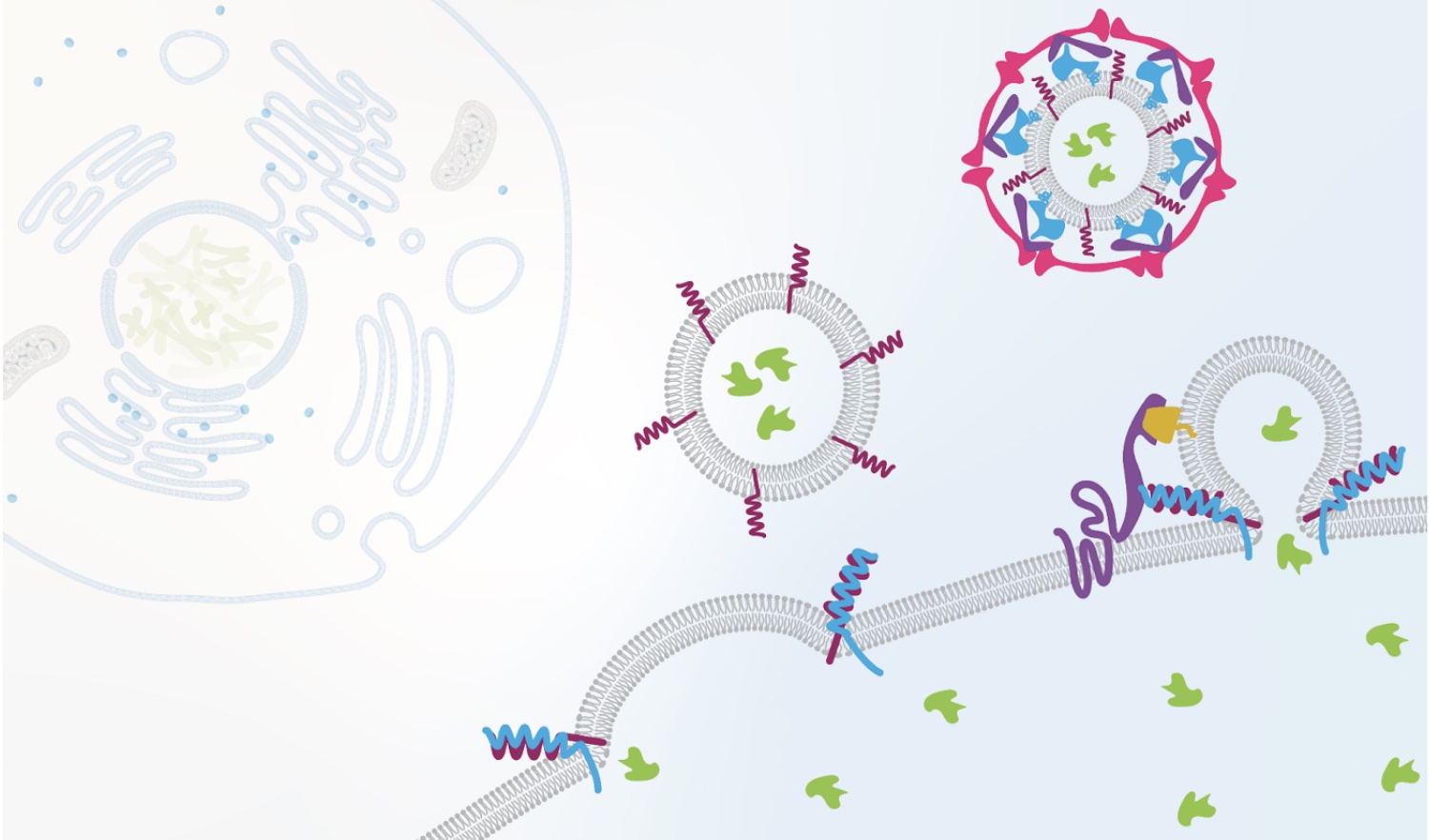
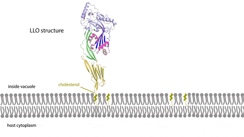
Combining different types of representations to illustrate how the protein, Listeriolysin O (LLO), forms a pore that allows the pathogen, Listeria monocytogenes, to establish infection.
Learning multimedia tools and theory
I became engrossed in learning how to use new tools that would help me make abstract concepts come to life, like modeling 3D protein structures and animating cellular processes. As I learned the techniques and skills necessary to represent subcellular biological processes, I also studied the rich body of scholarly work that addresses what makes visual representations compelling and accessible. Applications of how we use this multimedia learning theory and other evidence-based practices in our courses is better summarized elsewhere, but I found these ideas immensely helpful in creating visual resources for worldwide learners.
Gifs can help teach structural relationships in biochemistry. A 3D model of the enzyme, subtilisin (yellow), with an inhibitor, eglin C (blue). The active site residues are shown in red and calcium ion cofactors are in purple. PDB: 1SIB
Doing the work
Soon I was designing color palettes that maintained accessibility and cued learners to relationships between representations, simplifying schematics to convey essential processes and reduce extraneous details, and finding ways to emphasize important features through visually intuitive signaling. I spent hundreds of hours per course producing this content; drawing and animating biochemical reactions, editing the instructional videos captured during class, and revising the resulting media based on feedback.
Teaching as a way of understanding
Through the process of creating a couple thousand images, animations, and videos for MITx Biology, I have not only learned the technical and theoretical bases for multimedia design, but my understanding of core biological concepts has deepened. Updating my understanding of biological processes and conveying that understanding to others through multimedia, as well as sharing emerging best practices in teaching with technology, illustrates the iterative cycle of learning, doing, and teaching. Teaching others, whether it be about cellular signaling pathways or how to illustrate these pathways, has always been the best benchmark of where I am in my own learning journey.
The iterative cycle of learning, doing, and teaching is central to this work.
A career in learning engineering bridges my academic training in biology, instructional expertise, and technological skills to make experiences in the classroom, whether it be virtual or in-person, meaningful for all. I also see this profession as an ongoing meditation on lifelong learning. Teaching worldwide learners about biology, or my peers about best practices in educational multimedia is a dynamic process. New information and technology change the way we understand how people learn and how cells function, and I must integrate these changes into my work to create engaging learning experiences for global audiences. Through interactions with the same material, albeit on different sides of a learning management system, I also enjoy a philosophical kinship with our MITx Biology participants. At some level, we share the belief that no matter where you are or what you do, there is always more to learn.
Fellowship funds graduate studies at Stanford University.
Julia Mongo | Office of Distinguished Fellowships
May 11, 2022
MIT seniors Desmond Edwards, Michelle Lee, and Syamantak Payra; graduate student Tomás Guarna; and Pranav Lalgudi ’21 have been honored by this year’s Knight-Hennessy Scholars program. They will head to Stanford University this fall to commence their doctoral programs.
Knight-Hennessy Scholars receive full funding for up to three years of graduate studies in any field at Stanford University. Fellows, who hail from countries around the world, also participate in the King Global Leadership Program, which aims to prepare them to become inspiring and visionary leaders who are committed to the greater good.
MIT students seeking more information on the Knight-Hennessy Scholar program can contact Kim Benard, associate dean of distinguished fellowships in Career Advising and Professional Development.
Desmond Edwards
Desmond Edwards, from St. Mary, Jamaica, will graduate this May from MIT with bachelor’s degrees in biological engineering and biology, with a minor in French. As a Knight-Hennessy Scholar, he will embark on a PhD in microbiology and immunology at Stanford School of Medicine. Edwards is interested in infectious diseases — both in understanding their underlying mechanisms and devising novel therapeutics to fulfill unmet patient needs. He further aspires to blend this research with public policy, outreach, and education. He has investigated and engineered host-pathogen interactions in MIT’s Lamason lab and has evaluated AAV gene therapies in Caltech’s Gradinaru lab and at Voyager Therapeutics. Edwards is the first undergraduate to serve as MIT Biotech Group co-president, is president of MIT’s chapter of the Tau Beta Pi Engineering Honour Society, was co-president of MIT’s Biological Engineering Undergraduate Board, and vice-captained MIT’s Quidditch Team. Edwards is a recipient of MIT’s Whitehead Prize in Biology, MIT’s Peter J Eloranta Summer Undergraduate Research Fellowship, a 2022 NSF Graduate Research Fellowship, and a 2021 Amgen Scholars Fellowship.
Tomás Guarna
Tomás Guarna, from Buenos Aires, Argentina, will pursue a PhD in Stanford’s Communication Department. He graduated from Universidad Torcuato Di Tella with a degree in social sciences, and then worked in the Office of the President of Argentina’s digital communications team. He is currently completing his SM in comparative media studies at MIT. Guarna aims to explore the role of technology in our civic life, understanding the relations between governments, technology companies, and civil society. Guarna was a Human Rights and Technology Fellow at the MIT Center for International Studies and a fellow at MIT’s Priscilla King Gray Public Service Center. He will be joining Stanford as a Knight-Hennessy Scholar and as a Stanford EDGE Fellow.
Pranav Lalgudi
Pranav Lalgudi, from San Jose, California, graduated from MIT in 2021 with a bachelor’s degree in biology, a minor in data science, and a concentration in philosophy. He will pursue a PhD in genetics at Stanford School of Medicine. Lalgudi is keen to answer fundamental questions in biology to improve our understanding of human health. At MIT, he uncovered how cells regulate metabolism in response to nutrients, processes which are disrupted in cancer and diabetes. He previously worked at Stanford, creating new tools for studying the genetic diversity of cancers. Lalgudi aspires to make academic research more collaborative, rigorous, and accessible. He is also passionate about addressing inequities in access to education and has worked at schools in Spain and Italy to develop more interactive STEM curricula for students. Lalgudi’s research has been accepted for publication in several peer-reviewed journals, including Nature, and he was awarded the NSF GRFP and NDSEG Fellowships.
Michelle Lee
Michelle Lee, from Seoul, South Korea, is an MIT senior majoring in chemistry. She will continue on at Stanford for a PhD in chemistry as a Knight-Hennessy Scholar and NSF GRFP Fellow. Lee’s goal is to understand and precisely manipulate the cellular machinery with synthetic molecules, which will open a door for novel, efficient, and affordable therapeutic strategies, especially in curing genetic diseases. At MIT, she designed a small molecule “switch” to CRISPR activity, which can precisely manipulate the activity of CRISPR-Cas protein, increasing its efficacy and reducing off-target effects. She also designed an affordable, rapid “mix-and-read” Covid-19 diagnostics tool for use in low- and middle-income countries, the work for which she was a first author of a publication. Lee has pushed to increase the accessibility of education by leading multiple educational enrichment programs.
Syamantak Payra
Syamantak Payra, from Friendswood, Texas, will graduate this spring from MIT with a bachelor’s degree in electrical engineering and computer science, and minors in public policy and in entrepreneurship and innovation. He will pursue a PhD in electrical engineering at Stanford School of Engineering as a Knight-Hennessy Scholar and Paul and Daisy Soros Fellow. Alongside creating new biomedical devices that can help improve daily life for patients worldwide, Payra aspires to shape American educational and scientific ecosystems to better empower upcoming generations. At MIT, he conducted research creating digital sensor fibers that have been woven into health-monitoring garments and next-generation spacesuits. He has organized and led literacy and STEM outreach programs benefiting a thousand underprivileged students nationwide. Payra earned multiple first-place awards at International Science and Engineering Fairs, placed ninth in the 2018 Regeneron Science Talent Search, was inducted into the National Gallery of America’s Young Inventors, and was an Astronaut Scholar, Coca-Cola Scholar, and U.S. Presidential Scholar.
Family trees of lung cancer cells reveal how cancer evolves from its earliest stages to an aggressive form capable of spreading throughout the body.
Greta Friar | Whitehead Institute
May 5, 2022
Over time, cancer cells can evolve to become resistant to treatment, more aggressive, and metastatic — capable of spreading to additional sites in the body and forming new tumors. The more of these traits that a cancer evolves, the more deadly it becomes. Researchers want to understand how cancers evolve these traits in order to prevent and treat deadly cancers, but by the time cancer is discovered in a patient, it has typically existed for years or even decades. The key evolutionary moments have come and gone unobserved.
MIT Professor Jonathan Weissman and collaborators have developed an approach to track cancer cells through the generations, allowing researchers to follow their evolutionary history. This lineage-tracing approach uses CRISPR technology to embed each cell with an inheritable and evolvable DNA barcode. Each time a cell divides, its barcode gets slightly modified. When the researchers eventually harvest the descendants of the original cells, they can compare the cells’ barcodes to reconstruct a family tree of every individual cell, just like an evolutionary tree of related species. Then researchers can use the cells’ relationships to reconstruct how and when the cells evolved important traits. Researchers have used similar approaches to follow the evolution of the virus that causes Covid-19, in order to track the origins of variants of concern.
Weissman and collaborators have used their lineage-tracing approach before to study how metastatic cancer spreads throughout the body. In their latest work, Weissman; Tyler Jacks, the Daniel K. Ludwig Scholar and David H. Koch Professor of Biology at MIT; and computer scientist Nir Yosef, associate professor at the University of California at Berkeley and the Weizmann Institute of Science, record their most comprehensive cancer cell history to date. The research, published today in Cell, tracks lung cancer cells from the very first activation of cancer-causing mutations. This detailed tumor history reveals new insights into how lung cancer progresses and metastasizes, demonstrating the wealth of understanding that lineage tracing can provide.
“This is a new way of looking at cancer evolution with much higher resolution,” says Weissman, who is a professor of biology at MIT, a member of the Whitehead Institute for Biomedical Research, and an investigator with Howard Hughes Medical Institute. “Previously, the critical events that cause a tumor to become life-threatening have been opaque because they are lost in a tumor’s distant past, but this gives us a window into that history.”
In order to track cancer from its very beginning, the researchers developed an approach to simultaneously trigger cancer-causing mutations in cells and start recording the cells’ history. They engineered mice such that when their lung cells were exposed to a tailor-made virus, that exposure activated a cancer-causing mutation in the Kras gene and deactivated tumor suppressing gene Trp53 in the cells, as well as activating the lineage tracing technology. The mouse model, developed in Jacks’ lab, was also engineered so that lung cancer would develop in it very similarly to how it would in humans.
“In this model, cancer cells develop from normal cells and tumor progression occurs over an extended time in its native environment. This closely replicates what occurs in patients,” Jacks says. Indeed, the researchers’ findings closely align with data about disease progression in lung cancer patients.
The researchers let the cancer cells evolve for several months before harvesting them. They then used a computational approach developed in their previous work to reconstruct the cells’ family trees from their modified DNA barcodes. They also measured gene expression in the cells using RNA sequencing to characterize each individual cell’s state. With this information, they began to piece together how this type of lung cancer becomes aggressive and metastatic.
“Revealing the relationships between cells in a tumor is key to making sense of their gene expression profiles and gaining insight into the emergence of aggressive states,” says Yosef, who is a co-corresponding author on both the current work and the previous lineage tracing paper.
The results showed significant diversity between subpopulations of cells within the same tumor. In this model, cancer cells evolved primarily through inheritable changes to their gene expression, rather than through genetic mutations. Certain subpopulations had evolved to become more fit — better at growth and survival — and more aggressive, and over time they dominated the tumor. Genes that the researchers identified as commonly expressed in the fittest cells could be good candidates for possible therapeutic targets in future research. The researchers also discovered that metastases originated only from these groups of dominant cells, and only late in their evolution. This is different from what has been proposed for some other cancers, in which cells may gain the ability to metastasize early in their evolution. This insight could be important for cancer treatment; metastasis is often when cancers become deadly, and if researchers know which types of cancer develop the ability to metastasize in this stepwise manner, they can design interventions to stop the progression.
“In order to develop better therapies, it’s important to understand the fundamental principles that tumors adopt to develop,” says co-first author Dian Yang, a Damon Runyon Postdoctoral Fellow in Weissman’s lab. “In the future, we want to be able to look at the state of the cancer cells when a patient comes in, and be able to predict how that cancer’s going to evolve, what the risks are, and what is the best treatment to stop that evolution.”
The researchers also figured out important details of the evolutionary paths that cancer subpopulations take to become fit and aggressive. Cells evolve through different states, defined by key characteristics that the cell has at that point in time. In this cancer model the researchers found that early on, cells in a tumor quickly diversified, switching between many different states. However, once a subpopulation landed in a particularly fit and aggressive state, it stayed there, dominating the tumor from that stable state. Furthermore, the ultimately dominant cells seemed to follow one of two distinct paths through different cell states. Either of those paths could then lead to further progression that enabled cancers to enter aggressive “mesenchymal” cell states, which are linked to metastasis.
After the researchers thoroughly mapped the cancer cells’ evolutionary paths, they wondered how those paths would be affected if the cells experienced additional cancer-linked mutations, so they deactivated one of two additional tumor suppressors. One of these affected which state cells stabilized in, while the other led cells to follow a completely new evolutionary pathway to fitness.
The researchers hope that others will use their approach to study all kinds of questions about cancer evolution, and they already have a number of questions in mind for themselves. One goal is to study the evolution of therapeutic resistance, by seeing how cancers evolve in response to different treatments. Another is to study how cancer cells’ local environments shape their evolution.
“The strength of this approach is that it lets us study the evolution of cancers with fine-grained detail,” says co-first author Matthew Jones, a graduate student in the Weissman and Yosef labs. “Every time there is a shift from bulk to single-cell analysis in a technology or approach, it dramatically widens the scope of the biological insights we can attain, and I think we are seeing something like that here.”
The MARC U*STAR program is equipping students with the skills to thrive in graduate school and land competitive jobs as researchers
Gisela Valencia | Florida International University
May 1, 2022
When Alejandra Ramos was 15 years old, she emigrated from Cuba to Miami with her family. She re-started her life, learned a new language and worked to understand the education system in her new home.
Today, she is set to graduate with a bachelor’s in biochemistry and top-notch research experience under her belt — including a summer internship at Massachusetts Institute of Technology (MIT), which extended into an additional semester-long stay as a visiting research student at the invitation of that university. She will be graduating in the spring of 2022 and will begin her Ph.D. in biology at MIT in the fall.
What’s the force behind her success? Her tenacity and her secret weapon: FIU’s MARC U*STAR Program, which hosted an event where Ramos met representatives from MIT — all of which led to the internship. Funded by the National Institutes of Health and acting as a branch of the countrywide initiative, the MARC U*STAR program at FIU, housed within the College of Arts, Sciences & Education, prepares undergraduate fellows for careers in research through mentorship, internships and lab work.
“The greatest mission of the program is to increase diversity in biomedical research,” says Amy Reid, the program’s coordinator. “The idea is to provide opportunities for traditionally underrepresented students, so they are prepared to apply for graduate programs. They also make a two-year commitment to participate in a research lab. This all makes them very competitive.”
Becoming researchers
The fellows earn hands-on experience at FIU labs — currently, students are researching topics including ovarian cancer, lung cancer, melanoma and the sleep cycles of mosquitoes. Students receive a stipend for their research work and a partial tuition waiver, as well as funding for travel to present their research at top national conferences, including the Annual Biomedical Research Conference for Minority Students (ABRCMS), where various students including Ramos have won awards for their work.
The students also participate in professional development workshops at FIU and enjoy a variety of networking opportunities to help them connect with future grad schools or employers — or simply to help them practice their professional communication skills.
“The FIU research mentors combined with the MARC program really give the students everything they need to be ready for graduate school,” Reid says. “Critical thinking, learning how to develop and test a research question and all those extra soft skills, like learning how to interview.”
The program requires students to participate in a summer internship at a university or organization outside FIU, giving them wide-reaching experiences and a greater competitive edge.
For biochemistry senior Celeste Marin, the summer internship — and the conferences — proved critical to discovering her dream job. While showcasing her research at an ABRCMS conference in 2020 (hosted virtually due to the pandemic), she met a representative from the renowned pharmaceutical company Eli Lilly. She was invited to interview for an internship at the company.
She succeeded and was offered the internship. Yearning to get a taste of the industry side of research, she accepted — and found her calling.
“During the internship, I realized I really want to work in the pharmaceutical industry,” says Marin, who also won an award for her research at the 2021 ABRCMS conference. “I’m very interested in drug discovery or therapy. To me, that sense of discovery, of problem-solving, of being a scientist, that’s what I find the most fun in terms of the work that I’ve done.”
She decided to apply to pharmacology and biochemistry Ph.D. programs and was accepted into various institutions including the University of Pennsylvania, University of North Carolina at Chapel Hill and the University of Florida. She is graduating this week, and in the fall, she will begin her Ph.D. in biochemistry at Duke University.
She says MARC U*STAR’s emphasis on networking was a gamechanger for her, one that allowed her to find her career.
“Networking was something I was iffy about,” she says. “To introduce myself and my research, to go out of my way to do that, it seemed difficult before. Now that I’ve done it a few times, it’s easier to go network, to have those moments of connection.”
Making a difference
Thanks to the program, many Panthers like Marin and Ramos have gained connections, found career paths and landed crucial opportunities.
“Honestly, the MARC program has changed my life,” Ramos says. “The program provided me with the tools, confidence and support that I needed to succeed. It gave me the confidence to apply to top graduate programs.”
She applied and was accepted into MIT, Harvard, Stanford, UC Berkeley, University of Central Florida, Johns Hopkins, Columbia and UC San Francisco.
“I would have never thought of applying to those programs if it wasn’t for the MARC program and [hearing about] previous fellows that have made it to those places,” Ramos says. “I wouldn’t be where I am today without the MARC program.”
Graduate student Maria Jose Santiago agrees. Just a few years ago, Santiago worked full-time at Home Depot. Today, she’s a doctoral student in biochemistry conducting pioneering research at FIU.
A Cuban immigrant who worked hard to support her family, Santiago sometimes felt her dream — earning a degree in biology — was impossible. But she worked hard, earned her associate’s degree at Miami-Dade, and came to FIU ready to succeed. She heard about the MARC U*STAR program, and it opened her eyes to the career that was truly, completely possible to achieve.
“When I joined MARC U*STAR, I felt that I was part of the scientific community,” recalls Santiago, who is now a McKnight Fellow and an FIU Transdisciplinary Biomolecular and Biomedical Sciences Fellow. “I felt like I belonged to something.”
Santiago says that one of the greatest aspects of the program is Reid — who mentors, helps and guides students every step of the way — and the support system of the MARC staff and students.
“It wasn’t just my principal investigator [researcher]. It was Amy [Reid],” Santiago says. “She was so sweet and caring. She helped me with applications, with everything. I didn’t feel alone.”
For her part, Reid says the students she helps are stars.
“Our students are extremely gifted,” she says. “I love watching the students come in with little experience and by the end of two years they are so confident and their skill level is unbelievable. They do presentations at the caliber of graduate students. They are really amazing. I’m always floored by their capabilities and what they achieve.”
If you’re wondering whether you should try the program, Santiago has a message for you:
“You should apply,” she says. “It’s going to be the best experience of your life. You’re going to have to work hard, but it’s totally worth it. You shouldn’t miss this opportunity.”
Eva Frederick | Whitehead Institute
April 28, 2022
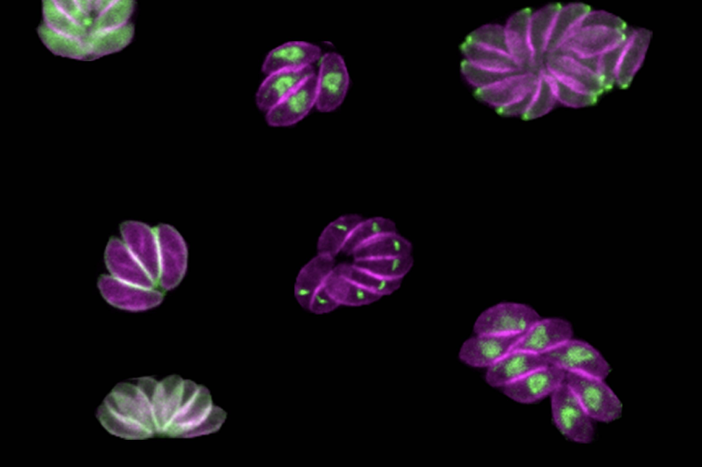
Whitehead Institute Member Sebastian Lourido and his lab members study the parasite Toxoplasma gondii. The parasite causes the disease toxoplasmosis, which can be dangerous for pregnant or immunocompromised patients.
As the parasite evolved over millennia, its phylum (the Apicomplexan parasites) split off from other branches of life, which poses a challenge to researchers hoping to understand its genetics. “Toxoplasma is very highly diverged from the organisms that we typically study, like mice, yeast and [nematodes],” said Lourido lab researcher and Massachusetts Institute of Technology (MIT) graduate student Tyler Smith. “Our lab focuses a lot on developing toolkits to probe and study the genomes of these parasites.”
Now, in a paper published in the journal Nature Microbiology on April 28, Smith and colleagues describe a new method for determining the role of genes within the genome of the parasite. The method can be conducted by a single investigator, and goes a step beyond simply assessing whether or not a given gene is essential for survival. By inserting specific sequences — such as those encoding fluorescent markers or sequences that can turn a gene on and off — throughout the Toxoplasma genome, the method allows the researchers to visualize where an individual gene’s product resides within the parasites and identify when in the life cycle important genes became essential, providing more detailed information than a traditional CRISPR screen.
Although the method could theoretically be used with any gene family, Smith and Lourido decided to first focus on a family of proteins called kinases, the genetic code for which comprises around 150 of Toxoplasma’s 8,000 total genes.
“Kinases are interesting from a basic biology perspective because they are signaling hubs of basic biological processes,” said Smith, who is first author of the study. “From a more translational perspective, kinases are really common drug targets. We have a lot of inhibitors that work with kinases. For some cancers that are linked to specific kinases, the inhibitors can be chemotherapies.”
Using the method, researchers discovered a gene encoding a previously unstudied kinase which they named SPARK. They were able to show that the SPARK kinase is involved in the process of the parasites entering and leaving host cells, and future research on inhibitors of SPARK could lead to new treatments for toxoplasmosis. “Identifying these kinases that are really vital for these critical decision points in a parasite’s life cycle could be really fruitful for developing new therapeutics,” said Lourido, who is also an associate professor of biology at MIT.
New dimensions of screening
Many CRISPR screens use gene editing technology to knock out genes throughout the genomes of a sample of cells, creating a population where every gene in the genome is mutated in at least one of the cells. Then, by looking at which mutations have detrimental effects on the cells, researchers can extrapolate which genes are essential for survival.
But the workings of a whole organism are infinitely more complicated than just survival or death, and researchers are often faced with a challenge when it comes to figuring out exactly what different gene products are doing in the cells. That’s why Smith and Lourido decided to design a method of screening for Toxoplasma genes that could provide more information about what the products of those genes do. “CRISPR screens can tell you which genes are important, but it doesn’t give you much information about why they’re important,” Smith said. “We were seeking to make a kind of platform that could look at other dimensions.”
Smith and Lourido used CRISPR technology to introduce small amounts of new DNA into the parasites’ genes that code for kinases. The new DNA included sequences encoding a fluorescent marker protein and sequences that could be used to manipulate gene expression levels.
After creating a population of parasites modified this way, the researchers then used imaging to determine where the fluorescently tagged proteins had ended up in the cells, and to observe what happened in the cells when the proteins were turned off. “Being able to see different cell division phenotypes — for instance parasites that either failed to replicate at all, or tried to replicate but would have some abnormalities — that gets us closer and allows us to generate hypotheses as to actually why these kinases are important, not just whether or not they are important,” Smith said.
The depletion of some proteins caused the parasites to die instantly, while others affected the parasites at a later point in their life cycles, so they would drop out of the population more slowly. “Cells with mutations in these kinases replicate fine, but a problem might arise when they need to leave their host cell and enter a new host cell later on down the line,” Smith said.
A “SPARK” of inspiration
After the screen, the researchers followed up on one of these kinases in particular, which they called SPARK (short for Store Potentiating/Activating Regulatory Kinase). Mutants depleted of SPARK died, but not until a later phase of the life cycle. Smith and Lourido conducted further experiments to understand SPARK’s role, and found evidence that the protein was involved in the release of calcium in the cell that is required for a parasite to enter or leave a host cell.
“The thing I found very interesting about SPARK is that it’s a kinase that’s very different from the analogous kinase in other model organisms, but is conserved throughout all of the apicomplexan phylum,” Smith said. “That’s the phylum that includes Toxoplasma and a bunch of other single-celled parasites like Plasmodium, which is the malaria parasite.”
Because SPARK is far different from its human analog and essential to the parasite’s life cycle, a SPARK-specific kinase inhibitor could be used to treat toxoplasmosis by killing the parasite without affecting the patient. “The hope would be that you can target SPARK and inhibit it without hitting mammalian kinases,” Smith said. “It’s easy enough to design something that kills a cell, but the trick is only killing parasites and not your own cells.”
In the future, the researchers hope to turn their new screening method to other families of genes, such as transcription factors, to understand their function in the parasites. “Our results have been quite encouraging in that we think this method will be scalable, and we can target larger gene sets in the future,” Smith said. “I think the ultimate end goal would be to do the whole genome.”
“There’s this whole universe of parasite proteins that we know so little about, where this type of analysis will be incredibly insightful.” Lourido said. “We’re really very excited about scaling it up further.
Greta Friar | Whitehead Institute
April 18, 2022
Drug overdose, mostly from opioid use, is the leading cause of accidental death in the United States. Prior studies of twins have revealed that genetics play a key role in opioid use disorder. Researchers know that a mixture of genetic and environmental risk factors contribute to heritability of the disorder, but identifying the specific risk factors is challenging. Opioid use disorder is complex, so instead of one or a few genes causing the disorder, there may be many contributing factors that can combine in different ways. Researchers want to understand which genes contribute to opioid use disorder because this will lead to a better understanding of its underlying biology and could help identify people who will be most at risk if exposed to opioids, enabling researchers, health care providers, and social services to develop strategies for prevention, treatment, and support.
The usual approach for finding genes associated with disease risk is to do a genome wide association study, which compares the genetics of many people to identify patterns in different gene versions occurring in association with a disease. This approach is being used to look at opioid use disorder, but requires many more patient samples than are currently available to reach clear conclusions. Researchers from multiple research universities and institutes, including Whitehead Institute Member Olivia Corradin and her former PhD advisor, Case Western Reserve University Professor Peter Scacheri; as well as Icahn School of Medicine Professor Schahram Akbarian; Eric O. Johnson, a distinguished fellow at RTI International; Dr. Kiran C. Patel College of Allopathic Medicine at Nova South Eastern University Professor Deborah C. Mash; and Richard Sallari of Axiotl, Inc., developed a shortcut for identifying genes that are associated with opioid use disorder and may contribute to it using only a small number of patient samples. Genome wide studies may require hundreds of thousands of samples, but this new method, described in their research published in the journal Molecular Psychiatry on March 17, uses only around 100 samples—51 cases and 51 controls—to narrow in on five candidate genes.
“With this work, we think we’re only seeing the tip of the iceberg of the complex, diverse factors contributing to opioid overdose,” says Corradin, who is also an assistant professor of biology at the Massachusetts Institute of Technology. “However, we hope our findings can help prioritize genes for further study, to speed up the identification of risk markers and possible therapeutic targets.”
In order to learn more about the underlying biology of opioid use disorder, the researchers analyzed brain tissue samples from people who had died of opioid overdoses and compared them with samples from people with no known opioid use history who died of other accidental causes. They specifically looked at neurons from the dorsolateral prefrontal cortex, an area of the brain known to play important roles in addiction. Instead of analyzing the genes in these cells directly, the researchers instead looked at the regulators of the genes’ activity, and searched for changes in these regulators that could point them to genes of interest.
To identify a gene, first map its community
Genes have DNA regions, often close to the gene, that can ratchet up and down the gene’s expression, or the strength of its activity in certain cells. Researchers have only recently been able to map the three-dimensional organization of DNA in a cell well enough to identify all of the regulators that are close to and acting upon target genes. Corradin and her collaborators call a gene’s collection of close regulatory elements its “plexus.” Their approach finds genes of interest by searching for patterns of variation across each gene’s entire plexus, which can be easier to spot with a small sample size.
The patterns that the researchers look for in a plexus are epigenetic changes: differences in the chemical tags that affect regulatory DNA and in turn, modify the expression of the regulators’ target gene. In this case, the researchers looked at a type of epigenetic tag called H3K27 acetylation, which is linked to increases in the activity of regulatory regions. They found nearly 400 locations in the DNA that consistently had less H3K27 acetylation in the brains of people who died of opioid overdose, which would lower activity of target genes. They also identified under-acetylated DNA locations that were often specific to individuals rather than uniform across all opioid overdose cases. The researchers then looked at how many of those locations belonged to regulatory elements in the same plexus. Surprisingly, these individual-specific changes often occurred within the same gene’s plexus. A gene whose plexus had been heavily affected as a collective was flagged as a possible contributor to opioid use disorder.
“We know that the factors that contribute to opioid use disorder are numerous, and that it’s an extremely complex disease that by definition is going to be extremely heterogeneous,” Scacheri says. “The idea was to figure out an approach that embraces that heterogeneity, and then try to spot the themes within it.”
Using this approach, the researchers identified five candidate genes, ASTN2, KCNMA1, DUSP4, GABBR2, and ENOX1. One of the genes, ASTN2, is related to pain tolerance, while KCNMA1, DUSP4, and GABBR2 are active in signaling pathways that have been linked more broadly to addiction. Follow up experiments can confirm whether these genes contribute to opioid use disorder.
The five genes and their plexi are also involved in the heritability of generalized anxiety disorder, metrics of tolerance for risk-taking, and educational attainment. Heritability of these traits and opioid use disorder have previously been found to coincide, and people with opioid use disorder often also have generalized anxiety. Furthermore, heritability of these traits and opioid use disorder all have been associated with early childhood adversity. These connections suggest the possibility that early childhood adversity could be contributing to the epigenetic changes observed by the researchers in the brains of people who died of opioid overdose—a useful hypothesis for further research.
The researchers hope that these results will provide some insights into the genetics and neurobiology of opioid use disorder. They are interested in moving their research forward in several ways: they would like to see if they can identify more candidate genes by increasing their sample number, examine different parts of the brain and different cell types, and further analyze the genes already identified. They also hope that their results demonstrate the potency of their approach, which was able to discern useful patterns and identify candidate genes from the neurons of only 51 cases.
“We’re trying a different approach here that relies on this idea of convergence and leverages our understanding of the three-dimensional architecture of DNA, and I hope this approach will be applied to further our understanding of all sorts of complex diseases,” Scacheri says.
Eva Frederick | Whitehead Institute
April 20, 2022
The Australian stinging tree (Dendrocnide moroides) is a plant that many peopleavoid at all costs. The tree, which is a member of the nettle family, is covered in thin silicon needles laced with one of nature’s most excruciating toxins, a compound called moroidin. “It’s notorious for causing extreme pain, which lingers for a very long time,” said Whitehead Institute Member Jing-Ke Weng.
There’s another side to moroidin, though; in addition to causing pain, the compound binds to cells’ cytoskeletons, preventing them from dividing, which makes moroidin a promising candidate for chemotherapy drugs.
Harvesting enough of the chemical to study has proven difficult, for obvious reasons. Now, in a paper published April 19 in the Journal of the American Chemical Society, Weng, who is also an associate professor of biology at the Massachusetts Institute of Technology (MIT) and former postdoc Roland Kersten, now an assistant professor at the University of Michigan College of Pharmacy, present the first published method to biosynthesize moroidin within the tissues of harmless plants such as tobacco, facilitating research on the compound’s utility for cancer treatments.
Taking a leaf out of plants’ book to create peptides
Moroidin is a bicyclic peptide — a type of molecule made up of building blocks called amino acids and circularized to contain two connected rings. For synthetic chemists, moroidin has proved nearly impossible to synthesize due to its complex chemical structure. Weng and Kersten wanted to dig deeper into what methods the plants were using to create this molecule.
In plant cells, cyclic peptides are made from specific precursor proteins synthesized by the ribosome, the macromolecular machine that produces proteins by translating messenger RNAs. After leaving the ribosome, these precursor proteins are further processed by other enzymes in the cell to give rise to the final cyclic peptides. In 2018, Weng and Kersten had elucidated the biosynthetic mechanism of another type of plant peptides called lyciumins, first found in the goji berry plant, which gave them some insight into how post-translational modifications might play a role in creating different types of plant peptide chemistry. “We learned a lot about the principal elements of this system by studying lyciumins,” said Weng.
When they began to look into how moroidin was synthesized, the researchers found a few other plants, such as Kerria japonica and Celosia argentea, also produce peptides with similar chemistry to moroidin. “That really gave us the very critical insight that this is a new class of peptides,” Weng said.
Weng and Kersten previously learned that the BURP domain, which is part of the precursor proteins for lyciumins and several other plant cyclic peptides, catalyzes key reactions involved in the peptide ring formation. They found that the BURP domain was present in the precursor proteins for moroidins in Kerria japonica, and seemed to be essential for creating the two-ring structure of the molecules. The BURP domain creates ring chemistry when in the presence of copper, and when the researchers incubated the moroidin precursor protein with copper chloride in the lab together with other downstream proteolytic enzymes, they were able to create moroidin-like peptides.
With this information, they were able to produce a variety of moroidin analogs in tobacco plants by transgenically expressing the moroidin precursor gene of Kerria japonica and varying the core motif sequence corresponding to moroidin peptides. “We show that you can produce the same moroidin chemistry in a different host plant,” Weng said. “Tobacco itself is easier to be farmed on a large scale, and we also think in the future we can derive a plant cell line from the existing tobacco cell lines that we put in the moroidin precursor peptide, then we can use the cell line to produce the molecule, which really enables us to scale up for medicine production.”
Future use of moroidin
Moroidin’s anti-cancer property is due, at least in part, to the compound’s unique structure that allows it to bind to a protein called tubulin. Tubulin forms a skeletal system for living cells, and provides the means by which cells separate their chromosomes as they prepare to divide. Currently, two existing anti-cancer drugs, vincristine and paclitaxel, work by binding tubulin. These two compounds are derived from plants as well (the Madagascar periwinkle and Pacific yew tree, respectively).
In their new work, Weng and Kersten synthesized a moroidin analog called celogentin C. They tested its anti-cancer activity against a human lung cancer cell line, and found that the compound was toxic to the cancer cells. Their new study also suggests potentially new anti-cancer mechanisms specific to this lung cancer cell line in addition to tubulin inhibition.
In the past, researchers have run into issues when trying to create effective drugs from peptides. “There are two major challenges for peptides as medicine,” Weng said. “For one thing they are not very stable in vivo, and for another they are not very bioavailable and don’t readily pass the membrane of a cell.”
But cyclic peptides like moroidin and its analogs are a bit different. “These peptides essentially evolve to be drug-like,” Weng said. “In the case of the Australian stinging tree, the peptides are present because the plants want to deter any animals that want to eat the leaves. So over millions of years of evolution these plants eventually figured out a way to construct these specific cyclic peptides that are stable, bioavailable and can get to the animal that is trying to eat the plants.”
It’s likely that the painful reaction that occurs when moroidin enters the body through a sting from the tree would not be an issue in traditional methods of administering chemotherapy. “The pain is really caused if you get injections of the compound into the skin,” Weng said. “If you take it orally or intravenously, your body will most likely not sense the pain.”
Somewhat counterintuitively, the compound could also be used as a pain reliever. “If something causes pain, you can sometimes use that as an anti-pain medicine,” Weng said. “You could essentially exhaust the pain receptors, or if you alter the structure a little bit, you could turn an agonist into an antagonist and potentially block the pain.”
On a more fundamental level, moroidin could help researchers study pain receptors. “We don’t know exactly why being stung by the stinging tree produces that enormous amount of pain, and there may be additional pain receptors people haven’t identified,” Weng said. “Being able to synthesize moroidin provides a chemical probe that allows us to study this unknown pain perception in humans.”
In the future, the researchers hope to create analogs of moroidin to study, and hopefully create an optimal version for use in cancer therapy. “We want to generate a library of moroidin-like peptides,” Weng said. “We’ve done this for lyciumins, and since the initial moroidins are anti-tubulin molecules, we can use this system to find an improved version that binds to tubulin even tighter and contains other pharmacological properties making it suitable to be used as a therapeutic.
Greta Friar | Whitehead Institute
April 20, 2022
Stem cells are the versatile building blocks from which every cell type in the body, from neurons, to skin cells, to blood cells, is ultimately descended. Researchers have also figured out how to turn stem cells into different cell types in the lab, which has been helpful for studying health and disease in their normal cellular contexts, and could be used to generate cells for medical transplants. Whitehead Institute Founding Member Rudolf Jaenisch not only uses these cells in his research, but has spent much of his career discovering and improving the methods for making accurate laboratory models out of stem cell-derived cells.
One challenge that Jaenisch’s lab is focusing on is how to eliminate the differences between cell types as they are found in the body and their stem cell-derived equivalents. In particular, they have found that stem-cell derived cells are often immature, more closely resembling the cells found in fetuses rather than in adults. These differences can make the cells less accurate research models and prevent them from being medically useful as functional transplant cells. Stem cells in the body receive complex cocktails of molecular signals as they transform into different cell types. The challenge for researchers lies in figuring out which of the many molecular signals in the body are relevant and then get the recipe exactly right in their recreations.
Postdoc Haiting Ma in the Jaenisch lab decided to tackle this problem for hepatocytes, the main type of cell in the liver. In work published in Cell Stem Cell on April 21, Jaenisch and Ma share their findings on why stem cell-derived liver cells resemble fetal liver cells, and what’s needed to make them mature—including an important role for a thyroid hormone.
The liver filters everything that enters the body through the digestive system. It helps to store and modify nutrients, safely break down toxins and waste, process medications, and more. There is still a lot to learn about how the liver functions, and what goes wrong in a number of liver-associated diseases, and accurate stem cell-derived models will help with that research. Liver cells are also needed to treat end-stage liver disease, and if researchers could mass produce stem cell-derived liver cells that can function safely in an adult liver, this could help to meet the demand for liver cell transfusions.
For this study, Jaenisch and Ma grew liver cells from stem cells in two setups: a typical 2D culture, in which the cells were grown in a dish, and a 3D spheroid, in which cells that started out in the normal culture were then allowed to grow into three-dimensional balls of cells. The spheroids can be designed to mimic some aspects of the cells’ natural environment in ways that a 2D culture cannot. In each case, the researchers exposed the cells to a carefully timed mixture of signals to prompt them to develop into liver cells. The researchers then analyzed cells from both the 2D and 3D cultures and compared them to primary liver cells, or cells from a body, using a variety of techniques to look for differences related to DNA and gene expression. They found that the cells cultivated in the 3D system were closer to cells from the adult body than those in the 2D system.
“The 3D culture not only contributes to maturation of the liver cells, but it can also be used to scale up production of the cells, which could be very useful for cell therapies in the future,” Ma says.
However, both sets of lab-derived cells lacked important features of adult liver cells. The analyses pointed to one important missing factor in particular: in the adult liver cells, a hormone receptor called Thyroid Hormone Receptor Beta (THRB) binds to a number of places in the DNA. THRB then senses the presence or absence of thyroid hormones, and regulates a variety of gene expression processes accordingly. However, the researchers found that while the stem cell-derived liver cells made the right amount of THRB, something was preventing it from binding where it should and performing its function.
Normally, THRB has a partner that helps it bind to DNA, the thyroid hormone T3. When the researchers added T3 to their 2D and 3D cultures, this led to more typical binding of THRB, which in turn made the cells—especially the cells from the 3D culture—more closely resemble adult liver cells in a number of ways. Improved THRB binding increased the expression of key liver genes, restored the activity of regulatory elements in the DNA that modify gene expression, and reduced the expression of a fetal liver gene. The researchers also gained insights into the molecules that THRB interacts with and the mechanisms by which it affects liver maturation, painting a more complete picture of its key roles in liver cells.
Altogether, this work led to a better recipe for making adult liver cells from stem cells in the lab–using the 3D spheroid culture and adding T3. When cells developed with this approach were incorporated into the livers of mice, the cells integrated successfully and the liver maintained normal function long term.
The new and improved stem cell-derived liver cells are still not a perfect match for adult liver cells—the researchers have ideas about which missing characteristics they could tackle next—but the current cells’ ability to seamlessly integrate into the liver, as well as indicators from the analyses that they would be good models for liver-associated diseases, suggest that they will be useful in a variety of projects.
“As we improve the authenticity of our stem cell-derived cell types, we open up new opportunities for research,” Jaenisch says. “We can build more accurate models in which to study high-impact diseases, such as liver diseases, diabetes, and chronic viral infections, and using those models we can develop strategies for treatment and prevention.”
Merrill Meadow | Whitehead Institute
April 20, 2022
This year’s Vanderbilt Prize in Biomedical Science will be awarded to Whitehead Institute director Ruth Lehmann. It recognizes women scientists with a stellar record of research accomplishments who also have made significant contributions to mentoring other women in science.
“Dr. Lehmann’s determination to solve the deepest mysteries of life while encouraging others at the beginning of their careers exemplifies the spirit of the Vanderbilt Prize in Biomedical Science,” says Jeff Balser, president and chief executive officer of Vanderbilt University Medical Center – which bestows the Prize – and dean of Vanderbilt University School of Medicine. “I’m honored to congratulate her as the 2022 Vanderbilt Prize recipient.”
“I’m thrilled to be receiving this honor, recognizing the importance of mentoring and empowering the next generation of scientists,” says Lehmann, who has mentored scores of students and research fellows during her career, and has developed a mentorship program specifically designed to encourage and empower junior faculty in science. Read more here.
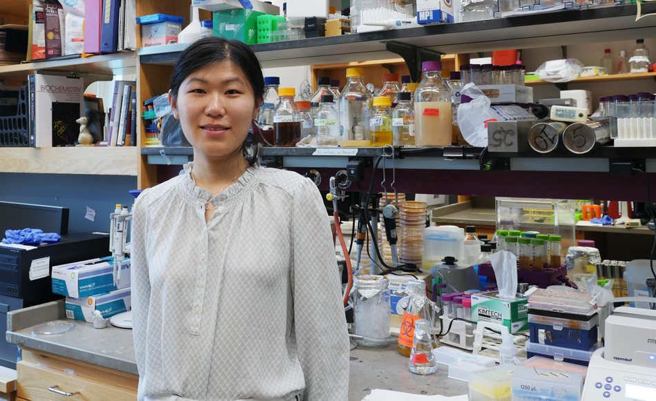
Graduate student En Ze Linda Zhong-Johnson is creating new methods to measure and enhance enzyme activity — which she hopes will help restore a plastic-choked world.
Grace van Deelen
April 21, 2022
After graduating with her undergraduate degree in molecular genetics from the University of Toronto in 2016, En Ze Linda Zhong-Johnson celebrated with a trip to Alaska. There, she saw a pristine landscape unlike the plastic-littered shores of the Toronto waterfront. “What I saw up there was so different from what I saw in the city,” Zhong-Johnson says. “I realized there shouldn’t be all this waste floating everywhere, in our water, in our environment. It’s not natural.”
As a trained biologist, Zhong-Johnson began to think about the problem of plastic pollution from a biological perspective. One solution, she thought, could be biological recycling: a process by which living organisms break down materials, using digestion or other metabolic processes to turn these materials into smaller pieces or new compounds. Composting, for example, is a type of biological recycling — microbes in the soil break down discarded food, speeding up the decomposition process. Zhong-Johnson wondered if there were any organisms on Earth that could use the carbon in polyethylene terephthalate (PET), a common plastic used in water bottle and food packaging, as an energy source.
Earlier that same year, Japanese scientists discovered that a bacterium, Ideonella sakaiensis, could do just that by producing enzymes that could break down PET. The two main PET-degrading enzymes, referred to as IsPETase and IsMHETase, are able to turn PET into two chemical compounds, terephthalic acid and ethylene glycol, which I. sakaiensis can use for food.
The discovery of these enzymes opened up many new questions and possible applications that scientists have continued to work on since. However, because there was — and still is — much to learn about PET-degrading enzymes, they are still not widely used to recycle consumer products. Zhong-Johnson figured that, in graduate school, she could build on the existing IsPETase research and help to accelerate their use at recycling facilities. Specifically, she wanted to engineer the enzyme to work faster at lower temperatures, and study how, fundamentally, the enzymes worked on the surface of PET plastic to degrade it.
“I hopped on the excitement train, along with the rest of the world,” she says.
A better enzyme
After receiving her acceptance to MIT to complete her PhD, Zhong-Johnson approached various professors, pitching her idea to speed up IsPETase activity. Christopher Voigt, the Daniel I. C. Wang Professor of Biological Engineering, and Anthony Sinskey, professor of biology, were interested, and formed a co-advisorship to support Zhong-Johnson’s project. Sinskey, in particular, was impressed by her idea to help solve the world’s plastic problem with PET-degrading enzymes.
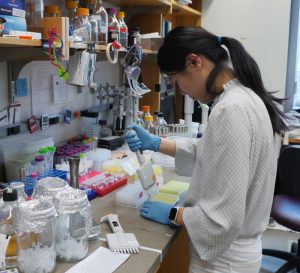
Zhong-Johnson screens for enzyme variants with improved activity. Credit: Grace van Deelen
“Plastic pollution is a big problem,” he says, “and that’s the kind of problem my lab likes to tackle.” Plus, he says, he feels “committed to helping graduate students who want to apply their science and technology learnings to the environment.”
While the idea of a plastic-degrading enzyme seems like a panacea, the enzyme’s practical applications have been limited by its biology. The wild-type IsPETase is a mesophilic enzyme, meaning the structure of the enzyme is only stable around ambient temperatures, and the enzyme loses its activity above that threshold. This restriction on temperature limits the number and types of facilities that can use IsPETase, as well as the rate of the enzyme reaction, and drives up the cost of their use.
However, Zhong-Johnson thinks that, with combined approaches of biological and chemical engineering, it’s possible to scale up the use of the enzymes by increasing their stability and activity. For example, an enzyme that’s highly active at lower temperatures could work in unheated facilities, or even be sprinkled directly into landfills or oceans to degrade plastic waste — a process called bioremediation. Increasing the activity of the enzyme at ambient temperatures could also expand the possible applications.
“Most of the environments where plastic is present are not above 50 degrees Celsius,” said Zhong-Johnson. “If we can increase enzyme activity at lower temperatures, that’s really interesting for bioremediation purposes.”
Now a fifth-year graduate student, Zhong-Johnson has honed her project, and is focusing on increasing the activity of IsPETase. To do so, she’s using directed evolution — creating random mutations in the IsPETase gene, and selecting for IsPETase variants that digest PET faster. When they do, she combines the beneficial mutations and uses that as template for the next round of library generation, to improve the enzyme even further. The evolution is “directed” because Zhong-Johnson herself, rather than nature, is picking out which gene sequences of enzyme proceed through to the next round of random mutagenesis, and which don’t. Her ultimate goal is to create a more efficient and hardier enzyme that will, hopefully, work faster at ambient temperatures.
A better protocol
Just as Zhong-Johnson was beginning her project, she ran into an obstacle: There wasn’t a standard way to measure whether her experiments were successful. In particular, no immediately applicable method existed to measure enzyme kinetics for IsPETases on solid substrates like plastic bottles and other plasticware. That was a problem for Zhong-Johnson because understanding enzyme activity was a crucial part of how she selected her enzymes in the directed evolution process.
Usually, enzyme activity is measured via product accumulation: When enzymes metabolize a substance, they create a new substance in return, called a product. Measuring the amount of product created by an enzyme after a certain amount of time gives the researcher a snapshot of that enzyme’s activity.
There are two problems with the product accumulation method, though. First, it is usually done using liquid or soluble substrates. In other words, the material that the enzyme is targeting is dissolved, like sugar dissolved in water. Then, the enzyme is added to that liquid concoction and mixed evenly throughout. However, the substrate Zhong-Johnson wanted to use — PET — was not soluble but solid, meaning it could not be evenly distributed like a soluble substrate. Second, the product accumulation measurement methods available were only practical for measuring less than a handful of timepoints for a few enzyme or substrate concentrations. As a result, many in the field opted to measure a single time point, late in the enzyme reaction, which doesn’t provide an indication of how an enzyme’s rate of digestion actually changes over the course of time — something that can be measured through kinetic measurements.
Taking kinetic measurements would help researchers like Zhong-Johnson illustrate the full pattern of enzyme activity and answer questions like: When is the enzyme most active? Does most product accumulation happen at the beginning of the reaction or the end? How does temperature impact the rate of these reactions over time? To answer these questions, she realized she would have to develop the method herself.
Through a serendipitous discussion with a group of chemical engineering undergraduate students that Zhong-Johnson was mentoring, she came up with a solution, which she published in a 2021 paper in Scientific Reports. The undergraduates brought to her attention many factors that she had overlooked about the enzyme, and she says she would not have realized the importance of kinetic measurements if it weren’t for the fact that she was trying to design an experiment that the undergraduates could perform over the course of three hours.
The paper outlined a new way to measure enzyme activity, which Zhong-Johnson calls “the bulk absorbance method.” Instead of measuring the final product accumulation at very late time points, the bulk absorbance method involves taking multiple kinetic measurements at early time intervals during the experiment. This technique informs Zhong-Johnson’s directed evolution approach: If she can find which enzymes are most active at low temperatures, she can select the best possible enzyme for the next round of analyses. She hasn’t yet engineered an enzyme she’s completely happy with, but she’s gotten much closer to her ultimate goal.
Solving big problems together
Zhong-Johnson’s discoveries have been made possible by the collaboration between her and her two co-advisors, Voigt and Sinskey, who have supported her independence throughout her five years at MIT.
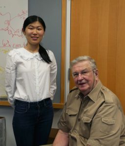
Zhong-Johnson and her advisor, professor Anthony Sinskey, in his office. Credit: Grace van Deelen
When she first started her graduate work, neither Voigt nor Sinskey had expertise in enzyme biochemistry involving solid substrates: Sinkey’s lab focuses on bacterial metabolism, while Voigt’s lab focuses on genetic engineering (though Voigt did have experience with directed evolution research). Additionally, Zhong-Johnson’s path to her project was rather unconventional. Most grad students do not come to potential advisors proposing entire dissertations, which posed a unique challenge for Zhong-Johnson.
Despite not having specific expertise in enzyme biochemistry involving solid substrates, Voigt and Sinskey have supported Zhong-Johnson in other ways: by helping her to develop critical thinking skills and connecting her to other people in her field, such as potential collaborators, who can help her project thrive in the future. Zhong-Johnson has supplemented her MIT experience by having enzyme experts as part of her dissertation committee as well.
Sinskey says that, in the future — once Zhong-Johnson has engineered the ideal enzyme — they would like to partner with industry, and work on making the enzyme into a product that waste companies might use to recycle plastic. Additionally, Sinskey says, the plastic problem and the IsPETase solution raise so many interesting questions that Zhong-Johnson’s project will probably live on in the Voigt and Sinskey labs even after she graduates. He’d like to see other graduate students working to understand the enzyme’s activity and progressing the directed evolution that Zhong-Johnson started.
Zhong-Johnson is already working on understanding the specifics of how IsPETase act on PET. “How does it eat a hole in a plastic bottle? How does it move along and make the hole bigger as it moves through the process? Does it jump around? Or does it keep degrading a single polymer chain until its completely broken down? We just don’t know the answers yet,” says Sinskey.
But Zhong-Johnson is up to the task. “My graduate students have to have three skills, in my opinion,” Sinskey says. “One, they have to be intelligent. Two, they have to be energetic, and three, they have to be of high integrity, in research and behavior.” Zhong-Johnson, he says, has all three qualities.