Eva Frederick | Whitehead Institute
January 21, 2021
Sex-specific chromosomes are a dangerous place to be, if you’re a gene. Because these chromosomes — Y chromosomes, in humans — do not have a matching chromosome with which to exchange genetic information, they are prone to losing non-essential genes left and right in a process called genetic decay.
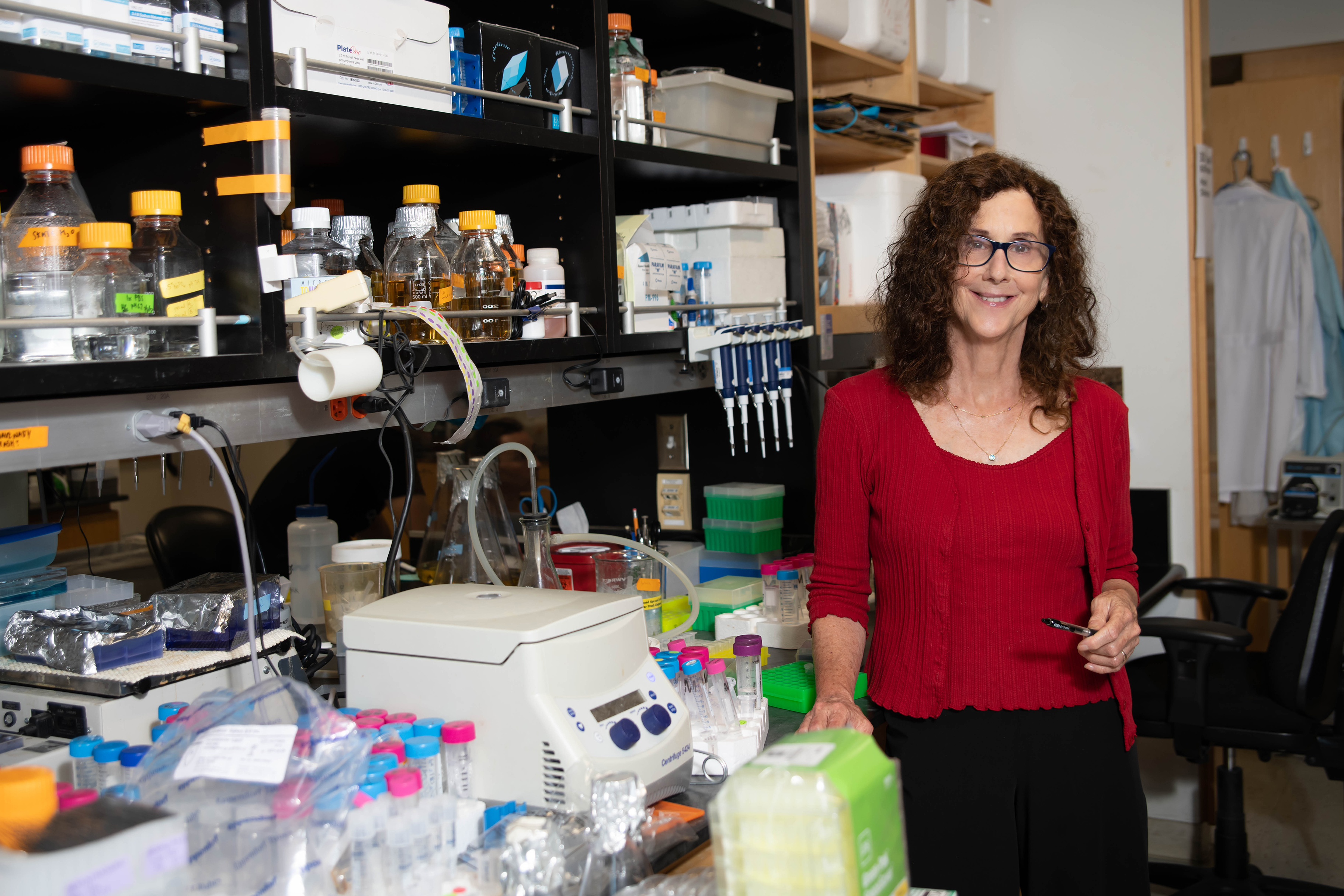
Now, a new study from research scientist Daniel Winston Bellott in the lab of Whitehead Institute Member David Page broadens our understanding of what makes a gene able to survive on a sex-specific chromosome by looking at one especially slithery branch of the evolutionary tree: snakes.
Comparing surviving genes on snake sex-specific chromosomes to those that are lost to the ravages of time can teach scientists about the evolutionary pressures that shaped sex chromosomes as we know them today. “You might think, ‘These are sex chromosomes, so the surviving genes should have something to do with sex, right?’” Bellott said. “But they don’t.”
Instead, many of these genes are essential to the survival of the animal, and take part in key developmental processes. “It turns out that these survivor genes on sex-specific chromosomes may play a very big role in governing how all of the genes across all the chromosomes are read, interpreted and expressed,” said Page, who is also a professor of biology at Massachusetts Institute of Technology (MIT) and an Investigator of the Howard Hughes Medical Institute. “Winston’s study is absolutely foundational to our understanding of what the sex chromosomes are, how the two sexes come to be, and how health and disease traits play out similarly or differently in males and females.”
What is a sex chromosome, anyway?
Over the course of evolution, all sex chromosomes start out as regular, matching chromosomes called autosomes. Then, somewhere along the line, a mutation happens, and one of the chromosomes gains a “switch,” that, when present, causes an embryo to to develop as a specific sex. “It’s actually really easy to make a sex chromosome,” said Bellott. “In most cases, you only need to change one or two genes and you’ve started the sex chromosome system.”
This process has happened numerous times during the course of evolution. It makes sense; sexual reproduction is an efficient way to ensure genetic diversity. But the whole thing is a bit mysterious; are, for example, certain chromosomes predisposed to become sex chromosomes?
That’s where Bellott thought snakes could be especially helpful. “Snakes have a relatively old system of sex chromosomes, where you have a lot of time for the chromosomes to diverge,” Bellott said. “Time has swept away all the genes that aren’t important, and you can see what kind of genes are left.”
Their sex chromosome system also evolved from different autosomes, some 100 million years after humans’, and thus would provide a useful vantage point from which to consider our own genomes.
To learn more about the evolution of these chromosomes, Bellott and Page first gathered a list of “ancestral genes,” which were likely on the chromosome from which the snake sex chromosomes evolved. New sequencing data for several species of animals distantly related to snakes meant that they had a more complete list of these genes — 1,648 to be exact.
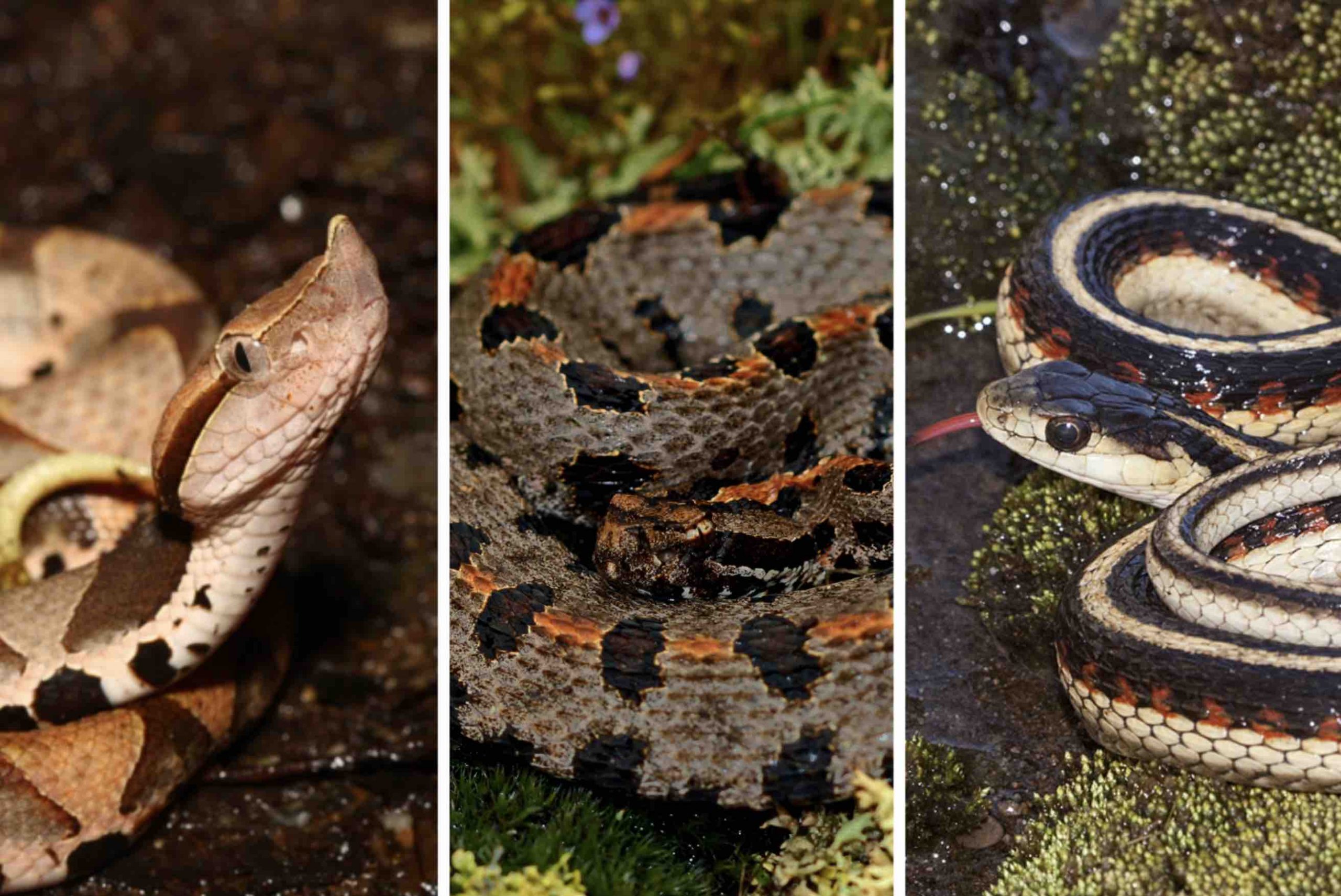
Bellott began painstakingly sifting through the genes that remained on the sex-specific chromosomes of three species of snake: the pygmy rattlesnake, mountain garter snake, and the five-pacer viper. He eventually identified 103 ancestral genes that had survived as long as 90 million years of evolution on the snakes’ sex chromosomes. With this list in hand, Bellott could then ask what these surviving genes had in common that set them apart from the hundreds of genes that were swept off the snakes’ sex chromosomes by genetic decay.
What makes a survivor?
To Bellott’s surprise, the genes that remained on the snakes’ sex-specific chromosome had nothing to do with sex determination; neither were they expressed more often in sex-specific tissues, or more often in one sex than the other.
Instead, Bellott and Page’s research identified three key properties that led to a gene’s survival on snake sex-specific chromosomes. First, the gene must be dosage sensitive. In other words, the snake’s body depends on its cells to produce an exact amount of that gene’s protein product. Any more, or any less, and the snake experiences illness or death. Second, a surviving gene is likely broadly expressed in different tissues across the body, not localized to one specific organ or area. And third, surviving genes are subject to strong purifying, or negative, selection. Simply put, this means that if something goes wrong with one of these genes, the snake has a slim chance of survival or producing offspring.
When Bellott dove deeper into the genes’ function, he discovered that for many of them the equivalent gene in humans played a role in key developmental processes such as the formation of the face. When these genes were mutated in humans, their faces — and other essential parts of the body — would not develop properly. “What Winston is seeing here is that the genes that were preserved on the sex specific chromosomes in snakes are disproportionately involved in birth defects in people,” Page said. “We think that nature is selecting for the survival of [sex chromosome] genes whose dosage in certain parts of embryonic development is especially critical.”
In time, Bellott said, this may allow scientists to predict genes whose role in developmental disorders is yet to be discovered. “In some sense, you get to the place where you’re starting to work the experiment backwards in your mind, and say, ‘Let’s take the set of genes that are on sex specific chromosomes in snakes and birds, but that have not yet been implicated in birth defects in humans,’” Page said. “They might be prime candidates to be responsible for heretofore unexplained birth defects.”
From snakes to humans
Next, the researchers sought to broaden their scope. They compared ancestral genes across the three species of snakes and 38 species of birds and mammals with a larger pool of genes that made it to the present day. Many of the surviving genes on bird and mammal chromosomes had different functions than those on snake chromosomes, but again, most had little to do with sex determination.
“Adding the snakes in with the birds and the mammals gave Winston enough data points to be able to see further and to see more precisely, and now for the first time, he was able to confirm something that we had been suspecting for a long time but really didn’t have sufficient data to pin down,” Page said. “And that is that the chromosomes that became sex chromosomes were not sort of inclined to function in sex differences. Before they got picked out of the crowd, they weren’t specialized towards differentiating between the sexes in much of any way.”
Then, as genes were lost over time, evolutionary pressures ensured that the same sort of genes survived. This idea that sex chromosomes — besides their key developmental switch — have little do do with sex determination challenges the common notion of what a sex chromosome actually is.
“I hope people will pick up on this idea that the chromosomes that became sex chromosomes weren’t in any way preordained,” Page said. “They were just ordinary chromosomes out for a walk in the park, and something happened.”
In the future, Bellott and Page plan to further broaden their scope to include other animals, toward the ultimate goal of understanding our own sex chromosomes. “We take these results, and we turn them into a lens through which we look at sex differences in health and disease in our own species,” Page said. “This research really refines our ideas about what it means to be a gene on the human X or Y chromosome, and how we should think about those genes that survive.”
Bellott, D.W. and Page, D.C. “Dosage-sensitive functions in embryonic development drove the survival of genes on sex-specific chromosomes in snakes, birds, and mammals.” Genome Research, Jan. 21, 2021. DOI: 10.1101/gr.268516.120.