Global perspectives on microscopic pathogens

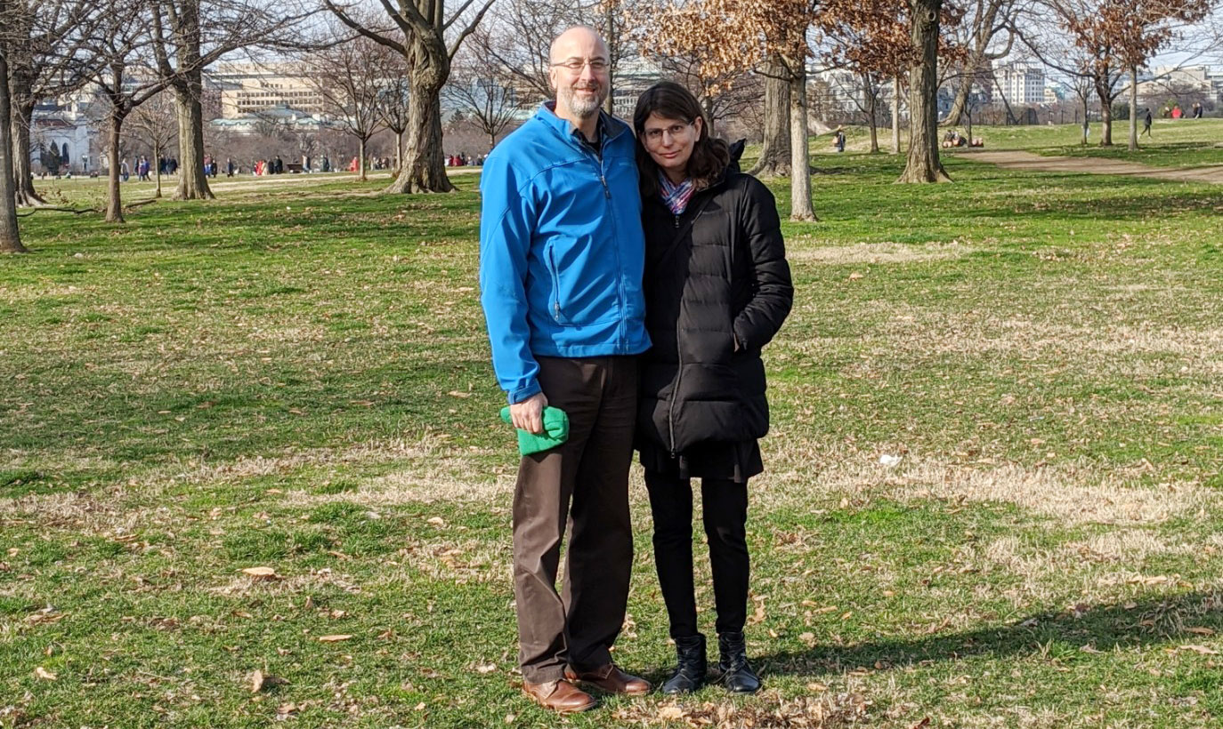
Junior Emily O’Rourke traveled to South Africa to investigate epidemics and returned with a broader outlook on her fundamental disease research.

Raleigh McElvery
March 31, 2020

Growing up in El Paso, Texas near the border of the U.S. and Mexico, Emily O’Rourke could venture across cultures in less time than it takes most people to commute to work. In fact, her dad would make this short trip each day for his job as a mechanical engineer. Watching him cross over so frequently reminded O’Rourke that “ideas and skills don’t stop at the border.” O’Rourke herself would visit Mexico to see relatives, and these experiences seeded aspirations to spearhead international scientific collaborations. Now a junior in Course 7 (Biology), O’Rourke is continuing to add stamps to her passport while exploring the global implications of disease research.

O’Rourke chose MIT because it offered a particularly wide array of study abroad programs, in addition to having top-tier research opportunities. One such study abroad program, MIT International Science and Technology Initiatives (MISTI), operates 25 regional programs, matching undergraduate and graduate students with fully-funded internship, research, and teaching opportunities in over 40 countries. The summer after her first year, O’Rourke participated in MISTI’s MIT-Italy Program in order to gain some research experience in the realm of urban planning. For six weeks, she investigated the urban effects of sea level rise while living in Venice.

When she returned to campus for her sophomore year, O’Rourke was intending to double major in physics and biology. But she ultimately opted to drop physics and pursue the life sciences once she started working in Becky Lamason’s lab in the Department of Biology.

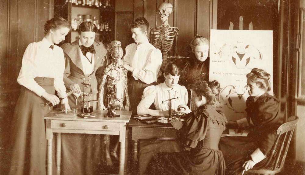
“I started to see how biology worked on a practical level,” she says. “I get to experience a hands-on connection by running DNA on a gel and doing other experiments. During our weekly lab meetings, I witness scientific stories as they unfold.”

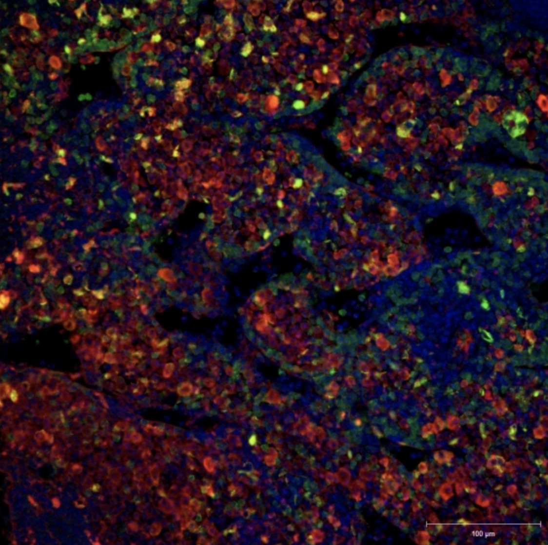
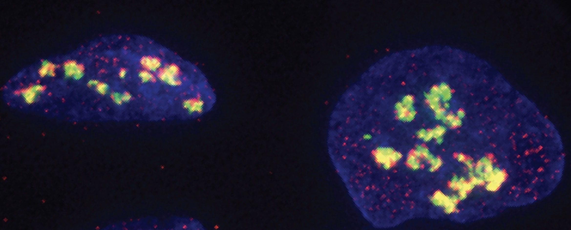
More recently, the duo has begun to examine how Sca4 may coopt another protein in the host cell, known as clathrin, for its own malicious means. “Sca4 is a really big protein and we still don’t know its entire structure,” O’Rourke says, “and we’re hoping to uncover some new functions.”The Lamason lab investigates how parasites hijack host cells processes in order to spread infection. O’Rourke is working with graduate student Cassandra Vondrak to probe the proteins that allow the tick-borne Rickettsia parkeri to migrate from one cell to the next. Their protein of interest, surface cell antigen 4 (Sca4), is secreted by the bacterium and binds to the host’s cell membrane, reducing the tension across the membrane and allowing Rickettsia to punch through to the neighboring cell. O’Rourke and Vondrak aim to determine how Rickettsia releases Sca4, in the hopes of piecing together a general mechanism by which pathogens propagate.

While O’Rourke was studying infectious disease on a cellular level, she heard about an opportunity to explore epidemics on a global scale. Each January, the Harvard-MIT Program in Health Sciences and Technology sponsors a two-week class in South Africa called Evolution of an Epidemic. The class, taught by Professor of the Practice Bruce Walker, covers the medical, scientific, and political responses to new diseases, focusing on the HIV/AIDS epidemic. Walker, who is also the director of the Ragon Institute of MGH, MIT and Harvard, is a world leader in the study of immune control and evasion in HIV infection. Since then, he’s developed strong connections and research partnerships in South Africa where the disease is most prevalent.

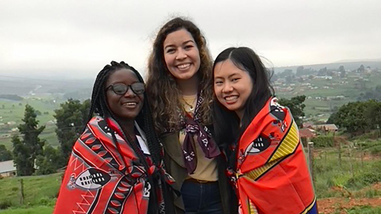
O’Rourke enrolled in Evolution of an Epidemic, and MISTI helped her to plan her trip. On January 16, she landed in Johannesburg, the first of three destinations. The cohort of students from MIT, Harvard, and the African Leadership Academy attended lectures, spoke with patients, and met medical professionals.

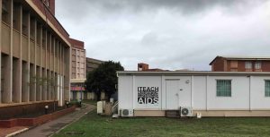
After Johannesburg, the class traveled to Durban where they visited traditional healers who were learning to administer HIV/AIDS tests as part of the iTeach program.

“We had the chance to ask these healers how they felt about interacting with Western medicine, and whether it clashed with their traditional values,” O’Rourke says. “They said HIV was so new that they couldn’t draw upon ancient wisdom from their ancestors to treat it. They were directing patients towards Western treatments because they’d seen the devastation the disease could cause.”

iTeach building
The iTEACH Program located in KwaZulu-Natal, South Africa.

At their third and final destination, the province of KwaZulu-Natal, O’Rourke toured the FRESH Program. Twice a week, as part of a clinical trial, healthy African women around O’Rourke’s age attend classes that address topics like self-esteem, gender-based violence, HIV prevention, career development, and computer training. Before each session, the women are tested for HIV/AIDS, so if they contract it the researchers can treat it early and learn more about the disease’s initial stages.

“I really liked going there because it helped me see a direct connection between science and social good,” O’Rourke says. “It showed the value of talking to patients and asking about their experiences, rather than just looking at study outcomes.”

After two weeks, O’Rourke returned to MIT Biology and the Lamason lab with a broader outlook on her parasite research. “I’m able to see how my works fits into a larger context,” she says, “and how it may eventually have far-reaching impacts on disease evolution and spread.”

O’Rourke still plans to pursue fundamental biological research, but intends to seek out international collaborations focused on global health as well. It’s hard to leave the MIT bubble, she says, but it’s worth it. “Traveling can really broaden your perspective as a scientist, and inform your research in unexpected ways.”

Photos courtesy of Emily O’Rourke
Posted 4.1.20
Women at the forefront of MIT Biology

In honor of Women’s History Month, meet two of the first female students to earn biology degrees at MIT.

Saima Sidik
March 19, 2020

In MIT’s 1887 annual report, former Institute President Francis A. Walker included a section titled, “Women as students in the Institute.” He predicted: “The number of young women attending the Institute of Technology is never likely to be large, considering the nature of the professions to which our courses lead, and the severity of our requirements for admission and for graduation.” More than 100 years later, it’s clear Walker was sorely mistaken; today, women comprise around 40% of the student body and 58% of the students in the Department of Biology. But, for decades, only about 5% of MIT’s students were female.

In 1958, decades after Walker’s report, Marilynn Bever was one of 28 women in her first-year class of 652 students. “I was aware that MIT women are often regarded with suspicion, as being somehow ‘different’ from normal coeds,” Bever later wrote. She was so fascinated by this attitude that she wrote a thesis for her bachelor’s degree in anthropology in which she catalogued early female MIT students and documented their experiences. Bever reported that Caroline Augusta Woodman was the first woman to obtain a Course 7 bachelor’s degree in 1889, and that Helen Louise Breed became the first woman to earn a PhD from MIT’s Department of Biology in 1937. In honor of Women’s History Month, meet these two women who helped set MIT on a path toward gender equality.

Caroline Augusta Woodman

Woodman (pictured above, second from the left) was born in 1844 in Minot, Maine near a textile manufacturing center. After graduating from high school in nearby Portland, Maine in 1866, Woodman taught at Portland’s Center Grammar School for Girls. By 1874, she’d moved from the coast of Maine to the banks of the Hudson River and earned a bachelor’s degree from Vassar College, which had recently opened as an all-women’s institution.

According to the Vassar Registrar’s Office, early Vassar students took a prescribed set of courses rather than declaring a major, so Woodman studied art, science, languages, and religion, among other topics. Her transcript indicates that she supplemented these classes with “special courses,” which the 1870-71 course catalogue described as “intended only for ladies of maturity,” as the college’s faculty felt that students should complete the usual curriculum before venturing on to these more complex topics. Vassar’s faculty must have seen Woodman as an accomplished scholar, as she was allowed to take extra classes in German, chemistry, math, astronomy, and geography.

The foreign language skills that Woodman learned in these special courses must have served her well after completing her degree, as she traveled around Europe before moving to the Finger Lakes in upstate New York, where she taught at a high school for girls for about twelve years. But in 1889 she found herself back in college and earning a second bachelor’s degree, this time at MIT.

Just as Woodman joined Vassar during its incipient years, she came to MIT when the Institute consisted of only a handful of buildings near Copley Square in Boston’s Back Bay. Graduate degrees had only been established a few years earlier, and the first dormitory wouldn’t be built for another decade. Today’s MIT undergraduates can choose interdisciplinary programs in Chemistry and Biology (Course 5-7) or Computer Science and Molecular Biology (Course 6-7). But in Woodman’s time, even the name “Biology” was new to the department, which had been previously called “Natural History” and re-named to encompass modern aspects of the expanding discipline such as techniques for preserving food safely.

Black and white image of lab
An MIT Biology lab photographed the year Woodman earned her degree. Credit: Photogravure Views of the Mass. Institute of Technology, Boston, 1889, by Henry Lewis Johnson

Course 7 was a small program when Woodman arrived. MIT’s yearbook, MIT Technique, shows that there were only about 10 biology undergraduate students. Although few women attended the Institute as a whole, MIT Biology had a strong female presence by comparison; there were at least five women in the program. However, many of these women were classified as “special” students — a category intended for students taking select classes rather than pursuing full degrees — and few of them were able to complete the classes necessary to earn a bachelor’s degree.

Woodman did earn her second bachelor’s degree, later becoming a physiology instructor in the Zoology Department at Wellesley College, where the student newspaper described her as “a teacher of experience.” A picture from the Wellesley Archives (above) depicts her overseeing a group of young women as they examine a model human body, concoct mixtures of chemicals, and take notes on their experiments.

In 1895, Woodman moved back to her home state and accepted a position as a librarian at Bates College in Lewiston, Maine. Five years later, a student publication reported that she had added substantially to the library’s collections, instituted the Dewey decimal system, and was constructing a card catalogue.

By the time Woodman died in 1912, she had experienced much more of the world than most people from the rural mill town where she was born. From studying at Vassar during the school’s first decade, to traveling through Europe and paving the way for women in MIT Biology, she strayed far from home, then returned to tell the tale.

Helen Louise Breed

Black and white yearbook photo
A picture of Helen Louise Breed when she earned a Bachelor of Arts in 1931. Credit: Wellesley College Archives, Library & Technology Services.

It would be 30 more years before Helen Louise Breed joined MIT in the fall of 1933 and became the first woman to earn a PhD from MIT Biology. The Great Depression was in full swing; although enrollment, staff, and funding had decreased, life went on relatively normally for those lucky enough to remain at the Institute. On September 25, 1933, the student newspaper The Tech reported that the first-year class had successfully captured the sophomore class president and dunked him in Lake Massapoag during their orientation. Several alumni were also returning to the Institute to offer career advice to current students, and the Architecture Department had added a course in city planning to its curriculum. “People didn’t talk much about the Depression,” said one MIT alumna whom Bever interviewed. “We were all busy studying.”

Breed had a strong interest in medicine before attending MIT. Her PhD thesis contains a short biography in which she wrote that after completing a bachelor’s of arts at Wellesley college, she took premedical courses at Harvard and at Radcliffe. But research caught her attention, and she ended up devoting the next few decades to studying the microbes that cause disease rather than treating patients in the clinic.

Breed’s interests were well in line with MIT’s research priorities, as Course 7 had a strong focus on microbiology, infectious disease, and food safety when she attended. In fact, MIT Biology was called Biology and Public Health in Breed’s time, having been re-named from simply Biology in 1911. Many graduate students took classes in bacteriology, planktonology, and fermentation, whereas today’s students might be more likely to study nucleic acid structure or the mutations that lead to cancer. The department’s student population had increased dramatically since Woodman’s time, and there were around 90 biology students when Breed attended, a quarter of which were in graduate programs. Female students were relatively rare, and Breed was one of around 10 female biology students.

Breed performed her PhD research in Murray Horwood’s lab in the Biology Department, where she assessed how efficiently bacteria use various organic molecules to generate the cellular energy that they use to live and reproduce.

Diagram
Breed’s automatic pipette. Credit: “The Comparative Availability of Monohydric Alcohols As Sole Sources of Carbon for Certain Bacteria,” 1937, by Helen Louise Breed, Massachusetts Institute of Technology Distinctive Collections, Cambridge Massachusetts.

Today, these types of experiments might be performed by high school students, but the techniques Breed employed were novel at the time. To quantify bacterial growth, she needed a device that could measure the amount of light absorbed by a liquid culture of bacteria. Such instruments are common in today’s labs, but Breed’s was custom-built by Marshall Jennison, a professor in the Department of Biology.

Breed used her own engineering skills to advance her studies as well. The long hours of pipetting required to grow many species of bacteria in many kinds of media motivated her to construct an automatic pipette (left) using a series of tubes and flasks. While today’s graduate students often use a much smaller version of this device, called a repeater pipette, Breed’s creation warranted an appendix in her thesis.

Job prospects were limited for all MIT graduates during the Depression, but Breed found work in her field after graduating. In 1940, census records showed that in addition to having married and changed her last name to Arnold, she was working as a bacteriologist. Similarly, her death certificate from 1999 shows that Breed had a career in scientific research at Harvard University and Massachusetts General Hospital.

For her thesis, Bever also interviewed several women who were Breed’s contemporaries at MIT during the 1930s. Attitudes towards the few women who studied at the Institute varied, and one student recalled a faculty member telling her, “We tolerate females around here, but we don’t encourage them.” In contrast, another student suspected the dean of the Architecture Department may have paid for her scholarship out of his own pocket because he was so determined to keep her at MIT.

Despite the hardships, Bever and her interviewees said they obtained a thorough education at MIT, and it’s likely that Breed and Woodman left the Institute with the same impression. “I had some marvelous teachers,” one woman said. “We were pushed, we were pushed, we were pushed.”

Top image: “Physiology Class: Woodman oversees a group of Wellesley’s physiology students as they perform lab experiments.” Credit: Wellesley College Archives, Library & Technology Services.
Special thanks to the MIT Libraries and Institute Archives, the Registrar’s Office at Vassar College, and Deb Smith.
Cathy Drennan earns Dorothy Crowfoot Hodgkin Award
The Protein Society
March 13, 2020

Protein Society Awards

The nominating process for the 2021 Protein Society Awards is now open. To learn more and submit your nomination for one of our seven awards, click here. Membership is required to submit a nomination, but the nominee does not have to be a member of the Society.

TPS awards recognize excellence across the diverse disciplines that collectively advance our understanding of proteins; their structure, function, design, and application. The Awards honor researchers who have distinguished themselves with significant achievements in protein research and those who have made outstanding contributions in leadership, teaching, and service. TPS members submit nominations, which are awarded by Executive Council, and recipients are honored at the Annual Symposium.

Catherine Drennan,
2020 Dorothy Crowfoot Hodgkin Award Winner

(Massachusetts Institute of Technology)

The 2020 recipient is Professor Catherine Drennan (Massachusetts Institute of Technology). Dr. Drennan  has made enormous contributions  by solving high-resolution structures of proteins and protein complexes that  enhance our understanding of the biology of metalloproteins. Dorothy Crowfoot Hodgkin was famous for using X-ray crystallography to determine the structure of Vitamin B12, and Dr. Drennan has provided monumental insights into the structure and function of proteins that bind to B12.  Dr. Drennan is known for going beyond single proteins and elucidating structures that illuminate entire pathways, capturing multiple snapshots of enzymes as they proceed through their reaction cycles. Among her many notable accomplishments, Dr. Drennan determined the first structure of the cobalamin-dependent ribonucleotide reductase, one of the three enzymes that catalyze the final step in production of deoxyribonucleotides in all organisms. Dr. Drennan’s insights are solidly etched into textbooks and the fabric of our field. Drennan is also an outstanding and widely recognized educator and a tireless advocate for inclusion and equity in science. 

Dorothy Crowfoot Hodgkin Award

Dorothy Crowfoot Hodgkin was a founder of protein crystallography as well as a Nobel laureate. The Dorothy Crowfoot Hodgkin Award, sponsored by Genentech, is granted in recognition of exceptional contributions in protein science which profoundly influence our understanding of biology.

Pioneering researcher Jonathan Weissman joins MIT Biology
Whitehead Institute
March 9, 2020

Whitehead Institute announced today that the globally respected cell biologist Jonathan Weissman has become the Institute’s newest Member and will be the inaugural Landon T. Clay Professor of Biology at Whitehead Institute. Weissman has also been appointed a Professor of Biology at Massachusetts Institute of Technology (MIT). Until joining the Institute, Weissman was Professor and Vice Chair of Cellular and Molecular Pharmacology at University of California, San Francisco (UCSF). He has also been—and continues to be—a Howard Hughes Medical Institute (HHMI) Investigator.

“Jonathan’s extraordinary scientific creativity and productivity, entrepreneurial spirit, and profound expertise will mesh perfectly with Whitehead Institute’s community of innovative, accomplished, and deeply knowledgeable investigators,” says David C. Page, Whitehead Institute Director and Member. “We are thrilled that he will join our quest for new knowledge that ultimately leads to improved human health.”

Weissman is globally renowned for both scientific discovery and building innovative research tools. At Whitehead Institute, he will continue to study the mechanisms used by cells to ensure the correct folding of proteins; develop experimental and analytical tools and approaches to investigate the organization of complex biological systems; and work to develop new applications of the CRISPR-Cas9 gene editing system for biological research and the development of new therapeutics.

“I am excited to be part of the extraordinary communities of Whitehead Institute, MIT, and greater Boston,” says Weissman, who has already built strong research connections in those communities. Those include robust collaborations with MIT’s Aviv Regev and Tyler Jacks; and service on the Science Advisory Board of the Klarman Cell Observatory at Broad Institute. Indeed, Weissman knows Cambridge well, having earned a B.A. in Physics from Harvard University in 1988 and a Ph.D. in Physics from MIT in 1993. At MIT, he conducted research in the lab of former Whitehead Institute Member Peter Kim, where he began studying protein folding. He went on to conduct postdoctoral research at Yale University from 1993 to 1996, working with Arthur Horwich to study the mechanism of GroEL, a molecule key to proper protein folding. Weissman was appointed to the UCSF faculty in 1996 and was named an HHMI Investigator in 2000.

“Jonathan is an outstanding scientist, mentor, and public citizen. He has done transformative work, has developed and applied powerful new technologies, and shared those technologies generously and broadly,” says Alan D. Grossman, Praecis Professor of Biology and Department Head. “Jonathan combines breadth and depth of analyses in ways that very few others can do and we are tremendously pleased that he will be part of the communities at the Whitehead Institute, the Biology Department, and MIT.”

Weissman has published more than 220 peer-reviewed studies, plus numerous research review articles and book chapters; and he has delivered scores of invited lectures and addresses around the world, including the 2019 TY Chen Lecture in Chemical Biology at MIT. A talented educator, Weissman has mentored many leading researchers—notably including Whitehead Fellow Silvi Rouskin and MIT Assistant Professor of biology Gene-wei Li.

Among his many scientific achievements, Weissman developed the ribosome profiling approach that has transformed researchers’ ability to probe the molecular mechanism of translation in vivo; and his lab has subsequently elucidated many fundamental aspects of translation. In addition, Weissman and colleagues—including Stanley Qi, Assistant Professor of Bioengineering and of Chemical and Systems Biology at Stanford University, and Jennifer Doudna, Professor of Biochemistry, Biophysics and Structural Biology at the University of California, Berkeley (UC Berkeley)—developed a CRISPR-associated catalytically inactive dCas9 protein as a general platform for RNA-guided DNA targeting. That work revealed the potential of using “CRISPR interference”—now known as CRISPRi—to precisely regulate gene expression and drive a new type of therapeutic discovery.

Earlier this year, Weissman and Doudna were named as two of the co-leaders of the new Laboratory for Genomic Research (LGR), a $67 million, five-year partnership funded by GlaxoSmithKline to drive development of CRISPR-based therapeutics. Weissman will retain his leadership role in LGR and will maintain close connection with the Innovative Genomics Institute, a joint initiative of UCSF and UC Berkeley that focuses on unraveling the mechanisms underlying CRISPR-based genome editing and applying this technology to improve human health.

In addition, Weissman continues to be a member of the Chan Zuckerberg BioHub’s President’s Advisory Group; to chair the Scientific Advisory Board (SAB) of the Stowers Institute of Medical Research; and to serve on SABs for Amgen and the Helen Hay Whitney Foundation.

An elected member of the National Academy of Sciences (NAS), Weissman received the Protein Society’s Irving Sigal Young Investigator Award in 2004, the Raymond and Beverly Sackler International Prize in 2008, and the NAS Award for Scientific Discovery in 2015. This coming April, Weissman will receive the Genetics Society of America’s Ira Herskowitz Award for outstanding contributions in the field of yeast research in the last 20 years.

Weissman holds five patents (with five more pending) and has been a founder of two biotech companies: Maze Therapeutics and Kendall Square-based KSQ Therapeutics.

Enduring connections

During graduate school, the Schmidts formed lasting ties to the MIT Biology community and one another, which continue to strengthen over time.

Raleigh McElvery
March 9, 2020

Eric and Tracy Schmidt arrived at the MIT Department of Biology in 1990, just two strangers excited to begin their PhDs. Six years later, they left with critical thinking skills that extended beyond the lab bench, and — perhaps more importantly — with each other, marrying in 1999. It’s been over two decades since the Schmidts graduated, but their ties to the department remain strong. Both continue to give back to the department, in order to support the mentorship opportunities and cutting-edge research they experienced as graduate students.

Before coming to MIT, Eric completed his degree in chemistry at the University of Pennsylvania. He aimed to understand life at a molecular level, and determined a PhD in biology would complement his chemistry background. He applied to MIT Biology because it was one of the first life science departments in the nation to embrace this molecular focus. “I was keen to immerse myself in the science and learn alongside the very best, from the very best,” he says. His father had also attended MIT for graduate school, which only made Eric more enthusiastic about joining the Institute.

Tracy (née Tracy Smith) received her bachelor’s degree from Carleton College in Minnesota, majoring in biology. She chose to pursue graduate work at MIT Biology because the department’s energy was, as she puts it, “palpable.” From collecting data in the lab to attending lectures and striking up casual conversations with colleagues, she can still recall the intense intellectual atmosphere. “MIT Biology was, and continues to be, a cutting-edge and collaborative research community,” she says.

Tracy worked in Robert Sauer’s lab studying cooperative DNA binding and transcriptional regulation, while Eric was in Paul Schimmel’s lab investigating transfer RNA synthetases. But they both learned more than just the scientific principles of their respective fields — they gleaned analytical skills, strong work ethics, and the ability to objectively assess the pros and cons of a given situation.

Eric remembers his academic experience as being rigorous, but also collaborative and fun. “The department had a nonhierarchical structure that helped to soften the intensity that is often associated with innovative research,” he says. He recalls playing intramural sports alongside accomplished professors, and discussing current events with senior faculty members over drinks.

Post-MIT, Eric co-founded Cambridge Biological Consultants, and went on to become vice president and research analyst at UBS Securities. Later, he served as the managing director and senior biotechnology analyst at Cowen and Company. In 2018, he joined Allogene Therapeutics as their chief financial officer, furthering their goal of creating off-the-shelf CAR-T therapies to treat cancer.

Tracy went on to complete her postdoc at the University of California, San Francisco under Cori Bargmann PhD ’87, and eventually became the chief editor at Nature Structural Biology (now Nature Structural and Molecular Biology). Since 2001, she’s been working as a freelance scientific writer and editor, and she is also an aspiring children’s book author.

“Both my graduate research at MIT and my work as a scientific editor and writer helped to hone my critical thinking skills, as well as my writing abilities,” she says. “These experiences helped me to develop patience in achieving long-term goals.”

Today, the Schmidts remain active members of the MIT Biology community from their home in New York. As part of the Visiting Committee, Eric returns to campus at least every other year to discuss important trends, provide an industry perspective, and help guide the trajectory of the department.

Both Eric and Tracy continue to assist the department, and serve as a resource for Department Head Alan Grossman. Since graduating, they have supported MIT Biology philanthropically on an annual basis, and will be presented with the 77 Society Medallion later this year for their leadership and generosity.

“Having benefited so much from MIT Biology, we feel fortunate to be able to give something back,” Eric says, and recommends that other alumni get involved with the department. “If you do become involved, I think you’ll find that while much has changed, the department’s goals and values have not, and it is easy to reconnect no matter how long you have been away.”

Their time at MIT allowed Eric and Tracy to form enduring ties to the community — and each other — which continue to strengthen to this day. “MIT has been largely responsible for our family,” Eric says. “We now have three wonderful children, who may or may not pursue a degree in biology.” Tracy agrees, “We have many friends that we met at MIT, and we will always have a bond through shared MIT experiences.”

Chimeras offer a new way to study childhood cancer in mice
Eva Frederick | Whitehead Institute
March 5, 2020

In a new paper published March 5 in the journal Cell Stem Cell, researchers in Whitehead Institute Member Rudolf Jaenisch’s lab introduce a new way to model human neuroblastoma tumors in mice using chimeras — in this case, mice that have been modified to have human cells in parts of their nervous systems. “This may serve as a unique model that you can use to study the dynamic of immune cells within human tumors,” says Malkiel Cohen, a postdoc in Jaenisch’s lab and the first author of the paper.

Neuroblastoma is a rare and unpredictable form of childhood cancer that affects around 800 young children in the US each year. Neuroblastoma tumors often occur in parts of the sympathetic nervous system, which includes the nerves that run parallel to the spinal cord and the adrenal medulla, part of the glands that produce hormones such as adrenaline. Neuroblastoma is notoriously hard to study primarily because of its disparate behavior: the tumors often shrink spontaneously in infants, while in toddlers they are highly aggressive and often fatal. “The seeds for the cancer are sown during fetal life,” says Rani George, MD, PhD, an associate professor of pediatrics at Harvard Medical School and a neuroblastoma researcher and physician at Dana-Farber Cancer Institute and Boston Children’s Hospital, and a co-senior author on the paper. “For obvious reasons, you can’t really study the development of these tumors in humans.”

Until now, researchers didn’t have many realistic ways to study these tumors in animal models, either. They could create transgenic mice with cancer-causing genes, but the resulting tumors were mouse tumors, not human ones, and had some key differences. Another method involved taking human tumor cells and implanting them in a mouse — a process called xenotransplantation — but that only worked in mice with compromised immune systems, and didn’t allow researchers to study how the tumors formed in the first place or how they interacted with a fully functioning immune system. “This is where we think the new model is a perfect fit,” said Stefani Spranger, PhD, an assistant professor of Biology at the Massachusetts Institute of Technology (MIT) and the Koch Institute for Integrative Cancer Research at MIT and a co-senior author on the paper.

Human-mouse chimeras have been used in the past to study Alzheimer’s disease and brain development. Jaenisch, who is also a professor of biology at MIT, and his lab had been working for years to create chimeric mice with human cells in the neural crest — the group of developing cells that go on to form parts of the sympathetic nervous system — and published their findings in 2016. “In this study, we hoped to use these mice with human neural crest cells to study how neuroblastoma tumors form and respond to immune system attacks,” Jaenisch says.

To create these chimeric mice, Cohen and coauthors at MIT’s Koch Institute and the Dana-Farber Cancer Institute first engineered human pluripotent stem cells to express two genes known to be abnormal in neuroblastoma, MYCN and mutated ALK, and modified them so they became neural crest cells, from which human neuroblastomas are derived. The genes could be turned on and off with the addition of doxycycline, an antibiotic. They also inserted the gene for eGFP, a brightly glowing fluorescent protein originally isolated from jellyfish. This would allow the team to tell whether the cells were spreading correctly through the bodies of the mice, and would cause any tumors originating from these human cells to be luminous under fluorescent light.

The researchers injected mouse embryos with these cells, and watched over the course of embryonic development as the cells proliferated and human tissues crept into the developing peripheral nervous systems of the tiny mice. To activate the two cancer-causing genes, researchers spiked the pregnant mice’ water with doxycycline, and over the next few days in utero — and in the weeks and months after the pups were born — the researchers inspected the chimeras to see whether tumors would appear.

Over the course of the next 15 months, 14% of the mice developed tumors — 29 mice out of 198 total. The tumors mostly appeared in the space behind the abdominal cavity close to the nerves along the spinal cord, although one mouse developed a tumor in its adrenal gland. Both locations are common places for human children to develop neuroblastoma. The researchers took samples of the tumors and found that they contained the glowing protein eGFP, which confirmed that they were of human origin.

When the team examined the growth patterns of the cancerous cells, they found that the tumors were remarkably similar to human neuroblastomas: they contained cell markers typical of human tumors, and some grew in characteristic rosette shapes — features that did not often appear in tumors implanted in immunocompromised mice through xenotransplantation.

Having successfully induced neuroblastoma tumors in the chimeric mice, the researchers took the opportunity to examine the communication between immune cells and tumors — and specifically, how the tumors evaded destruction by anti-cancer immune cells called T cells. One factor that makes human neuroblastomas and many other cancers dangerous is their sophisticated strategy for avoiding being destroyed by T cells. “The cancer tricks the immune system,” Cohen says.  By activating chemical signals that exhaust the T cells, the tumors effectively weaken their attack. The tumors in the chimeric mice, Cohen found, use a similar method to human neuroblastomas to evade immune responses.

Cohen and others plan to test the new system’s potential for modeling other cancers such as melanoma, and to use it to investigate potential treatments for neuroblastoma patients. “The obvious next step is to study how treatment of these tumors will allow these chimeric mice to be cured,” he says. “This is a model that will allow us to approach not only how to get rid of the tumor, but also to fix the immune system and recover those exhausted T cells, allowing them to fight back and deplete the tumor.”

This research was funded by the National Institutes of Health, as well as grants from the Emerald Foundation, the LEO Foundation, the Melanoma Research Foundation, and the St. Baldrick’s Foundation.

Citation: Cohen, M., et al. Formation of Human Neuroblastoma in Mouse-Human Neural Crest Chimeras. Cell Stem Cell. March 5, 2020. DOI: https://doi.org/10.1016/j.stem.2020.02.001

***

Written by Eva Frederick

***

Exploring How Cells Repair and Tolerate DNA Damage
National Institute of Environmental Health Sciences
March 2, 2020

Graham Walker, Ph.D., studies the processes cells use to repair and tolerate DNA damage from environmental pollutants. For more than 40 years, he has worked to understand how cells respond to DNA damage, and how these processes can introduce mutations that lead to cancer and other human diseases.

His current NIEHS-funded work focuses on translesion synthesis (TLS). This damage tolerance process allows specialized enzymes that copy DNA, called TLS DNA polymerases, to replicate past lesions in damaged DNA. The process can help cells tolerate environmental DNA damage, but because TLS polymerases frequently insert the wrong DNA base, they can also lead to DNA mutations.

“The TLS process is critically important to human health because it helps cells survive DNA damage, but it can come at a cost,” said Walker. “It isn’t the kind of repair system you would think we would want because it makes a lot of mistakes. However, as we drill into these details, we are finding that there is so much more to be learned than just the strict biochemistry.”

In 2017, Walker was one of eight environmental health scientists to receive an inaugural Revolutionizing Innovative, Visionary Environmental Health Research (RIVER) Outstanding Investigator Award from NIEHS. The grant, which funds researchers rather than specific projects, provides Walker with flexibility to explore novel directions in his research.

From the Ames Test to TLS

Walker was drawn into the world of DNA repair and mutagenesis as a postdoctoral fellow at the University of California, Berkeley, under the guidance of Bruce Ames, Ph.D. Ames’ group created the Ames test, still used today, to determine whether a given chemical is likely to cause cancer. The Ames test uses bacterial strains that include a derivative of a naturally occurring drug-resistant plasmid, a small circular DNA molecule, known as pKM101. This molecule significantly increases the mutation rate of bacterial genes in response to chemical exposures, playing an important role in this quick and convenient test to estimate carcinogenic potential.

“I decided there must be something really interesting on that plasmid because it led to much higher mutation rates in bacteria for the same amount of damage,” said Walker.

After arriving at the Massachusetts Institute of Technology, his current employer, Walker continued to study the mechanisms behind these mutations.

Walker and his research team discovered the specific genes of pKM101 that are needed for it to produce more mutations. They showed that these genes are orthologs, or genes that evolved from a common ancestral gene, in the Escherichia coli (E. coli) chromosome that are required for the bacteria to mutate in response to DNA damage. This work helped lay the groundwork for the discovery of TLS DNA polymerases and how they are controlled.

“When we first sequenced these genes, nothing like them had been previously reported, but subsequently more and more related genes were discovered in all domains of life,” said Walker. “After decades of work by many labs, we now know that these are all TLS DNA polymerases and that the pKM101 plasmid encodes a polymerase that is responsible for the increased mutations.”

Using Bacteria to Understand DNA Damage

Walker’s prior research on the mutagenesis-enhancing function of pKM101 also led him to analyze E. coli’s SOS system, a set of biological responses that are activated to rescue cells from severe DNA damage. Walker and his team identified genes turned on by DNA damage that are regulated as part of E. coli’s SOS response. Many of the genes encode functions involved in DNA repair or mutagenesis. This work on the SOS response of E. coli was the first to directly demonstrate, in any organism, that DNA damage from environmental sources can change gene expression.

By further exploring TLS DNA polymerases in E. coli, he also identified the biological role of one of the most conserved DNA-damage response enzymes, DinB, which encodes a TLS DNA polymerase, and reported that the gene is required for resistance to some DNA-damaging agents. His work on DinB also suggested an additional mechanism by which antibiotics can become toxic to bacterial cells.

Blocking TLS in Cancer

“While a postdoc in the mid 1970’s with Bruce Ames, my ambitious hope was that by studying pKM101, I would learn something about the fundamental mechanism of how mutations arose in bacteria and humans, and might even learn how to control it,” said Walker. “That is now happening with my current, NIEHS-funded work.”

Some tumors can withstand damage from chemotherapy drugs by relying on TLS, which allows them to survive by replicating past damaged DNA caused by the drugs. In eukaryotes, including humans, mutagenic TLS is carried by two TLS DNA polymerases known as Rev1 and Pol zeta.

In addition to his innovative research, Walker is devoted to improving education and helping undergraduate students. In 2002, Walker became a Howard Hughes Medical Institute Professor and used his funding to establish a science education group modeled on his laboratory research group.

“I feel that training the next generations of scientists is as important as the science itself, and I have been incredibly lucky to have a spectacular set of grad students and post docs work with me over the years,” said Walker. “I have tried to focus as much on training, through teaching and mentoring, as on advancing the science.”

“Not only are these TLS polymerases responsible for introducing a lot of mutations that cause cancer, they also help cancer cells survive in the face of chemotherapy drugs that introduce DNA damage that would otherwise kill them,” said Walker.

Recently, Walker and his colleagues discovered that a small molecule and compound known as JH-RE-06 can block the Rev1-Pol zeta mutagenic TLS pathway by interfering with the ability of the Rev1 domain to recruit Pol zeta. The researchers tested the molecule in human cancer cell lines and showed that it enhanced the ability of several different types of chemotherapy to kill cancer cells, while also suppressing their ability to mutate in the presence of DNA-damaging drugs. In a mouse model of human melanoma, they found that not only did the tumors stop growing in mice treated with a combination of the chemotherapy drug cisplatin and JH-RE-06, those mice also survived longer.

“I am able to take more chances and try more high-risk experiments with the RIVER award,” said Walker. “The flexibility and extra resources are now allowing me to identify TLS inhibitors, which are offering startlingly unexpected mechanistic insights and also show potential to improve chemotherapy.”

Unusual labmates: Biology all-stars
Greta Friar | Whitehead Institute
February 25, 2020

Meet the stars

Zak Swartz, a postdoctoral researcher in Whitehead Institute Member Iain Cheeseman’s lab, gets an unusual delivery a few times a year. It comes in a cardboard box a few feet in length on each side with a “perishable” label on top. When the most recent box arrived, Swartz took it to a small, chilly room across from Cheeseman’s lab. He cut and tore his way through several layers of packaging, insulation, and cold packs to get to his prize: a bunch of plastic bags half-filled with water, the sort of thing that might contain a goldfish at a county fair. Instead of goldfish, these makeshift aquaria each contained two or three bat stars (Patiria miniata), a hardy species of starfish seemingly unphased by their transcontinental trip in a cardboard box.

Bat stars are so named because the thick webbing between their arms—of which they typically have five, though they can have up to nine—gives their short limbs a bat-wing-like appearance. The stars are most often some shade of red or orange, but come in a variety of colors and patterns. Each star that Swartz pulled out of the box had a unique design coating its body.

Swartz untied the plastic bags and took the bat stars out one by one. He gently dropped each star into an aquarium in the corner of the room, where it would sink slowly to the bottom, then crawl to the sides and inch its way up, clinging to the glass. Although their movements are barely perceptible to a human watching, within minutes bat stars coated the walls of the aquarium, the tiny tube feet on the underside of each arm sticking them firmly in place.

As the first round of bat stars settled onto their chosen perches, Swartz returned to the box. He took another bat star out of its bag and held it in his hand for a moment.

“This one’s heavy,” he said, with satisfaction; heavy stars are more likely to be full of eggs, and that’s what Swartz is interested in. He’s researching how cells, such as immature egg cells, that remain dormant or non-dividing for a long time retain their ability to divide. The proteins necessary for cell division degrade over time, like parts of a machine rusting and breaking down, and yet many cells remain able to divide long after their unused cellular machinery should have become useless. In the case of humans, precursor egg cells can spring back into action after decades of dormancy in the ovaries, and can go on to perform that most impressive feat of cell division: the creation of a whole new organism from one cell. But human eggs are not the most accessible or readily available research material, and so Swartz has turned to the bat stars, an excellent source of reproductive cells, to help answer his questions.

Fertile ground for discovery

Bats stars reproduce by spawning. The females release millions of eggs into the ocean through pores in between their arms, while at the same time males release clouds of sperm. The reproductive characteristics of bat stars make them ideal research animals for Swartz. They have a long breeding season during which they can ovulate, they produce millions of eggs at a time, and they release these eggs out into their environment, where they develop externally.

In the lab, Swartz must extract the immature egg cells before they are released, so he can study the processes that take place in the cells during their development. This is much easier than extracting cells from mammalian ovaries; all it requires is a minimally invasive procedure from which the starfish quickly recover. Once Swartz has extracted the eggs in their “hibernating” pre-spawn state, he can control and observe all of the steps of their development, from their re-activation through to fertilization and beyond.

In the wild, fertilized bat star eggs develop into embryos, which quickly become tiny, transparent larvae that swim freely and live as plankton. These larvae have strong regenerative capabilities: if a bat star larva is cut in half, each half can regrow the missing parts of its body. Some species of starfish maintain robust regenerative capacity into their adult stage.
Unlike the adult stage of the bat star, the larvae have bilateral symmetry, with mirroring left and right sides, just like humans. Bat stars develop through several stages as bilateral larva, becoming a bipinnaria and then a brachiolaria. These stages are transparent, their insides easily visible—a good feature for research subjects. Only when the stars metamorphose into their juvenile and then adult forms do they assume their familiar, five-point radially symmetric shape.
Bat stars share a common ancestor with mammals, and bat star and human embryonic development are similar enough for the former to be a useful model for the latter in research like Swartz’s. In fact, bat stars belong to the phylum Echinodermata, a group of animals also including sea urchins, sea cucumbers, sand dollars and others, that have historically proved to be important tools for research into reproduction. The first observation of sperm fertilizing an egg occurred in transparent sea urchin eggs, which helped solve the mystery of how each sex’s gametes contribute to sexual reproduction.

One reason Swartz has chosen bat stars over other starfish species is because of their hardiness. Bat stars fare well in a laboratory aquarium—they even do well being shipped in a cardboard box—whereas other species that Swartz considered using are less adaptable to these conditions. So, while it may have been more convenient to use a species from local Atlantic waters, in the interest of maintaining a healthy lab population of specimens, Swartz has had to procure his research subjects long distance. The geographical range of bat stars is the stretch of Pacific Ocean along the coast of North America.

How are the animals getting from the Pacific coast to Whitehead Institute? They are collected by a contact Swartz made several years ago in California.

Scuba diving for science

Josh Ross runs a research specimen procurement company based in San Pedro, California called South Coast Bio-Marine. Ross has been collecting starfish for Swartz since 2015, and he also collects a variety of marine animals, from sea urchins to limpets to nudibranchs, for researchers at other institutions. Specimens from South Coast Bio-Marine have been used in research on, among other topics, fertilizationmemory formationsleep, and shape changes in oocytes.

Most mornings, Ross and his employees load up a boat with scuba gear and equipment and head to their chosen dive spots, where they collect specimens for the first half of the day.

“I love the fact that I get to dive and work in the ocean every day. When Monday comes, all the weekend boaters and fishermen go back to their jobs, and we have the ocean almost all to ourselves. We are out in the wilderness with truly wild ocean creatures,” Ross says—though sometimes those wild creatures can interfere with their collection plans.

The team then take what they have collected back to Ross’ lab, where the specimens are kept in chilled seawater tanks for a few days to acclimate before Ross ships them to researchers. Ross takes care to harvest specimens with sustainability in mind. When finding starfish for Swartz, he will only take animals that have had time to grow large and spawn several times in their native habitat before being collected.

Ross searches for bat stars at dive sites in 55- to 70-foot deep water, either out in the open, on rocky shelf reefs, or on the sand near the reefs. Bat stars’ habitat ranges from the low intertidal zone, the part of the seashore that’s covered with water except at low tide, into the mild depths of the subtidal zone. They live among kelp and surf grass forest, and use the numerous tube feet lining the underside of each arm to crawl across the sandy ocean floor and cling to rocks. Instead of blood, starfish pump sea water through a water vascular system to circulate nutrients through their bodies and control their limbs. They pump water into and out of their tube feet to make them extend and contract, allowing the stars to move. The tube feet can also release glue-like chemicals that help the stars adhere to rocks even in the strong currents of the ocean—or cling to glass walls in aquariums.

Bat stars are voracious eaters—scavengers as well as predators—and Ross often finds them in the middle of a meal. Bat stars eat by extending their stomachs out of their bodies to dissolve their prey in digestive juices, then drink it up. This system allows them to eat larger prey than their small mouths would otherwise allow. A second stomach that remains inside the star further digests the food.

Ross tries to select starfish for Swartz that feel “ripe,” meaning they are ready or nearly ready to spawn. Indicators that Ross uses include ripe stars having larger “shoulders” and being puffier than unripe stars. After Ross has collected enough bat stars, he ships them from California to Cambridge, Massachusetts, where their role in Swartz’ research begins. Swartz is interested in female specimens, but there is no good way to identify a starfish’s sex on sight, so the bat stars that Ross sends to Swartz tend to be fifty-fifty female and male. Swartz must examine a small biopsy of the gonad to find out which of them contain oocytes, the immediate precursor cells to fertilizable eggs. Once identified, the animals are separated into two aquariums by sex, ready to provide oocytes for experiments. Swartz feeds the starfish a steady diet primarily consisting of raw, peeled shrimp, which keeps them developing new oocytes. When the starfish have completed their time in the lab, Swartz tries to donate them to local aquariums.

Eggs with answers: What we’ve learned from bat stars

Swartz is using the bat stars to investigate how cells divide—specifically, how cells retain the ability to divide after long periods without doing so, and how cell division processes are adapted to the context of animal reproduction and development. Whitehead Member Iain Cheeseman’s lab, where Swartz is a postdoc, investigates the cellular machinery required for cell division. In particular, Cheeseman’s team studies the kinetochore, a complex of proteins involved in orchestrating the precise segregation of chromosomes during cell division, and the centromere, the region in the middle of the chromosome where the kinetochore assembles. The centromere is not defined by its DNA sequence, but by proteins that attach there and signal the kinetochore to assemble at that location. One of the necessary proteins that marks the centromere is called CENP-A. Without CENP-A, the centromere won’t function properly, so chromosomes won’t be correctly distributed into the two new cells created during cell division. However, as with other proteins, there was an open question whether CENP-A degrades over time. Once it is lost at the centromere the cell cannot get it back, and loses the ability to divide. This fact caused Cheeseman and Swartz to wonder how cells that spend long periods of time without dividing can start up again. What sort of maintenance do cells need to do to keep their cell division machinery operational?

Eggs and oocytes, the precursor cells that will develop into eggs, are a great test case because they remain non-dividing for a very long time. Swartz harvested the bat stars’ oocytes and used a fluorescent tag to track the quantity of CENP-A inside of the cells as they progressed through their cell cycle stages. To get a close look at what happens to the CENP-A in oocytes during their dormancy, Swartz maintained the cells in a state of arrested development in petri dishes by putting them in a mixture he calls “starfish juice,” a blend of culture fluids, antibiotics, and some of the bat stars’ own natural fluids.

With the help of the fecund bat stars, Swartz and Cheeseman found the answer to their questions about CENP-A. In research published in Developmental Cell in 2019 [4], the scientists discovered that cells slowly replace their CENP-A over time, swapping out the old protein at risk of breaking down with new functional protein. This finding upended the previous understanding of CENP-A as a static protein that was placed on the centromere once and then remained as long as it could. The researchers also tested a human cell line that can enter dormancy and divide later, and found that, like the sea star oocytes, those cells gradually exchanged CENP-A. In contrast, the researchers discovered that cell types that never need to divide again, like muscle cells or other specialized cells, let most of their CENP-A degrade and so permanently lose the capacity to divide. This finding means that the presence of CENP-A may be a good indicator for use in determining whether any given cell retains the ability to divide in the future. The question of a specialized, or terminally differentiated, cell’s potential for renewed cell division is of great interest in regenerative medicine research. Indeed, this work sparked collaboration between Cheeseman, Swartz and Whitehead Institute Fellow Kristin Knouse, who studies regeneration in mouse and human cells.

The findings could also explain why tissues like muscle rarely develop cancers; the cells cannot replicate and so cannot grow tumors. Furthermore, Swartz thinks that their findings could prove valuable for assisted fertility research.

“Understanding the natural biology that keeps eggs in good shape, able to resume and finish their development after long dormancies, could provide insight into what goes wrong when eggs do not remain viable,” Swartz says.

The possibilities for future research spawning from Swartz’ work are many. The advances that may come, whether in regenerative medicine, assisted fertility, or elsewhere, will all be owed in part to a group of bat stars that travelled across a continent, from ocean to ocean, in a chilled cardboard box to help unravel the mysteries of cell division.

Researchers discover an RNA-related function for a DNA repair enzyme
Raleigh McElvery
February 26, 2020

After decades of speculation, researchers have demonstrated that a classical DNA repair enzyme also binds to RNA, affecting blood cell development.

The DNA-dependent protein kinase, otherwise known as DNA-PK, is one of the most important enzymes that binds DNA and repairs double-stranded breaks. This mode of repair is essential for generating receptors that help the immune system fight off intruders. But DNA-PK doesn’t just bind DNA; it also binds RNA. Although researchers have known this for decades, they didn’t fully understand what kinds of RNAs DNA-PK bound in mammalian cells, or the physiological consequences of this binding.

In a new study published on February 26 in Nature, researchers from MIT and Columbia University have uncovered a mechanism whereby DNA-PK binds to the RNA involved in ribosome assembly. Ribosomes — the cell’s protein synthesis machinery — ensure that stem cells give rise to enough red blood cells. The researchers found that mutating DNA-PK prevents the ribosomes from being built properly, which prevents blood cells from doing their job and leads to blood disorders.

“This is the first biochemical evidence of DNA-PK assembly and activation by RNA inside cells,” says Eliezer Calo, a co-senior author and assistant professor in MIT’s Department of Biology. “We’re still trying to determine the mechanisms that regulate protein synthesis in stem cells, and this study reveals one of them.”

Co-senior author, Shan Zha from Columbia University, had previously studied DNA-PK’s role in DNA repair by generating a mouse model that carried enzymatically-dead versions of DNA-PK. While using this model to investigate tumorigenesis, Zha’s lab found these mutant mice developed a form of blood cancer known as myeloid disease. At the same time, another research group showed that mutations in DNA-PK also led to anemia, which occurs when the body does not have enough healthy red blood cells

Neither myeloid disease nor anemia could be easily explained by DNA repair defects alone. However, the two blood disorders did share some similarities to diseases caused by ribosome defects. Because DNA-PK resides in the same organelle where ribosomes are made, the Zha and Calo labs began to wonder whether DNA-PK could bind to the RNA there and control ribosome biogenesis.

In this new study, the Zha lab found that DNA-PK mutations impaired protein translation in red blood cell progenitors, which might contribute to anemia. In parallel, the Calo lab was investigating ribosomal RNA processing and was surprised to find that DNA-PK seemed to be implicated in ribosome assembly. The Calo lab then mapped all the RNAs in cells that bind DNA-PK. The enzyme unexpectedly attached to U3, a small RNA that helps assemble one of the subunits comprising the ribosome. Once it binds U3, DNA-PK can transfer a phosphate group to several specific sites on one of its own subunits. If DNA-PK is defective and cannot transfer the phosphate group, protein synthesis in blood stem cells is impaired, eventually causing anemia.

DNA-PK is essential for cellular viability in nearly all human cell lines, including cancer cell lines, while many other proteins involved in same DNA repair pathway are dispensable. Several studies, including one published by the Zha lab, showed that DNA-PK protein levels are 50-fold higher in common human cell lines than in rodent cell lines. The researchers do not yet know why the enzyme is so critical, but they suspect it might have to do with its ability to bind RNA. “We are interested in exploring whether this new role for DNA-PK could provide clues to this puzzle,” Zha says.

Calo says their findings could also have important implications for cancer treatment, because DNA-PK has emerged as a promising target for cancer therapy. Drugs that inhibit DNA-PK could prevent cancer cells from repairing their DNA and replicating successfully, but he warns these same remedies could also impact stem cell function. The next step is to explore DNA-PK’s other RNA binding targets and the related molecular pathways.

“We’ve demonstrated that DNA-PK has an entirely separate role that has nothing to do with DNA repair,” Calo says. “In the future, we’re excited to learn what additional RNA-related duties it may have beyond stem cell maintenance.”

Top Image: Ribosomes are assembled in the nucleoli (shown here in human cells).

Citation:
“DNA-PKcs has KU-dependent function in rRNA processing and haematopoiesis”
Nature, online February 26, 2020, DOI: 10.1038/s41586-020-2041-2
Zhengping Shao, Ryan A. Flynn, Jennifer L. Crowe, Yimeng Zhu, Jialiang Liang, Wenxia Jiang, Fardin Aryan, Patrick Aoude, Carolyn R. Bertozzi, Verna M. Estes, Brian J. Lee, Govind Bhagat, Shan Zha, and Eliezer Calo