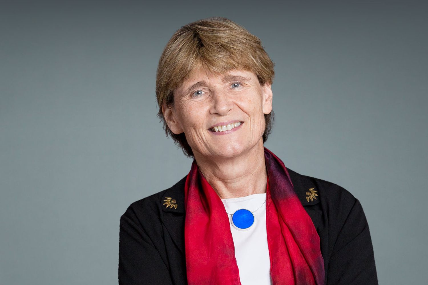
Study furthers radically new view of gene control

Along the genome, proteins form liquid-like droplets that appear to boost the expression of particular genes.

Anne Trafton | MIT News Office
August 8, 2019

In recent years, MIT scientists have developed a new model for how key genes are controlled that suggests the cellular machinery that transcribes DNA into RNA forms specialized droplets called condensates. These droplets occur only at certain sites on the genome, helping to determine which genes are expressed in different types of cells.

In a new study that supports that model, researchers at MIT and the Whitehead Institute for Biomedical Research have discovered physical interactions between proteins and with DNA that help explain why these droplets, which stimulate the transcription of nearby genes, tend to cluster along specific stretches of DNA known as super enhancers. These enhancer regions do not encode proteins but instead regulate other genes.

“This study provides a fundamentally important new approach to deciphering how the ‘dark matter’ in our genome functions in gene control,” says Richard Young, an MIT professor of biology and member of the Whitehead Institute.

Young is one of the senior authors of the paper, along with Phillip Sharp, an MIT Institute Professor and member of MIT’s Koch Institute for Integrative Cancer Research; and Arup K. Chakraborty, the Robert T. Haslam Professor in Chemical Engineering, a professor of physics and chemistry, and a member of MIT’s Institute for Medical Engineering and Science and the Ragon Institute of MGH, MIT, and Harvard.

Graduate student Krishna Shrinivas and postdoc Benjamin Sabari are the lead authors of the paper, which appears in Molecular Cell on Aug. 8.

“A biochemical factory”

Every cell in an organism has an identical genome, but cells such as neurons or heart cells express different subsets of those genes, allowing them to carry out their specialized functions. Previous research has shown that many of these genes are located near super enhancers, which bind to proteins called transcription factors that stimulate the copying of nearby genes into RNA.

About three years ago, Sharp, Young, and Chakraborty joined forces to try to model the interactions that occur at enhancers. In a 2017 Cell paper, based on computational studies, they hypothesized that in these regions, transcription factors form droplets called phase-separated condensates. Similar to droplets of oil suspended in salad dressing, these condensates are collections of molecules that form distinct cellular compartments but have no membrane separating them from the rest of the cell.

In a 2018 Science paper, the researchers showed that these dynamic droplets do form at super enhancer locations. Made of clusters of transcription factors and other molecules, these droplets attract enzymes such as RNA polymerases that are needed to copy DNA into messenger RNA, keeping gene transcription active at specific sites.

“We had demonstrated that the transcription machinery forms liquid-like droplets at certain regulatory regions on our genome, however we didn’t fully understand how or why these dewdrops of biological molecules only seemed to condense around specific points on our genome,” Shrinivas says.

As one possible explanation for that site specificity, the research team hypothesized that weak interactions between intrinsically disordered regions of transcription factors and other transcriptional molecules, along with specific interactions between transcription factors and particular DNA elements, might determine whether a condensate forms at a particular stretch of DNA. Biologists have traditionally focused on “lock-and-key” style interactions between rigidly structured protein segments to explain most cellular processes, but more recent evidence suggests that weak interactions between floppy protein regions also play an important role in cell activities.

In this study, computational modeling and experimentation revealed that the cumulative force of these weak interactions conspire together with transcription factor-DNA interactions to determine whether a condensate of transcription factors will form at a particular site on the genome. Different cell types produce different transcription factors, which bind to different enhancers. When many transcription factors cluster around the same enhancers, weak interactions between the proteins are more likely to occur. Once a critical threshold concentration is reached, condensates form.

“Creating these local high concentrations within the crowded environment of the cell enables the right material to be in the right place at the right time to carry out the multiple steps required to activate a gene,” Sabari says. “Our current study begins to tease apart how certain regions of the genome are capable of pulling off this trick.”

These droplets form on a timescale of seconds to minutes, and they blink in and out of existence depending on a cell’s needs.

“It’s an on-demand biochemical factory that cells can form and dissolve, as and when they need it,” Chakraborty says. “When certain signals happen at the right locus on a gene, the condensates form, which concentrates all of the transcription molecules. Transcription happens, and when the cells are done with that task, they get rid of them.”

A new view

Weak cooperative interactions between proteins may also play an important role in evolution, the researchers proposed in a 2018 Proceedings of the National Academy of Sciences paper. The sequences of intrinsically disordered regions of transcription factors need to change only a little to evolve new types of specific functionality. In contrast, evolving new specific functions via “lock-and-key” interactions requires much more significant changes.

“If you think about how biological systems have evolved, they have been able to respond to different conditions without creating new genes. We don’t have any more genes that a fruit fly, yet we’re much more complex in many of our functions,” Sharp says. “The incremental expanding and contracting of these intrinsically disordered domains could explain a large part of how that evolution happens.”

Similar condensates appear to play a variety of other roles in biological systems, offering a new way to look at how the interior of a cell is organized. Instead of floating through the cytoplasm and randomly bumping into other molecules, proteins involved in processes such as relaying molecular signals may transiently form droplets that help them interact with the right partners.

“This is a very exciting turn in the field of cell biology,” Sharp says. “It is a whole new way of looking at biological systems that is richer and more meaningful.”

Some of the MIT researchers, led by Young, have helped form a company called Dewpoint Therapeutics to develop potential treatments for a wide variety of diseases by exploiting cellular condensates. There is emerging evidence that cancer cells use condensates to control sets of genes that promote cancer, and condensates have also been linked to neurodegenerative disorders such as amyotrophic lateral sclerosis (ALS) and Huntington’s disease.

The research was funded by the National Science Foundation, the National Institutes of Health, and the Koch Institute Support (core) Grant from the National Cancer Institute.

How brain cells pick which connections to keep

Novel study shows protein CPG15 acts as a molecular proxy of experience to mark synapses for stabilization.

David Orenstein | Picower Institute for Learning and Memory
August 7, 2019

Brain cells, or neurons, constantly tinker with their circuit connections, a crucial feature that allows the brain to store and process information. While neurons frequently test out new potential partners through transient contacts, only a fraction of fledging junctions, called synapses, are selected to become permanent.

The major criterion for excitatory synapse selection is based on how well they engage in response to experience-driven neural activity, but how such selection is implemented at the molecular level has been unclear. In a new study, MIT neuroscientists have identified the gene and protein, CPG15, that allows experience to tap a synapse as a keeper.

In a series of novel experiments described in Cell Reports, the team at MIT’s Picower Institute for Learning and Memory used multi-spectral, high-resolution two-photon microscopy to literally watch potential synapses come and go in the visual cortex of mice — both in the light, or normal visual experience, and in the darkness, where there is no visual input. By comparing observations made in normal mice and ones engineered to lack CPG15, they were able to show that the protein is required in order for visual experience to facilitate the transition of nascent excitatory synapses to permanence.

Mice engineered to lack CPG15 only exhibit one behavioral deficiency: They learn much more slowly than normal mice, says senior author Elly Nedivi, the William R. (1964) and Linda R. Young Professor of Neuroscience in the Picower Institute and a professor of brain and cognitive sciences at MIT. They need more trials and repetitions to learn associations that other mice can learn quickly. The new study suggests that’s because without CPG15, they must rely on circuits where synapses simply happened to take hold, rather than on a circuit architecture that has been refined by experience for optimal efficiency.

“Learning and memory are really specific manifestations of our brain’s ability in general to constantly adapt and change in response to our environment,” Nedivi says. “It’s not that the circuits aren’t there in mice lacking CPG15, they just don’t have that feature — which is really important — of being optimized through use.”

Watching in light and darkness

The first experiment reported in the paper, led by former MIT postdoc Jaichandar Subramanian, who is now an assistant professor at the University of Kansas, is a contribution to neuroscience in and of itself, Nedivi says. The novel labeling and imaging technologies implemented in the study, she says, allowed tracking key events in synapse formation with unprecedented spatial and temporal resolution. The study resolved the emergence of “dendritic spines,” which are the structural protrusions on which excitatory synapses are formed, and the recruitment of the synaptic scaffold, PSD95, that signals that a synapse is there to stay.

The team tracked specially labeled neurons in the visual cortex of mice after normal visual experience, and after two weeks in darkness. To their surprise, they saw that spines would routinely arise and then typically disappear again at the same rate regardless of whether the mice were in light or darkness. This careful scrutiny of spines confirmed that experience doesn’t matter for spine formation, Nedivi said. That upends a common assumption in the field, which held that experience was necessary for spines to even emerge.

By keeping track of the presence of PSD95 they could confirm that the synapses that became stabilized during normal visual experience were the ones that had accumulated that protein. But the question remained: How does experience drive PSD95 to the synapse? The team hypothesized that CPG15, which is activity dependent and associated with synapse stabilization, does that job.

CPG15 represents experience

To investigate that, they repeated the same light-versus-dark experiences, but this time in mice engineered to lack CPG15. In the normal mice, there was much more PSD95 recruitment during the light phase than during the dark, but in the mice without CPG15, the experience of seeing in the light never made a difference. It was as if CPG15-less mice in the light were like normal mice in the dark.

Later they tried another experiment testing whether the low PSD95 recruitment seen when normal mice were in the dark could be rescued by exogenous expression of CPG15. Indeed, PSD95 recruitment shot up, as if the animals were exposed to visual experience. This showed that CPG15 not only carries the message of experience in the light, it can actually substitute for it in the dark, essentially “tricking” PSD95 into acting as if experience had called upon it.

“This is a very exciting result, because it shows that CPG15 is not just required for experience-dependent synapse selection, but it’s also sufficient,” says Nedivi, “That’s unique in relation to all other molecules that are involved in synaptic plasticity.”

A new model and method

In all, the paper’s data allowed Nedivi to propose a new model of experience-dependent synapse stabilization: Regardless of neural activity or experience, spines emerge with fledgling excitatory synapses and the receptors needed for further development. If activity and experience send CPG15 their way, that draws in PSD95 and the synapse stabilizes. If experience doesn’t involve the synapse, it gets no CPG15, very likely no PSD95, and the spine withers away.

The paper potentially has significance beyond the findings about experience-dependent synapse stabilization, Nedivi says. The method it describes of closely monitoring the growth or withering of spines and synapses amid a manipulation (like knocking out or modifying a gene) allows for a whole raft of studies in which examining how a gene, or a drug, or other factors affect synapses.

“You can apply this to any disease model and use this very sensitive tool for seeing what might be wrong at the synapse,” she says.

In addition to Nedivi and Subramanian, the paper’s other authors are Katrin Michel and Marc Benoit.

The National Institutes of Health and the JPB Foundation provided support for the research.

A new way to block unwanted genetic transfer

Researchers identify a strategy to prevent mobile genetic elements from breaching the bacterial cell wall.

Raleigh McElvery | Department of Biology
August 6, 2019

We receive half of our genes from each biological parent, so there’s no avoiding inheriting a blend of characteristics from both. Yet, for single-celled organisms like bacteria that reproduce by splitting into two identical cells, injecting variety into the gene pool isn’t so easy. Random mutations add some diversity, but there’s a much faster way for bacteria to reshuffle their genes and confer evolutionary advantages like antibiotic resistance or pathogenicity.

Known as horizontal gene transfer, this process permits bacteria to pass pieces of DNA to their peers, in some cases allowing those genes to be integrated into the recipient’s genome and passed down to the next generation.

The Grossman lab in the MIT Department of Biology studies one class of mobile DNA, known as integrative and conjugative elements (ICEs). While ICEs contain genes that can be beneficial to the recipient bacterium, there’s also a catch — receiving a duplicate copy of an ICE is wasteful, and possibly lethal. The biologists recently uncovered a new system by which one particular ICE, ICEBs1, blocks a donor bacterium from delivering a second, potentially deadly copy.

“Understanding how these elements function and how they’re regulated will allow us to determine what drives microbial evolution,” says Alan Grossman, department head and senior author on the study. “These findings not only provide insight into how bacteria block unwanted genetic transfer, but also how we might eventually engineer this system to our own advantage.”

Former graduate student Monika Avello PhD ’18 and current graduate student Kathleen Davis are co-first authors on the study, which appeared online in Molecular Microbiology on July 30.

Checks and balances

Although plasmids are perhaps the best-known mediators of horizontal transfer, ICEs not only outnumber plasmids in most bacterial species, they also come with their own tools to exit the donor, enter the recipient, and integrate themselves into the recipient’s chromosome. Once the donor bacterium makes contact with the recipient, the machinery encoded by the ICE can pump the ICE DNA from one cell to the other through a tiny channel.

For horizontal transfer to proceed, there are physical barriers to overcome, especially in so-called Gram-positive bacteria, which boast thicker cell walls than their Gram-negative counterparts, despite being less widely studied. According to Davis, the transfer machinery essentially has to “punch a hole” through the recipient cell. “It’s a rough ride and a waste of energy for the recipient if that cell already contains an ICE with a specific set of genes,” she says.

Sure, ICEs are “selfish bits of DNA” that persist by spreading themselves as widely as possible, but in order to do so they must not interfere with their host cell’s ability to survive. As Avello explains, ICEs can’t just disseminate their DNA “without certain checks and balances.”

“There comes a point where this transfer comes at a cost to the bacteria or doesn’t make sense for the element,” she says. “This study is beginning to get at the question of when, why, and how ICEs might want to block transfer.”

The Grossman lab works in the Gram-positive Bacillus subtilis, and had previously discovered two mechanisms by which ICEBs1 could prevent redundant transfer before it becomes lethal. The first, cell-cell signaling, involves the ICE in the recipient cell releasing a chemical cue that prohibits the donor’s transfer machinery from being assembled. The second, immunity, initiates if the duplicate copy is already inside the cell, and prevents the replicate from being integrated into the chromosome.

However, when the researchers tried eliminating both fail-safes simultaneously, rather than re-instating ICE transfer as they expected, the bacteria still managed to obstruct the duplicate copy. ICEBs1 seemed to have a third blocking strategy, but what might it be?

The third tactic

In this most recent study, they’ve identified the mysterious blocking mechanism as a type of “entry exclusion,” whereby the ICE in the recipient cell encodes molecular machinery that physically prevents the second copy from breaching the cell wall. Scientists had observed other mobile genetic elements capable of exclusion, but this was the first time anyone had witnessed this phenomenon for an ICE from Gram-positive bacteria, according to Avello.

The Grossman lab determined that this exclusion mechanism comes down to two key proteins. Avello identified the first protein, YddJ, expressed by the ICEBs1 in the recipient bacterium, forming a “protective coating” on the outside of the cell and blocking a second ICE from entering.

But the biologists still didn’t know which piece of transfer machinery YddJ was blocking, so Davis performed a screen and various genetic manipulations to pinpoint YddJ’s target. YddJ, it turned out, was obstructing another protein called ConG, which likely forms part of the transfer channel between the donor and recipient bacteria. Davis was surprised to find that, while Gram-negative ICEs encode a protein that’s quite similar to ConG, the Gram-negative YddJ equivalent is actually much different.

“This just goes to show that you can’t assume the transfer machinery in Gram-positive ICEs like ICEBs1 are the same as the well-studied Gram-negative ICEs,” she says.

The team concluded that ICEBs1 must have three different mechanisms to prevent duplicate transfer: the two they’d previously uncovered plus this new one, exclusion.

Cell-cell signaling allows a cell to spread the word to its neighbors that it already has a copy of ICEBs1, so there’s no need to bother assembling the transfer machinery. If this fails, exclusion kicks in to physically block the transfer machinery from penetrating the recipient cell. If that proves unsuccessful and the second copy enters the recipient, immunity will initiate and prevent the second copy from being integrated into the recipient’s chromosome.

“Each mechanism acts at a different step, because none of them alone are 100 percent effective,” Grossman says. “That’s why it’s helpful to have multiple mechanisms.”

They don’t know all the details of this transfer machinery just yet, he adds, but they do know that YddJ and ConG are key players.

“This initial description of the ICEBs1 exclusion system represents the first report that provides mechanistic insights into exclusion in Gram-positive bacteria, and one of only a few mechanistic studies of exclusion in any conjugation system,” says Gary Dunny, a professor of microbiology and immunology at the University of Minnesota who was not involved in the study. “This work is significant medically because ICEs can carry “cargo” genes such as those conferring antibiotic resistance, and also of importance to our basic understanding of horizontal gene transfer systems and how they evolve.”

As researchers continue to probe this blocking mechanism, it might be possible to leverage ICE exclusion to design bacteria with specific functions. For instance, they could engineer the gut microbiome and introduce beneficial genes to help with digestion. Or, one day, they could perhaps block horizontal gene transfer to combat antibiotic resistance.

“We had suspected that Gram-positive ICEs might be capable of exclusion, but we didn’t have proof before this,” Avello says. Now, researchers can start to speculate about how pathogenic Gram-positive species might control the movement of ICEs throughout a bacterial population, with possible ramifications for disease research.

This work was funded by research and predoctoral training grants from the National Institute of General Medical Sciences of the National Institutes of Health.

Speeding up drug discovery for brain diseases

Whitehead Institute team finds drugs that activate a key brain gene; initial tests in cells and mice show promise for rare, untreatable neurodevelopmental disorder.

Nicole Davis
August 2, 2019

A research team led by Whitehead Institute scientists has identified 30 distinct chemical compounds — 20 of which are drugs undergoing clinical trial or have already been approved by the FDA — that boost the protein production activity of a critical gene in the brain and improve symptoms of Rett syndrome, a rare neurodevelopmental condition that often provokes autism-like behaviors in patients. The new study, conducted in human cells and mice, helps illuminate the biology of an important gene, called KCC2, which is implicated in a variety of brain diseases, including autism, epilepsy, schizophrenia, and depression. The researchers’ findings, published in the July 31 online issue of Science Translational Medicine, could help spur the development of new treatments for a host of devastating brain disorders.

“There’s increasing evidence that KCC2 plays important roles in several different disorders of the brain, suggesting that it may act as a common driver of neurological dysfunction,” says senior author Rudolf Jaenisch, a founding member of Whitehead Institute and professor of biology at MIT. “These drugs we’ve identified may help speed up the development of much-needed treatments.”

KCC2 works exclusively in the brain and spinal cord, carrying ions in and out of specialized cells known as neurons. This shuttling of electrically charged molecules helps maintain the cells’ electrochemical makeup, enabling neurons to fire when they need to and to remain idle when they don’t. If this delicate balance is upset, brain function and development go awry.

Disruptions in KCC2 function have been linked to several human brain disorders, including Rett syndrome (RTT), a progressive and often debilitating disorder that typically emerges early in life in girls and can involve disordered movement, seizures, and communication difficulties. Currently, there is no effective treatment for RTT.

Jaenisch and his colleagues, led by first author Xin Tang, devised a high-throughput screen assay to uncover drugs that increase KCC2 gene activity. Using CRISPR/Cas9 genome editing and stem cell technologies, they engineered human neurons to provide rapid readouts of the amount of KCC2 protein produced. The researchers created these so-called reporter cells from both healthy human neurons as well as RTT neurons that carry disease-causing mutations in the MECP2 gene. These reporter neurons were then fed into a drug-screening pipeline to find chemical compounds that can enhance KCC2 gene activity.

Tang and his colleagues screened over 900 chemical compounds, focusing on those that have been FDA-approved for use in other conditions, such as cancer, or have undergone at least some level of clinical testing. “The beauty of this approach is that many of these drugs have been studied in the context of non-brain diseases, so the mechanisms of action are known,” says Tang. “Such molecular insights enable us to learn how the KCC2 gene is regulated in neurons, while also identifying compounds with potential therapeutic value.”

The Whitehead Institute team identified a total of 30 drugs with KCC2-enhancing activity. These compounds, referred to as KEECs (short for KCC2 expression-enhancing compounds), work in a variety of ways. Some block a molecular pathway, called FLT3, which is found to be overactive in some forms of leukemia. Others inhibit the GSK3b pathway that has been implicated in several brain diseases. Another KEEC acts on SIRT1, which plays a key role in a variety of biological processes, including aging.

In followup experiments, the researchers exposed RTT neurons and mouse models to KEEC treatment and found that some compounds can reverse certain defects associated with the disease, including abnormalities in neuronal signaling, breathing, and movement. These efforts were made possible by a collaboration with Mriganka Sur’s group at the Picower Institute for Learning and Memory, in which Keji Li and colleagues led the behavioral experiments in mice that were essential for revealing the drugs’ potency.

“Our findings illustrate the power of an unbiased approach for discovering drugs that could significantly improve the treatment of neurological disease,” says Jaenisch. “And because we are starting with known drugs, the path to clinical translation is likely to be much shorter.”

In addition to speeding up drug development for Rett syndrome, the researchers’ unique drug-screening strategy, which harnesses an engineered gene-specific reporter to unearth promising drugs, can also be applied to other important disease-related genes in the brain. “Many seemingly distinct brain diseases share common root causes of abnormal gene expression or disrupted signaling pathways,” says Tang. “We believe our method has broad applicability and could help catalyze therapeutic discovery for a wide range of neurological conditions.”

Support for this work was provided by the National Institutes of Health, the Simons Foundation Autism Research Initiative, the Simons Center for the Social Brain at MIT, the Rett Syndrome Research Trust, the International Rett Syndrome Foundation, the Damon Runyon Cancer Foundation, and the National Cancer Institute.

MIT “Russian Doll” tech lands $7.9M international award to fight brain tumors

Researchers from MIT will work with teams in the U.K. and Europe to use nanoparticles to carry multiple drug therapies to treat glioblastoma.

Koch Institute
July 30, 2019

Tiny “Russian doll-like” particles that deliver multiple drugs to brain tumors, developed by researchers at MIT and funded by Cancer Research UK, are at the center of a new international collaboration.

Professor Paula Hammond from the Department of Chemical Engineering developed the nanoparticle technology, which will be used in an effort to treat glioblastoma — the most aggressive and deadly type of brain tumor.

Hammond will be working with Professor Michael Yaffe from the Department of Biological Engineering to determine the combinations of drugs placed within the particles, and the order and timing in which the drugs are released.

The nanoparticles — 1,000 times smaller than a human hair — are coated in a protein called transferrin, which helps them cross the blood-brain barrier. This is a membrane that keeps a tight check on anything trying to get in to the brain, including drugs.

Not only are the nanoparticles able to access hard-to-reach areas of the brain, they have also been designed to carry multiple cancer drugs at once by holding them inside layers, similarly to the way Russian dolls fit inside one another.

To make the nanoparticles even more effective, they will carry signals on their surface so that they are only taken up by brain tumor cells. This means that healthy cells should be left untouched, which will minimize the side effects of treatment.

The researchers, who are based at the Koch Institute for Integrative Cancer Research, are also working with Professor Forest White from the Department of Biological Engineering. The group are one of three international teams to have been given Cancer Research UK Brain Tumor Awards — in partnership with The Brain Tumour Charity — receiving $7.9 million of funding. The awards are designed to accelerate the pace of brain tumor research. Altogether, teams were awarded a total of $23 million.

Just last year, around 24,200 people in the United States were diagnosed with brain tumors. With around 17,500 deaths from brain tumors in the same year, survival remains tragically low.

Brain tumors represent one of the hardest types of cancer to treat because not enough is known about what starts and drives the disease, and current treatments are not effective enough.

The researchers from MIT will now work with teams in the U.K. and Europe to use the nanoparticles to carry multiple drug therapies to treat glioblastoma.

Early research carried out in the lab has already shown that nanoparticles loaded with two different drugs were able to shrink glioblastomas in mice. The team has also demonstrated that the nanoparticles can kill lymphoma cells grown in the lab, and they are also exploring their use in ovarian cancer.

The Cancer Research UK Brain Tumor Award will now allow the researchers and their collaborators to use different drug combinations to find the best parameters to tackle glioblastomas.

Drugs that have already been approved, as well as experimental drugs that have passed initial safety testing in people, will be used. Because of this, if an effective drug combination is found, the team won’t have to navigate the initial regulatory hurdles needed to get them into clinical testing, which could help get promising treatments to patients faster.

“Glioblastoma is particularly challenging because we want to get highly effective but toxic drug combinations safely across the blood-brain barrier, but also want our nanoparticles to avoid healthy brain cells and only target the cancer cells,” Hammond says. “We are very excited about this alliance between the MIT Koch Institute and our colleagues in Edinburgh to address these critical challenges.”

Biologists and mathematicians team up to explore tissue folding

An algorithm developed to study the structure of galaxies helps explain a key feature of embryonic development.

Anne Trafton | MIT News Office
July 25, 2019

As embryos develop, they follow predetermined patterns of tissue folding, so that individuals of the same species end up with nearly identically shaped organs and very similar body shapes.

MIT scientists have now discovered a key feature of embryonic tissue that helps explain how this process is carried out so faithfully each time. In a study of fruit flies, they found that the reproducibility of tissue folding is generated by a network of proteins that connect like a fishing net, creating many alternative pathways that tissues can use to fold the right way.

“What we found is that there’s a lot of redundancy in the network,” says Adam Martin, an MIT associate professor of biology and the senior author of the study. “The cells are interacting and connecting with each other mechanically, but you don’t see individual cells taking on an all-important role. This means that if one cell gets damaged, other cells can still connect to disparate parts of the tissue.”

To uncover these network features, Martin worked with Jörn Dunkel, an MIT associate professor of physical applied mathematics and an author of the paper, to apply an algorithm normally used by astronomers to study the structure of galaxies.

Hannah Yevick, an MIT postdoc, is the lead author of the study, which appears today in Developmental Cell. Graduate student Pearson Miller is also an author of the paper.

A safety net

During embryonic development, tissues change their shape through a process known as morphogenesis. One important way tissues change shape is to fold, which allows flat sheets of embryonic cells to become tubes and other important shapes for organs and other body parts. Previous studies in fruit flies have shown that even when some of these embryonic cells are damaged, sheets can still fold into their correct shapes.

“This is a process that’s fairly reproducible, and so we wanted to know what makes it so robust,” Martin says.

In this study, the researchers focused on the process of gastrulation, during which the embryo is reorganized from a single-layered sphere to a more complex structure with multiple layers. This process, and other morphogenetic processes similar to fruit fly tissue folding, also occur in human embryos. The embryonic cells involved in gastrulation contain in their cytoplasm proteins called myosin and actin, which form cables and connect at junctions between cells to form a network across the tissue. Martin and Yevick had hypothesized that the network of cell connectivity might play a role in the robustness of the tissue folding, but until now, there was no good way to trace the connections of the network.

To achieve that, Martin’s lab joined forces with Dunkel, who studies the physics of soft surfaces and flowing matter — for example, wrinkle formation and patterns of bacterial streaming. For this study, Dunkel had the idea to apply a mathematical procedure that can identify topological features of a three-dimensional structure, analogous to ridges and valleys in a landscape. Astronomers use this algorithm to identify galaxies, and in this case, the researchers used it to trace the actomyosin networks across and between the cells in a sheet of tissue.

“Once you have the network, you can apply standard methods from network analysis — the same kind of analysis that you would apply to streets or other transport networks, or the blood circulation network, or any other form of network,” Dunkel says.

Among other things, this kind of analysis can reveal the structure of the network and how efficiently information flows along it. One important question is how well a network adapts if part of it gets damaged or blocked. The MIT team found that the actomyosin network contains a great deal of redundancy — that is, most of the “nodes” of the network are connected to many other nodes.

This built-in redundancy is analogous to a good public transit system, where if one bus or train line goes down, you can still get to your destination. Because cells can generate mechanical tension along many different pathways, they can fold the right way even if many of the cells in the network are damaged.

“If you and I are holding a single rope, and then we cut it in the middle, it would come apart. But if you have a net, and cut it in some places, it still stays globally connected and can transmit forces, as long as you don’t cut all of it,” Dunkel says.

Folding framework

The researchers also found that the connections between cells preferentially organize themselves to run in the same direction as the furrow that forms in the early stages of folding.

“We think this is setting up a frame around which the tissue will adopt its shape,” Martin says. “If you prevent the directionality of the connections, then what happens is you can still get folding but it will fold along the wrong axis.”

Although this study was done in fruit flies, similar folding occurs in vertebrates (including humans) during the formation of the neural tube, which is the precursor to the brain and spinal cord. Martin now plans to apply the techniques he used in fruit flies to see if the actomyosin network is organized the same way in the neural tube of mice. Defects in the closure of the neural tube can lead to birth defects such as spina bifida.

“We would like to understand how it goes wrong,” Martin says. “It’s still not clear whether it’s the sealing up of the tube that’s problematic or whether there are defects in the folding process.”

The research was funded by the National Institute of General Medical Sciences and the James S. McDonnell Foundation.

Genetic study takes research on sex differences to new heights

Differences in male and female gene expression, including those contributing to height differences, found throughout the body in humans and other mammals.

Greta Friar | Whitehead Institute
July 19, 2019

Throughout the animal kingdom, males and females frequently exhibit sexual dimorphism: differences in characteristic traits that often make it easy to tell them apart. In mammals, one of the most common sex-biased traits is size, with males typically being larger than females. This is true in humans: Men are, on average, taller than women. However, biological differences among males and females aren’t limited to physical traits like height. They’re also common in disease. For example, women are much more likely to develop autoimmune diseases, while men are more likely to develop cardiovascular diseases.

In spite of the widespread nature of these sex biases, and their significant implications for medical research and treatment, little is known about the underlying biology that causes sex differences in characteristic traits or disease. In order to address this gap in understanding, Whitehead Institute Director David Page has transformed the focus of his lab in recent years from studying the X and Y sex chromosomes to working to understand the broader biology of sex differences throughout the body. In a paper published in Science, Page, a professor of biology at MIT and a Howard Hughes Medical Institute investigator; Sahin Naqvi, first author and former MIT graduate student (now a postdoc at Stanford University); and colleagues present the results of a wide-ranging investigation into sex biases in gene expression, revealing differences in the levels at which particular genes are expressed in males versus females.

The researchers’ findings span 12 tissue types in five species of mammals, including humans, and led to the discovery that a combination of sex-biased genes accounts for approximately 12 percent of the average height difference between men and women. This finding demonstrates a functional role for sex-biased gene expression in contributing to sex differences. The researchers also found that the majority of sex biases in gene expression are not shared between mammalian species, suggesting that — in some cases — sex-biased gene expression that can contribute to disease may differ between humans and the animals used as models in medical research.

Having the same gene expressed at different levels in each sex is one way to perpetuate sex differences in traits in spite of the genetic similarity of males and females within a species — since with the exception of the 46th chromosome (the Y in males or the second X in females), the sexes share the same pool of genes. For example, if a tall parent passes on a gene associated with an increase in height to both a son and a daughter, but the gene has male-biased expression, then that gene will be more highly expressed in the son, and so may contribute more height to the son than the daughter.

The researchers searched for sex-biased genes in tissues across the body in humans, macaques, mice, rats, and dogs, and they found hundreds of examples in every tissue. They used height for their first demonstration of the contribution of sex-biased gene expression to sex differences in traits because height is an easy-to-measure and heavily studied trait in quantitative genetics.

“Discovering contributions of sex-biased gene expression to height is exciting because identifying the determinants of height is a classic, century-old problem, and yet by looking at sex differences in this new way we were able to provide new insights,” Page says. “My hope is that we and other researchers can repeat this model to similarly gain new insights into diseases that show sex bias.”

Because height is so well studied, the researchers had access to public data on the identity of hundreds of genes that affect height. Naqvi decided to see how many of those height genes appeared in the researchers’ new dataset of sex-biased genes, and whether the genes’ sex biases corresponded to the expected effects on height. He found that sex-biased gene expression contributed approximately 1.6 centimeters to the average height difference between men and women, or 12 percent of the overall observed difference.

The scope of the researchers’ findings goes beyond height, however. Their database contains thousands of sex-biased genes. Slightly less than a quarter of the sex-biased genes that they catalogued appear to have evolved that sex bias in an early mammalian ancestor, and to have maintained that sex bias today in at least four of the five species studied. The majority of the genes appear to have evolved their sex biases more recently, and are specific to either one species or a certain lineage, such as rodents or primates.

Whether or not a sex-biased gene is shared across species is a particularly important consideration for medical and pharmaceutical research using animal models. For example, previous research identified certain genetic variants that increase the risk of Type 2 diabetes specifically in women; however, the same variants increase the risk of Type 2 diabetes indiscriminately in male and female mice. Therefore, mice would not be a good model to study the genetic basis of this sex difference in humans. Even when the animal appears to have the same sex difference in disease as humans, the specific sex-biased genes involved might be different. Based on their finding that most sex bias is not shared between species, Page and colleagues urge researchers to use caution when picking an animal model to study sex differences at the level of gene expression.

“We’re not saying to avoid animal models in sex-differences research, only not to take for granted that the sex-biased gene expression behind a trait or disease observed in an animal will be the same as that in humans. Now that researchers have species and tissue-specific data available to them, we hope they will use it to inform their interpretation of results from animal models,” Naqvi says.

The researchers have also begun to explore what exactly causes sex-biased expression of genes not found on the sex chromosomes. Naqvi discovered a mechanism by which sex-biased expression may be enabled: through sex-biased transcription factors, proteins that help to regulate gene expression. Transcription factors bind to specific DNA sequences called motifs, and he found that certain sex-biased genes had the motif for a sex-biased transcription factor in their promoter regions, the sections of DNA that turn on gene expression. This means that, for example, a male-biased transcription factor was selectively binding to the promoter region for, and so increasing the expression of, male-biased genes — and likewise for female-biased transcription factors and female-biased genes. The question of what regulates the transcription factors remains for further study — but all sex differences are ultimately controlled by either the sex chromosomes or sex hormones.

The researchers see the collective findings of this paper as a foundation for future sex-differences research.

“We’re beginning to build the infrastructure for a systematic understanding of sex biases throughout the body,” Page says. “We hope these datasets are used for further research, and we hope this work gives people a greater appreciation of the need for, and value of, research into the molecular differences in male and female biology.”

This work was supported by Biogen, Whitehead Institute, National Institutes of Health, Howard Hughes Medical Institute, and generous gifts from Brit and Alexander d’Arbeloff and Arthur W. and Carol Tobin Brill.

Unmasking mutant cancer cells

A new dosing regimen for an old cancer drug shows new promise as an immunotherapy.

Bendta Schroeder | Koch Institute
July 16, 2019

As cancer cells progress, they accumulate hundreds and even thousands of genetic and epigenetic changes, resulting in protein expression profiles that are radically different from that of healthy cells. But despite their heavily mutated proteome, cancer cells can evade recognition and attack by the immune system.

Immunotherapies, particularly checkpoint inhibitors that reinvigorate exhausted T cells, have revolutionized the treatment of certain forms of cancer. These breakthrough therapies have resulted in unprecedented response rates for some patients. Unfortunately, most cancers fail to respond to immunotherapies and new strategies are therefore needed to realize their full potential.

A team of cancer biologists including members of the laboratories of David H. Koch Professor of Biology Tyler Jacks, director of the Koch Institute for Integrative Cancer Research at MIT, and fellow Koch Institute member Forest White, the Ned C. and Janet Bemis Rice Professor and member of the MIT Center for Precision Cancer Medicine, took a complementary approach to boosting the immune system.

Although cancer cells are rife with mutant proteins, few of those proteins appear on a cell’s surface, where they can be recognized by immune cells. The researchers repurposed a well-studied class of anti-cancer drugs, heat shock protein 90 (HSP90) inhibitors, that make cancer cells easier to recognize by revealing their mutant proteomes.

Many HSP90 inhibitors have been studied extensively for the past several decades as potential cancer treatments. HSP90 protects the folded structure of a number of proteins when cells undergo stress, and in cancer cells plays an important role in stabilizing protein structure undermined by pervasive mutations. However, despite promising preclinical evidence, HSP90 inhibitors have produced discouraging outcomes in clinical trials, and none have achieved FDA approval.

In a study appearing in Clinical Cancer Research, the researchers identified a potential reason behind those disappointing results. HSP90 inhibitors have only been clinically tested at bolus doses — intermittent, large doses — that often result in unwanted side effects in patients.

RNA profiling of human clinical samples and cultured cancer cell lines revealed that this bolus-dosing schedule results in the profound suppression of immune activity as well as the activation of heat shock factor 1 protein (HSF1). Not only does HSF1 activate the cell’s heat shock response, which counteracts the effect of the HSP90 inhibitor, but it is known to be a powerful enabler of cancer cell malignancy.

In striking contrast, the researchers used cancer mouse models with intact immune systems to show that sustained, low-level dosing of HSP90 inhibitors avoids triggering both the heat shock response and the immunosuppression associated with high doses.

Using a method devised by the White lab that combines mass spectrometry-based proteomics and computational modeling, the researchers discovered that the new dosing regimen increased the number and diversity of peptides (protein fragments) on the cell surface. These peptides, which the team found to be released by HSP90 during sustained low-level inhibition, were then free to be taken up by the cell’s antigen-presenting machinery and used to flag patrolling immune cells.

“These results connect a fundamental aspect of cell biology — protein folding — to anti-tumor immune responses” says lead author Alex Jaeger, a postdoctoral fellow in the Jacks lab and a former member of the laboratory of the late MIT biologist and Professor Susan Lindquist, whose work inspired the study’s HSP90 dosing scheule. “Hopefully, our findings can reinvigorate interest in HSP90 inhibition as a complementary approach for immunotherapy.”

Using the new dosing regimen, the researchers were able to clear tumors in mouse models at drug concentrations that are 25-50 times lower than those used in clinical trials, significantly reducing the risk for toxic side effects in patients. Importantly, because several forms of HSP90 inhibitors have already undergone extensive clinical testing, the new dosing regimen can be tested in patients quickly.

This work was supported in part by the Damon Runyon Cancer Research Foundation, the Takeda Pharmaceuticals Immune Oncology Research Fund, and an MIT Training Grant in Environmental Science; foundational work on HSF1 was supported by the Koch Institute Frontier Research Program.

Meet the 2019 tenured professors in the School of Science

Eight faculty members are granted tenure in five science departments.

School of Science
July 10, 2019

MIT granted tenure to eight School of Science faculty members in the departments of Biology; Chemistry; Earth, Atmospheric and Planetary Sciences; Mathematics; and Physics.

William Detmold’s research within the area of theoretical particle and nuclear physics incorporates analytical methods, as well as the power of the world’s largest supercomputers, to understand the structure, dynamics, and interactions of particles like protons and to look for evidence of new physical laws at the sub-femtometer scale probed in experiments such as those at the Large Hadron Collider. He joined the Department of Physics in 2012 from the College of William and Mary, where he was an assistant professor. Prior to that, he was a research assistant professor at the University of Washington. He received his BS and PhD from the University of Adelaide in Australia in 1996 and 2002, respectively. Detmold is a researcher in the Center for Theoretical Physics in the Laboratory for Nuclear Science.

Semyon Dyatlov explores scattering theory, quantum chaos, and general relativity by employing microlocal analytical and dynamical system methods. He came to the Department of Mathematics as a research fellow in 2013 and became an assistant professor in 2015. He completed his doctorate in mathematics at the University of California at Berkeley in 2013 after receiving a BS in mathematics at Novosibirsk State University in Russia in 2008. Dyatlov spent time after finishing his PhD as a postdoc at the Mathematical Sciences Research Institute before moving to MIT.

Mary Gehring studies plant epigenetics. By using a combination of genetic, genomic, and molecular biology, she explores how plants inherit and interpret information that is not encoded in their DNA to better understand plant growth and development. Her lab focuses primarily on Arabidopsis thaliana, a small flowering plant that is a model species for plant research. Gehring joined the Department of Biology in 2010 after performing postdoctoral research at the Fred Hutchinson Cancer Research Center. She received her BA in biology from Williams College in 1998 and her doctorate from the University of California at Berkeley in 2005. She is also a member of the Whitehead Institute for Biomedical Research.

David McGee performs research in the field of paleoclimate, merging information from stalagmites, lake deposits, and marine sediments with insights from models and theory to understand how precipitation patterns and atmospheric circulation varied in the past. He came to MIT in 2012, joining the Department of Earth, Atmospheric and Planetary Sciences after completing a NOAA Climate and Global Change Postdoctoral Fellowship at the University of Minnesota. Before that, he attended Carleton College for his BA in geology in 1993-97, Chatham College for an MA in teaching from 1999 to 2003, Tulane University for his MS from 2004 to 2006, and Columbia University for his PhD from 2006 to 2009. McGee is the director of the MIT Terrascope First-Year Learning Community, a role he has held for the past four years.

Ankur Moitra works at the interface between theoretical computer science and machine learning by developing algorithms with provable guarantees and foundations for reasoning about their behavior. He joined the Department of Mathematics in 2013. Prior to that, he received his BS in electrical and computer engineering from Cornell University in 2007, and his MS and PhD in computer science from MIT in 2009 and 2011, respectively. He was a National Science Foundation postdoc at the Institute for Advanced Study until 2013. Moitra was a 2018 recipient of a School of Science Teaching Prize. He is also a principal investigator in the Computer Science and Artificial Intelligence Laboratory (CSAIL) and a core member of the Center for Statistics.

Matthew Shoulders focuses on integrating biology and chemistry to understand how proteins function in the cellular setting, including proteins’ shape, quantity, and location within the body. This research area has important implications for genetic disorders and neurodegenerative diseases such as Alzheimer’s, diabetes, cancer, and viral infections. Shoulders’ lab works to elucidate, at the molecular level, how cells solve the protein-folding problem, and then uses that information to identify how diseases can develop and to provide insight into new targets for drug development. Shoulders joined the Department of Chemistry in 2012 after earning a BS in chemistry and minor in biochemistry from Virginia Tech in 2004 and a PhD in chemistry from the University of Wisconsin at Madison in 2009. He is also an associate member of the Broad Institute of MIT and Harvard, and a member of the MIT Center for Environmental Health Sciences.

Tracy Slatyer researches fundamental aspects of theoretical physics, answering questions about both visible and dark matter by searching for potential indications of new physics in astrophysical and cosmological data. She has developed and adapted novel techniques for data analysis, modeling, and calculations in quantum field theory; her work has also inspired a range of experimental investigations. The Department of Physics welcomed Slatyer in 2013 after she completed a three-year postdoctoral fellowship at the Institute for Advanced Study. She majored in theoretical physics as an undergraduate at the Australian National University, receiving a BS in 2005, and completed her PhD in physics at Harvard University in 2010. In 2017, Slatyer received the School of Science Prize in Graduate Teaching and was also named the first recipient of the school’s Future of Science Award. She is a member of the Center for Theoretical Physics in the Laboratory for Nuclear Science.

Michael Williams uses novel experimental methods to improve our knowledge of fundamental particles, including searching for new particles and forces, such as dark matter. He also works on advancing the usage of machine learning within the domain of particle physics research. He joined the Department of Physics in 2012. He previously attended Saint Vincent College as an undergraduate, where he double majored in mathematics and physics. Graduating in 2001, Williams then pursued a doctorate at Carnegie Mellon University, which he completed in 2007. From 2008 to 2012 he was a postdoc at Imperial College London. He is a member of the Laboratory for Nuclear Science.

Cancer biologists identify new drug combo

Two drugs that block cell division synergize to kill tumor cells.

Anne Trafton | MIT News Office
July 10, 2019

When it comes to killing cancer cells, two drugs are often better than one. Some drug combinations offer a one-two punch that kills cells more effectively, requires lower doses of each drug, and can help to prevent drug resistance.

MIT biologists have now found that by combining two existing classes of drugs, both of which target cancer cells’ ability to divide, they can dramatically boost the drugs’ killing power. This drug combination also appears to largely spare normal cells, because cancer cells divide differently than healthy cells, the researchers say. They hope a clinical trial of this combination can be started within a year or two.

“This is a combination of one class of drugs that a lot of people are already using, with another type of drug that multiple companies have been developing,” says Michael Yaffe, a David H. Koch Professor of Science and the director of the MIT Center for Precision Cancer Medicine. “I think this opens up the possibility of rapid translation of these findings in patients.”

The discovery was enabled by a new software program the researchers developed, which revealed that one of the drugs had a previously unknown mechanism of action that strongly enhances the effect of the other drug.

Yaffe, who is also a member of the Koch Institute for Integrative Cancer Research, is the senior author of the study, which appears in the July 10 issue of Cell Systems. Koch Institute research scientists Jesse Patterson and Brian Joughin are the first authors of the paper.

Unexpected synergy

Yaffe’s lab has a longstanding interest in analyzing cellular pathways that are active in cancer cells, to find how these pathways work together in signaling networks to create disease-specific vulnerabilities that can be targeted with multiple drugs. When the researchers began this study, they were looking for a drug that would amplify the effects of a type of drug known as a PLK1 inhibitor. Several PLK1 inhibitors, which interfere with cell division, have been developed, and some are now in phase 2 clinical trials.

Based on their previous work, the researchers knew that PLK1 inhibitors also produce a type of DNA and protein damage known as oxidation. They hypothesized that pairing PLK1 inhibitors with a drug that prevents cells from repairing oxidative damage could make them work even better.

To explore that possibility, the researchers tested a PLK1 inhibitor along with a drug called TH588, which blocks MTH1, an enzyme that helps cells counteract oxidative damage. This combination worked extremely well against many types of human cancer cells. In some cases, the researchers could use one-tenth of the original doses of each drug, given together, and achieve the same rates of cell death of either drug given on its own.

“It’s really striking,” Joughin says. “It’s more synergy than you generally see from a rationally designed combination.”

However, they soon realized that this synergy had nothing to do with oxidative damage. When the researchers treated cancer cells missing the gene for MTH1, which they thought was TH588’s target, they found that the drug combination still killed cancer cells at the same high rates.

“Then we were really stuck, because we had a good combination, but we didn’t know why it worked,” Yaffe says.

To solve the mystery, they developed a new software program that allowed them to identify the cellular networks most affected by the drugs. The researchers tested the drug combination in 29 different types of human cancer cells, then fed the data into the software, which compared the results to gene expression data for those cell lines. This allowed them to discover patterns of gene expression that were linked with higher or lower levels of synergy between the two drugs.

This analysis suggested that both drugs were targeting the mitotic spindle, a structure that forms when chromosomes align in the center of a cell as it prepares to divide. Experiments in the lab confirmed that this was correct. The researchers had already known that PLK1 inhibitors target the mitotic spindle, but they were surprised to see that TH588 affected the same structure.

“This combination that we found was very nonobvious,” Yaffe says. “I would never have given two drugs that both targeted the same process and expected anything better than just additive effects.”

“This is an exciting paper for two reasons,” says David Pellman, associate director for basic science at Dana-Farber/Harvard Cancer Center, who was not involved in the study. “First, Yaffe and colleagues make an important advance for the rational design of drug therapy combinations. Second, if you like scientific mysteries, this is a riveting example of molecular sleuthing. A drug that was thought to act in one way is unmasked to work through an entirely different mechanism.”

Disrupting mitosis

The researchers found that while both of the drugs they tested disrupt mitosis, they appear to do so in different ways. TH588 binds to microtubules, which form the mitotic spindle, and slows their assembly. Many similar microtubule inhibitors are already used clinically to treat cancer. The researchers showed that some of those microtubule inhibitors also synergize with PLK1 inhibitors, and they believe those would likely be more readily available for rapid use in patients than TH588, the drug they originally tested.

While the PLK1 protein is involved in multiple aspects of cell division and spindle formation, it’s not known exactly how PLK1 inhibitors interfere with the mitotic spindle to produce this synergy. Yaffe said he suspects they may block a motor protein that is necessary for chromosomes to travel along the spindle.

One potential benefit of this drug combination is that the synergistic effects appear to specifically target cancer cell division and not normal cell division. The researchers believe this could be because cancer cells are forced to rely on alternative strategies for cell division because they often have too many or too few chromosomes, a state known as aneuploidy.

“Based on the work we have done, we propose that this drug combination targets something fundamentally different about the way cancer cells divide, such as altered cell division checkpoints, chromosome number and structure, or other structural differences in cancer cells,” Patterson says.

The researchers are now working on identifying biomarkers that could help them to predict which patients would respond best to this drug combination. They are also trying to determine the exact function of PLK1 that is responsible for this synergy, in hopes of finding additional drugs that would block that interaction.

The research was funded by the National Institutes of Health, the Charles and Marjorie Holloway Foundation, the Ovarian Cancer Research Fund, the MIT Center for Precision Cancer Medicine, the Koch Institute Dana Farber/Harvard Cancer Center Bridge Project, an American Cancer Society Postdoctoral Fellowship, the Koch Institute Support (core) Grant from the National Cancer Institute, and the Center for Environmental Health Support Grant.