In a study from the lab of Whitehead Institute Member David Bartel, researchers have identified genetic sequences that can lead to the degradation of cellular regulators called microRNAs in the fruit fly Drosophila melanogaster.
Eva Frederick | Whitehead Institute
September 26, 2022
In a study from the lab of Whitehead Institute Member David Bartel, researchers have identified genetic sequences that can lead to the degradation of cellular regulators called microRNAs in the fruit fly Drosophila melanogaster. The findings were published September 22 in Molecular Cell.
“This is an exciting study that paves the way for a deeper understanding of the microRNA degradation pathway,” says Bartel, who is also a professor of biology at the Massachusetts Institute of Technology and investigator with the Howard Hughes Medical Institute. “Finding these ‘trigger’ sequences will allow us to more precisely probe the workings of this pathway in the lab, which is likely critical for flies — and possibly other species — to survive to adulthood.”
In order to produce new proteins, cells transcribe their DNA into messenger RNAs (or mRNAs), which provide information required to make the proteins . When a given mRNA has served its purpose, it’s degraded. The process of degradation is often led by tiny RNA sequences called microRNAs.
In previous work, researchers showed that certain mRNA or non coding RNA transcripts, rather than being degraded by microRNAs, can instead turn the tables on the microRNAs and lead to their destruction through a pathway called target-directed microRNA degradation, or TDMD. “This pathway leads to rapid turnover of certain microRNAs within the cell,” says former Bartel Lab graduate student Elena Kingston.
Kingston wanted to further understand the functions of the TDMD pathway in cells. “I wanted to get at the ‘why,’” she said. “Why are microRNAs regulated in this way, and why does it matter in an organism?”
Previous work on the TDMD pathway was primarily conducted in cultured cells. For the new study, the researchers decided to use the fruit fly Drosophila melanogaster. A fly model could provide more insight into how the pathway worked in a live organism — including whether or not it had an effect on the organism’s fitness or was essential for survival.
The researchers created a model to study TDMD by using flies with mutations in an essential TDMD pathway gene called Dora (the equivalent human gene is called ZSWIM8, as detailed in this paper). Very few flies with mutations in Dora were seen to make it to adulthood. Most died early in development, suggesting the TDMD pathway was likely important for their embryonic viability.
Putting a finger on the triggers of the TDMD pathway
While microRNAs don’t need many complementary base pairs to bind and regulate their mRNA targets, the opposite is true in the TDMD pathway. In order to work properly, the TDMD pathway needs a highly specific trigger, which can either be a mRNA that codes for proteins, or a non-coding RNA. “What’s unique about a trigger is it has a site that the microRNA can bind to that has a lot of complementarity to the microRNA,” Kingston said.
During the isolation of the early Covid-19 pandemic, Kingston set out to write a program that could pick out probable triggers of microRNA degradation in Drosophila based on their sequences. The program returned thousands of hits, and the researchers set to work narrowing down which sites were the best candidates to test in flies.
“As soon as we were able to get back into lab [after the lockdown], I took our top 10 or so candidates and tried perturbing them in flies,” she said. “Fortunately for me, about half of them ended up working out.”
These six new triggers more than double the list of known RNA sequences that can direct degradation of microRNAs. To take this finding a step further, the researchers conducted an analysis of what happened to the flies when a trigger was disrupted.
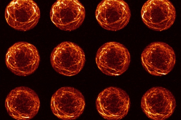
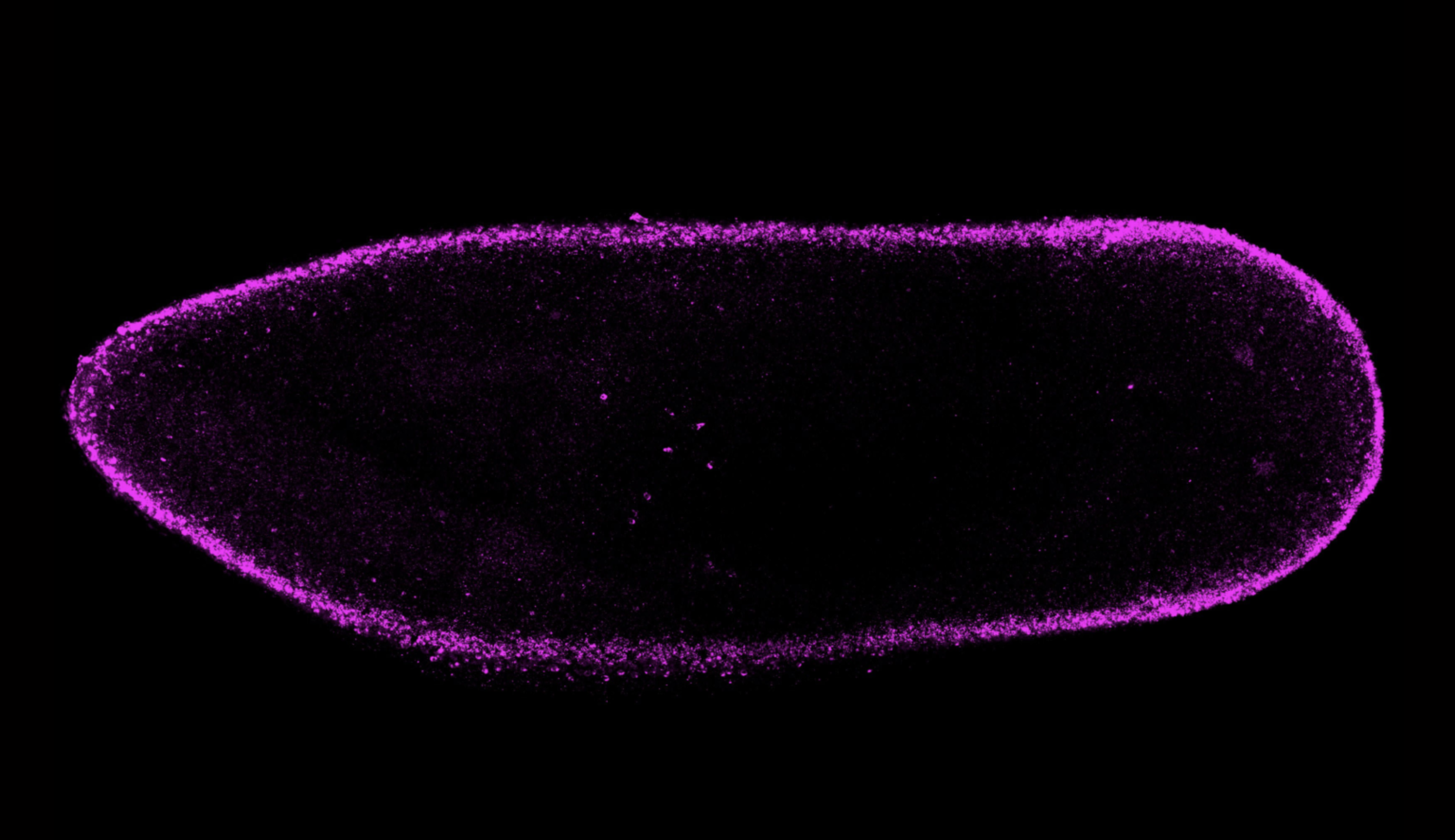
The researchers found that one of the triggers — a long non-coding RNA — plays a role in proper development of the cuticle, or the waterproof outer shell of a fly embryo. “We noticed that when we perturbed this trigger, the cuticles of fly embryos had altered elasticity,” Kingston said. “When we popped the embryos out of their egg shells, we could see these cuticles expand up and bloat.”
Because of the bloated phenotype, Kingston decided to name the long non-coding RNA marge after Aunt Marge, a character in the Harry Potter series. In “Harry Potter and the Prisoner of Azkaban”, Aunt Marge’s taunts lead Harry to accidentally perform magic on her, causing her to inflate and float away.
In the future, Kingston, who has since graduated and begun a career in the biotech industry, hopes researchers will pick up the torch on learning the roles of other TDMD triggers. “We still have several other triggers [from this paper] where there’s no known biological role for them in the fly,” she said. “I think this opens up the field for others to go in and to ask the questions, ‘Where are these triggers acting? What are they doing? And what’s the phenotype when you lose them?’”
Elena Kingston, Lianne Blodgett and David Bartel. “Endogenous transcripts direct microRNA degradation in Drosophila, and this targeted degradation is required for proper embryonic development.” Molecular Cell, September 22, 2022. DOI: https://doi.org/10.1016/j.molcel.2022.08.029