Pairing mismatch helps impaired fish RNA cleavage proceed swimmingly
December 21, 2017

Beyond tending to its multitudes of genetic, metabolic, and developmental processes, eukaryotic cells must additionally be vigilant against invasion by parasitic sequences such as viruses and transposons. RNA interference (RNAi) is a defense used by eukaryotic cells to protect themselves from such threats to their genomic harmony. Cellular RNAi components slice and destroy invading double-stranded RNA sequences and also help snip and process microRNAs, RNA sequences encoded by the genome that play key roles in gene regulation. An important process that occurs naturally in our cells, RNAi has also been harnessed by scientists as a tool to study gene function in common models such as worms, fruit flies, and mice. While many researchers have been using RNAi to tease apart gene function for over a decade, those using zebrafish, a powerful vertebrate model, have been forced to use other approaches because RNAi just did not seem to work well in these animals. Now, researchers at Whitehead Institute have uncovered how small changes in the fish Argonaute (Ago) protein, an RNA slicing protein, that happened in its lineage an estimated 300 million years ago greatly diminished the efficiency of RNAi in these animals, while another ancestral feature, in a critical pre-microRNA, was retained that enabled the microRNA to still be produced despite the fish’s impaired Ago protein.

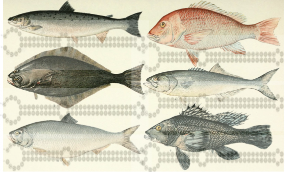
In an article published December 21 in the journal Molecular Cell, graduate student Grace Chen, along with both Whitehead Member David Bartel, also a professor of biology at Massachusetts Institute of Technology (MIT) and investigator with the Howard Hughes Medical Institute, and Whitehead Member and MIT professor of biology Hazel Sive, describe their discovery of a roughly 300 million-year-old, two amino acid substitutions in the fish Ago protein. The substitution is present in the ancestor all teleost fish, the class of fish which includes not only zebrafish but also the vast majority of fish species spanning those populating the ocean, aquarium, and supermarket. These two changes reside in and near the protein’s catalytic site and greatly decrease the ability of the fish Ago to perform its RNA slicing function, offering an explanation for why RNAi has not been a useful tool in zebrafish.

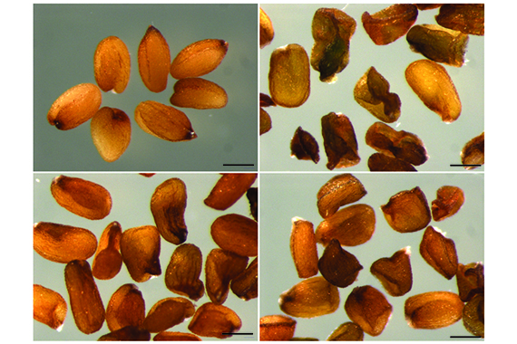
Despite the zebrafish’s deficiencies in RNAi, it is still able to produce the microRNA miR-451, an important regulator of red blood cell maturation and the only microRNA processed by Ago (the rest are produced with another protein called Dicer). MicroRNAs are short stretches of RNA that can regulate gene expression by inhibiting translation of mRNA into a protein and directing the destruction of mRNA before it can be used to make more protein. Since Chen had discovered that zebrafish lack an efficient Ago protein, it was mysterious as to how are fish were able to produce Ago cleavage-dependent miR-451. The Ago protein must process miR-451 by slicing the sequence out of a longer strand of RNA that has folded up on itself, forming a hairpin structure. What they determined was that in the pre-miR-451 hairpin in zebrafish, at a critical position in the miRNA, they found a “G–G” pairing mismatch that actually appears to facilitate cleavage by the impaired zebrafish Ago. No mismatch, no efficient cleavage.

Exploring the effects of a seed sequence mismatch on Ago-catalyzed cleavage kinetics further, they then tested its ability to slice other bound transcripts. The researchers discovered that while, as might be expected, a G–G mismatch slows Ago binding, it significantly enhances both slicing efficiency as well as the release of the bound product, more than off-setting the slower binding reaction kinetics and suggesting that non- “Watson–Crick” base pairing creates an exceptionally favorable geometry for the cleavage and release parts of the reaction.

These findings offer interesting insights into how animals can survive and thrive without an efficient RNAi system and suggest how the Ago protein could be “repaired” in order to allow zebrafish researchers to use RNAi in their experiments. Restoring a function that a lineage hasn’t had for 300 million years might also fuel additional findings into how the teleost class has diverged over time.

Written by Lisa Girard
***
David Bartel’s primary affiliation is with Whitehead Institute for Biomedical Research, where his laboratory is located and all his research is conducted. He is also a professor of biology at Massachusetts Institute of Technology and investigator with the Howard Hughes Medical Institute.
***
Paper cited:
Chen GR, Sive H, and Bartel DP. A Seed Mismatch Enhances Argonaute2-Catalyzed Cleavage and Partially Rescues Severely Impaired Cleavage Found in Fish. Molecular Cell, Dec 21 2017 DOI: 10.1016/j.molcel.2017.11.032.
Small RNA mediates genetic parental conflict in seed endosperm
December 19, 2017

CAMBRIDGE, MA–When it comes to gene expression in the endosperm of seeds, gene provenance matters. In this specialized tissue, plants actively strive to keep the expression of genes inherited from the mother versus the father in balance, according to Whitehead Institute scientists.

The endosperm, the starchy part of a seed that envelopes and nourishes the developing embryo, comprises two-thirds of the calories in a typical human diet. It is the meat of a coconut and the sweet part of the corn on the cob we eat.  In a paper published online December 19 in the journal Cell Reports, Whitehead Member Mary Gehring, first author and former Gehring graduate student Robert Erdmann, and colleagues reveal that the endosperm is also the site where the plant must actively orchestrate a delicate balance between expression of genes inherited from the mother and those of the father.  If this critical balance errs toward one parent or the other, seeds can be too small or even abort.

Unlike most plant cells, which have two copies of the genome, cells within the endosperm have three copies: one inherited from the father, and two inherited from the mother. This ratio is established when a sperm cell in the fertilizing pollen grain fuses with the central cell associated with the egg cell in a flower’s ovule. Unlike most cells, the central cell has two nuclei, so when the sperm’s nucleus merges with the central cell, the resulting endosperm is triploid.

 The 2-to-1 ratio of maternal to paternal gene expression is crucial, and deviation can have dire consequences:  If maternal gene expression is too high, the seeds are too small; if paternal gene expression is too high, the seeds abort. Although plant biologists have known the importance of this ratio for seed viability, the balance was assumed to be passively maintained for the majority of genes.  Previously, Gehring determined that a subset of genes expressed in the endosperm are imprinted—their expression is inherited from their parent. But what about the remaining majority of the genome?

Now Gehring and colleagues have discovered a role for small RNAs—snippets of RNA that interfere with and can reduce gene expression—in actively maintaining this 2-to-1 balance in those genes that are not imprinted.  This the first time scientists have documented small RNAs maintaining such a ratio. Using Arabadopsis thaliana and Arabadopsis lyrata plants, Gehring and her lab determined that these small RNAs tamp down the expression of maternally inherited genes. When the enzyme that creates the small RNAs is mutated, fewer small RNAs are produced, and the plant’s carefully balanced gene expression is thrown off. The resulting seeds have excessive maternal gene expression. To understand the significance of this elevated maternal gene expression, Satyaki Rajavasireddy, a postdoctoral researcher in Gehring’s lab and an author of the Cell Reports paper, turned to plants with seeds that abort  because they have additional copies of paternal genes. When these plants with extra paternal DNA had their small-RNA-producing enzyme mutated, the outcome was striking: The seeds were rescued and developed to maturity.

Although the research analyzed this phenomenon in A. thaliana and A. lyrata, Gehring expects it to be a widespread manifestation of the tug-of-war between maternal and paternal genetic contributions.

“Maintaining this maternal/paternal balance is crucial for seed development, including in crop plants,” says Gehring, who is also an associate professor of biology at Massachusetts Institute of Technology.  “We’ve looked at two species that are separated by 10 million years of evolution, and I anticipate we will find this mechanism in other species as well.”

This work was supported by the National Science Foundation (NSF CAREER grant 1453459).

Written by Nicole Giese Rura
* * *
Mary Gehring’s primary affiliation is with Whitehead Institute for Biomedical Research, where her laboratory is located and all her research is conducted. She is also an associate professor of biology at Massachusetts Institute of Technology.
* * *
Full Citation:
“A small RNA pathway mediates allelic dosage in endosperm”
Cell Reports, online December 19, 2017.
Robert M. Erdmann (1,2), P.R. V. Satyaki (1), Maja Klosinska (1), Mary Gehring (1,2).
1. Whitehead Institute for Biomedical Research, Cambridge, MA 02142 USA
2. Department of Biology, Massachusetts Institute of Technology, Cambridge, MA 02139 USA
Jacqueline Lees

Education

  • PhD, 1990, University of London
  • BSc, 1986, Biochemistry, University of York

Research Summary

We identify the proteins and pathways involved in tumorigenicity — establishing their mechanism of action in both normal and tumor cells. To do so, we use a combination of molecular and cellular analyses, mutant mouse models and genetic screens in zebrafish.

Rethinking transcription factors and gene expression

Study shows that, like proteins, genomes must fold appropriately to function properly and that some transcription factors provide the structural support.

Nicole Giese Rura | Whitehead Institute
December 7, 2017

Transcription — the reading of a segment of DNA into an RNA template for protein synthesis — is fundamental for nearly all cellular processes, including growth, responding to stimuli, and reproduction. Now, Whitehead Institute researchers have upended our understanding of how transcription is controlled and the role of transcription factors in the process.

The paradigm shift, described in an article online on Dec. 7 in the journal Cell, hinges on a small protein that plays a key role in genome structure and gives us new insights into how changes in the control of transcription and gene expression can lead to disease.

Transcription has several important players that must all be in the right place at the right time: the transcription machinery, transcription factors, promoters, and enhancers.  According to the existing model, transcription factors are proteins that bind to enhancer regions of the genome and recruit the transcription machinery to the promoter DNA regions, which then initiate the genes’ transcription.

“We’ve always assumed that the role of transcription factors was to recruit the transcription machinery to genes to turn them on or turn them off,” says Richard Young, a Whitehead Insistute member and professor of biology at MIT. “But we never imagined that the transcription factors we’ve studied for three decades actually contribute to the genome’s structure. And as a consequence, they regulate genes. So we now look at genomes like proteins: They have to fold up appropriately in order to control genes.”

Scientists have known that the genome’s structure — how it bends and folds — is essential for efficiently compressing two meters of DNA into each human cell, which is the equivalent of packing a strand ten football fields long into a space the size of a marble. Yet until recently, researchers have not had the tools necessary to appreciate this architecture’s importance in fine control of gene expression or study the genome’s structure at sites ready for transcription.

In 2014, Young and his lab determined that portions of the genome reside in loop-based structures, creating insulated neighborhoods that bring enhancers, promoters, and genes into close proximity. Each loop is tied at the top by a pair of molecules, called CTCF, that are bound together. This structure is essential for proper gene control: If the loop structure is broken, gene expression is altered, and cells can become diseased or die.

In the current research, Young along with co-first authors Abraham Weintraub and Charles Li took a closer look at a protein that is well known but not well understood: Yin Yang 1 (YY1). Hundreds of scientific papers have linked YY1 dysfunction to diseases such as viral infections, cancer, and arthritis, and yet the studies produced seemingly contradictory observations of YY1’s function.

According to Young and colleagues, YY1 is a unique transcription factor that occupies both enhancers and promoters, is essential for cell survival, and is found in almost every cell type in humans and mice. Like CTCF, YY1 can also pair with itself and bind to DNA to form loops that enhance DNA transcription.

“YY1 is expressed broadly, and it is necessary for establishing enhancer-promoter loops in multiple cell types,” says Weintraub. “That’s its job, not recruiting the transcription apparatus. When the structure created by YY1 is removed, the genome is no longer folded properly, gene control is lost and transcription of the affected genes is significantly diminished, which can cause dysfunction.”

This model of YY1’s function could account for its association with a number of disparate diseases. Earlier this year, scientists reported YY1 syndrome — a genetic syndrome causing cognitive disabilities in people with mutations in their YY1 gene.

According to Young, YY1 is probably not the only transcription factor with this loop-forming role, and his lab will be searching for additional factors with similar functions.

“YY1 is most likely just the first one, and there are probably a bunch of collaborators that have similar roles,” says Young. “Instead of the classic function that we thought these transcription factors had — interacting with the transcription apparatus and giving instructions on how much or how little of a gene’s transcript to produce — they are bringing together regulatory elements with the gene. The whole job of these transcription factors is just making structure. We are realizing that the things that form physical structures are much more important than we had appreciated.”

The researchers’ work was supported by the National Institutes of Health, the Ludwig Graduate Fellowship funds, the National Science Foundation, the American Cancer Society, a Margaret and Herman Sokol Postdoctoral Award, the Damon Runyon Cancer Research Foundation, and the Cancer Research Institute. The Whitehead Institute has filed a patent application based on this study.

Laurie A. Boyer

Education

  • PhD, 2001, University of Massachusetts Medical School
  • BS, 1990, Biomedical Science, Framingham State University

Research Summary

We investigate how complex circuits of genes are regulated to produce robust developmental outcomes particularly during heart development. A main focus is to determine how DNA is packaged into chromatin, and how ATP-dependent chromatin remodelers modify this packaging to control lineage commitment. We are now applying these principles to develop methods to stimulate repair of damaged cardiac tissue (e.g., regeneration). Our ability to combine genomic, genetic, biochemical, and cell biological approaches both in vitro and in vivo as well as ongoing efforts to use tissue engineering to model the 3D architecture of the heart will ultimately allow us to gain a systems level and quantitative understanding of the regulatory circuits that promote normal heart development and how faulty regulation can lead to disease.

Awards

  • Medicine by Design Distinguished Lecture, 2017
  • Cardiovascular Rising Star Distinguished Lecture, 2017
  • American Heart Association Innovative Research Award, 2013
  • Irvin and Helen Sizer Career Development Award, 2012
  • Smith Family Award for Excellence in Biomedical Science, 2009
  • Massachusetts Life Sciences Center New Investigator Award, 2008
  • Pew Scholars Award in the Biomedical Sciences, 2008
  • Honorary Doctorate, Framingham State College, 2007
  • The Scientific American World’s 50 Top Leaders in Research, Business or Policy, 2006
Robert A. Weinberg

Education

  • PhD, 1969, MIT
  • SB, 1964, Biology, MIT

Research Summary

We investigate three broad questions related to the origin and spread of cancer. First, how do cancer cells within a primary tumor acquire the ability to invade and metastasize? Second, how are the stem-cell state and the epithelial-mesenchymal transition interrelated? Third, how are the regulators of the epithelial-mesenchymal transition able to activate this profound change in cell phenotype?

Awards

  • Japan Prize, Japan Prize Foundation, 2021
  • Salk Institute Medal for Research Excellence, 2016
  • Breakthrough Prize in Life Sciences, 2013
  • Wolf Foundation Prize, 2004
  • Institute of Medicine, Member, 2000
  • Keio Medical Science Foundation Prize, 1997
  • National Science Foundation, National Medal of Science, 1997
  • Harvey Prize, 1994
  • American Academy of Arts and Sciences, Fellow, 1987
  • Sloan Prize, GM Cancer Research Foundation, 1987
  • National Academy of Sciences, Member, 1985
  • Robert Koch Foundation Prize, 1983
Omer H. Yilmaz

Education

  • PhD, 2008, University of Michigan; MD, 2008, University of Michigan Medical School
  • BS, 1999, Biochemistry and Physics, University of Michigan

Research Summary

The adult intestine is maintained by stem cells that require a cellular neighborhood, or niche, consisting in part of Paneth cells. Our laboratory will investigate the molecular mechanisms of how intestinal stem cells and their Paneth cell niche respond to diverse diets to coordinate intestinal regeneration with organismal physiology and its impact on the formation and growth of intestinal cancers.  By better understanding how intestinal stem cells adapt to diverse diets, we hope to identify and develop new strategies that prevent and reduce the growth of cancers involving the intestinal tract that includes the small intestine, colon, and rectum.

Awards

  • AAAS Martin and Rose Wachtel Cancer Research Award, 2018
  • Pew-Stewart Trust Scholar, 2016-2020
  • Sidney Kimmel Scholar, 2016-2020
  • V Foundation Scholar, 2014-2017
  • Harold M. Weintraub Award, 2007
H. Robert Horvitz

Education

  • PhD, 1974, Harvard University
  • BS, 1968, Mathematics and Economics, MIT

Research Summary

Our lab examines how genes control animal development and behavior. We use the experimentally tractable nematode Caenorhabditis elegans to identify and analyze molecular and cellular pathways involved in these important areas of biology. Ultimately, we hope to clarify these fundamental biological mechanisms and provide further insight into human disease.

Awards

  • U.S. National Academy of Inventors, Member, 2015
  • American Association for Cancer Research Academy, Fellow, 2013
  • Royal Society of London, Foreign Member, 2009
  • Genetics Society (U.K.), Mendel Medal, 2007
  • Eli Lilly Lecturer Award, 2007
  • Massachusetts Institute of Technology, James R Killian Jr Faculty Achievement Award, 2006
  • National Academy of Medicine, Member, 2003
  • American Cancer Society, Medal of Honor, 2002
  • The Nobel Foundation, Nobel Prize in Physiology or Medicine, 2002
  • Bristol-Myers Squibb, Award for Distinguished Achievement in Neuroscience, 2001
  • March of Dimes, Developmental Biology, 2000
  • Gairdner Foundation, Gairdner Foundation International Award, 1999
  • National Academy of Sciences, Member, 1991
  • American Academy of Arts and Sciences, Fellow, 1989
  • American Association for the Advancement of Science, Fellow, 1989
  • Howard Hughes Medical Institute, HHMI Investigator, 1988
Peter Reddien

Education

  • PhD, 2002, MIT
  • SB, 1996, Molecular Biology, University of Texas at Austin

Research Summary

We investigate how stem cells are regulated to regenerate missing tissues. We study the cellular events involved in this process and the attendant roles for regulatory genes that control regeneration steps. We utilize an array of methodologies, including high-throughput sequencing, RNA interference (RNAi) screening, and numerous assays and tools for phenotypic analysis to characterize regeneration regulatory genes.

Awards

  • Howard Hughes Medical Institute, HHMI Investigator, 2013
Richard A. Young

Education

  • PhD, 1979, Yale University
  • BS, 1975, Biological Sciences, Indiana University

Research Summary

We use experimental and computational technologies to determine how signaling pathways, transcription factors, chromatin regulators and small RNAs regulate gene expression in healthy and diseased cells. Our interests range from the basic molecular mechanisms behind gene control to drug development for cancer and other diseases caused by gene misregulation.

Awards

  • National Academy of Medicine, Member, 2019
  • National Academy of Sciences, Member, 2012