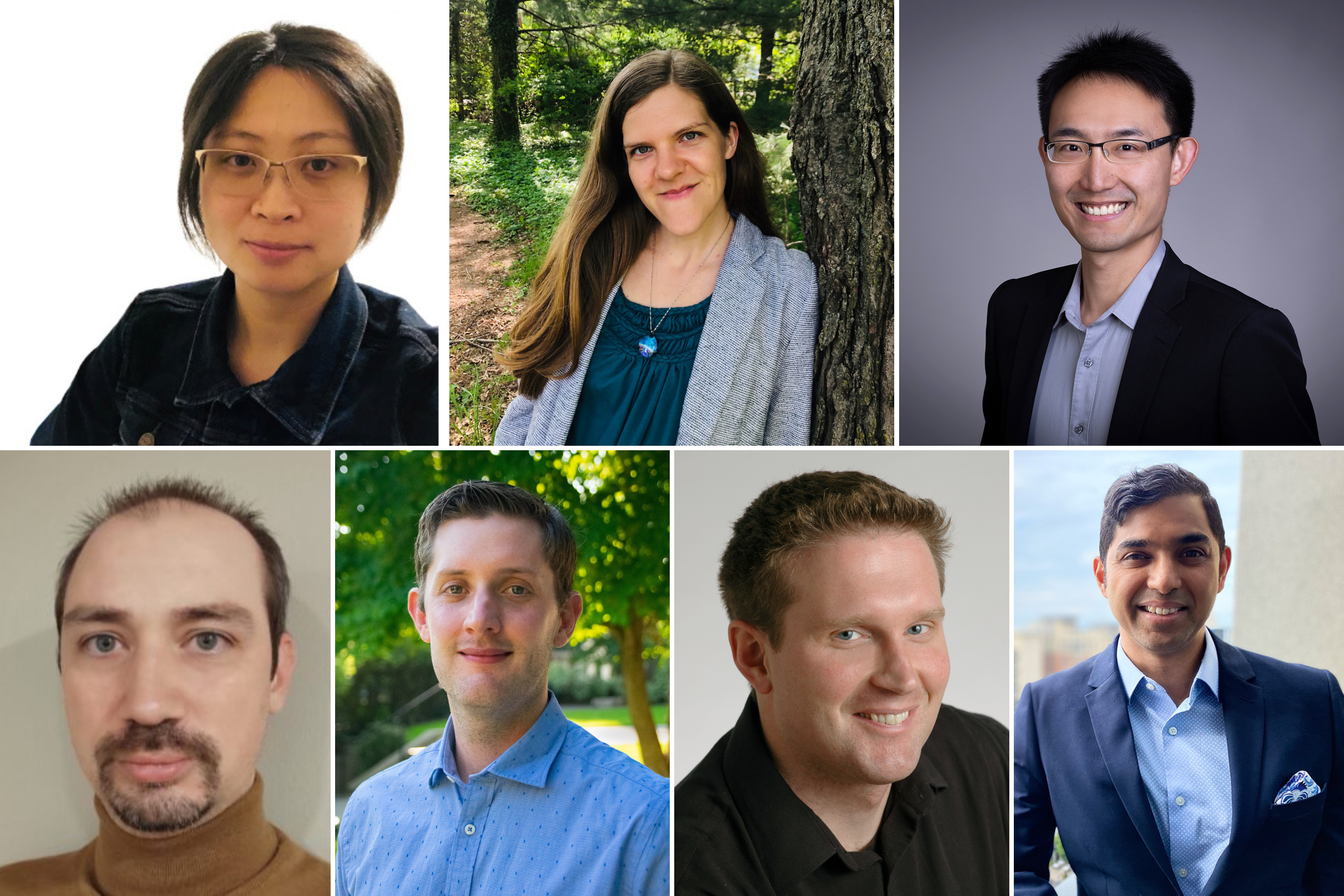
Seven professors join the departments of Biology; Chemistry; Earth, Atmospheric and Planetary Sciences; Mathematics; and Physics.
School of Science
November 17, 2022
This fall, the MIT School of Science welcomes seven new faculty to the departments of Biology; Chemistry; Earth, Atmospheric and Planetary Studies (EAPS); Mathematics; and Physics.
Wanying Kang researches large-scale atmospheric and oceanic dynamics, and their effects on the climate of Earth and other planetary bodies. She hopes to bridge multiple geoscience fields by applying tools from climate science on Earth to planetary science questions. Currently, Kang is looking into the atmospheric circulation on superhot lava worlds and the ocean circulation on icy moons, given the potential to observe them in more detail in the near future.
Kang earned an undergraduate degree in physics from Peking University and a PhD in applied math from Harvard University. She first joined the Department of Earth, Atmospheric and Planetary Sciences as a distinguished postdoc through the Houghton-Lorenz Fellowship. Now, Kang has been appointed an assistant professor in climate science in EAPS.
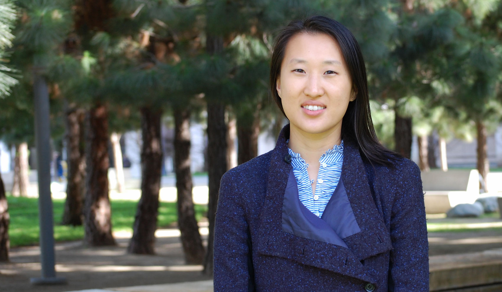
Sarah Millholland explores the demographics and diversity of extrasolar planetary systems. Using orbital dynamics and theory, she investigates how gravitational interactions like tides, resonances, and spin dynamics influence the formation and evolution of planetary systems and shape observable exoplanet properties.
Millholland obtained bachelor’s degrees in physics and applied mathematics from the University of Saint Thomas in 2015. She spent her first year of graduate school at the University of California at Santa Cruz before transferring to Yale University, earning her PhD in astronomy from Yale in 2020. She then moved to Princeton University, where she was a NASA Sagan Postdoctoral Fellow from 2020-22. Millholland joins MIT as an assistant professor in the Department of Physics and a member of the Kavli Institute for Astrophysics and Space Research.
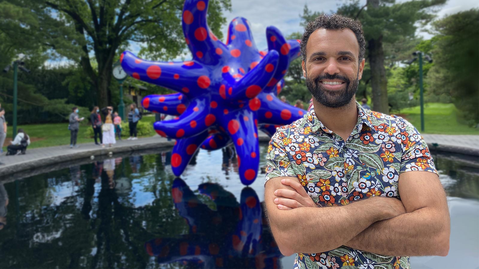
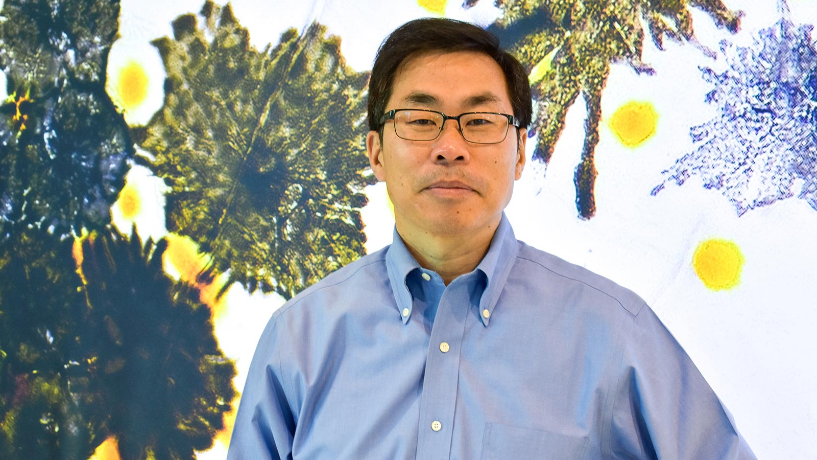
Sam Peng PhD ’14 aims to develop novel probes and microscopy techniques to visualize the dynamics of individual molecules in living cells, which will improve the understanding of molecular mechanisms underlying human diseases. Peng’s group will focus on studying molecular dynamics, protein-protein interactions, and cellular heterogeneity involved in neurobiology and cancer biology. Their long-term goal is to translate these mechanistic insights into drug discovery.
Peng received his bachelor’s degree in chemistry from the University of California at Berkeley, and his PhD from MIT in physical chemistry. Most recently, he completed postdoctoral research at Stanford University. He returns to MIT as an assistant professor in the Department of Chemistry and a core member of the Broad Institute of MIT and Harvard.
Julien Tailleur is a physicist focusing on the emerging properties of active materials, which encompass systems made of large assemblies of units able to exert propelling forces on their environment. From molecular motors to cells and animal groups, active systems are found at all scales in nature. Most recently, Tailleur combined the development of theoretical frameworks to describe active systems with their applications to the study of microbiological systems.
Tailleur completed his undergraduate studies in mathematics at Université Pierre et Marie Curie (UPMC) and in physics at Université d’Orsay. He earned his PhD in physics in 2007 from UPMC. After becoming an Engineering and Physical Sciences Research Council postdoc at the University of Edinburgh, Tailleur joined French National Centre for Scientific Research (CNRS) and Université Paris Diderot in 2011, then becoming a CNRS Director of Research in 2018. Tailleur joins the Department of Physics as an associate professor.
Richard Teague works to understand the earliest stages of planetary systems, specifically, where, when, and how they can form. A major component of his research is the development of new techniques to detect examples of planets while they are still embedded in their parental protoplanetary disks, a period of the planet’s growth phase which is currently hidden from view. Teague is also leading the exoALMA collaboration, searching for the youngest exoplanets with one of the largest telescopes in the world, the Atacama Large (sub-) Milimeter Array (ALMA).
Teague earned a master’s degree from the University of Edinburgh and a PhD from the Max-Planck-Institute for Astronomy. Previously, he was a Submillimeter Array fellow at the Harvard-Smithsonian Center for Astrophysics and a postdoc at the University of Michigan and the Max-Planck-Institute for Astronomy. Teague joins MIT as an assistant professor in the Department of Earth, Atmospheric and Planetary Sciences.
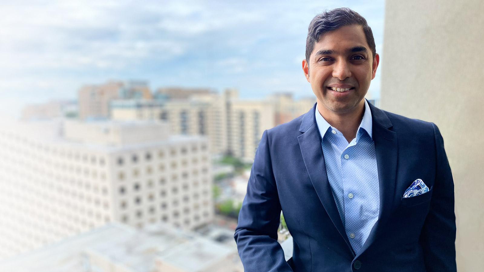
Research interests of Martin Wainwright PhD ’02 include high-dimensional statistics, statistical machine learning, information theory, and optimization theory. One focus is algorithms and Markov random fields, a class of probabilistic model based on graphs used to capture dependencies in multivariate data: for example, image models, data compression, and computational biology. He also studies the effect of decentralization and communication constraints in statistical inference problems. A final area of interest is methodology and theory for high-dimensional inference problems.
Wainwright received a bachelor’s degree in mathematics from University of Waterloo followed by a PhD in electrical engineering and computer science (EECS) from MIT. Most recently, he was the Chancellor’s Professor at the University of California at Berkeley with a joint appointment between the departments of Statistics and EECS. Wainwright returns to MIT as a professor of mathematics and electrical engineering and computer science.
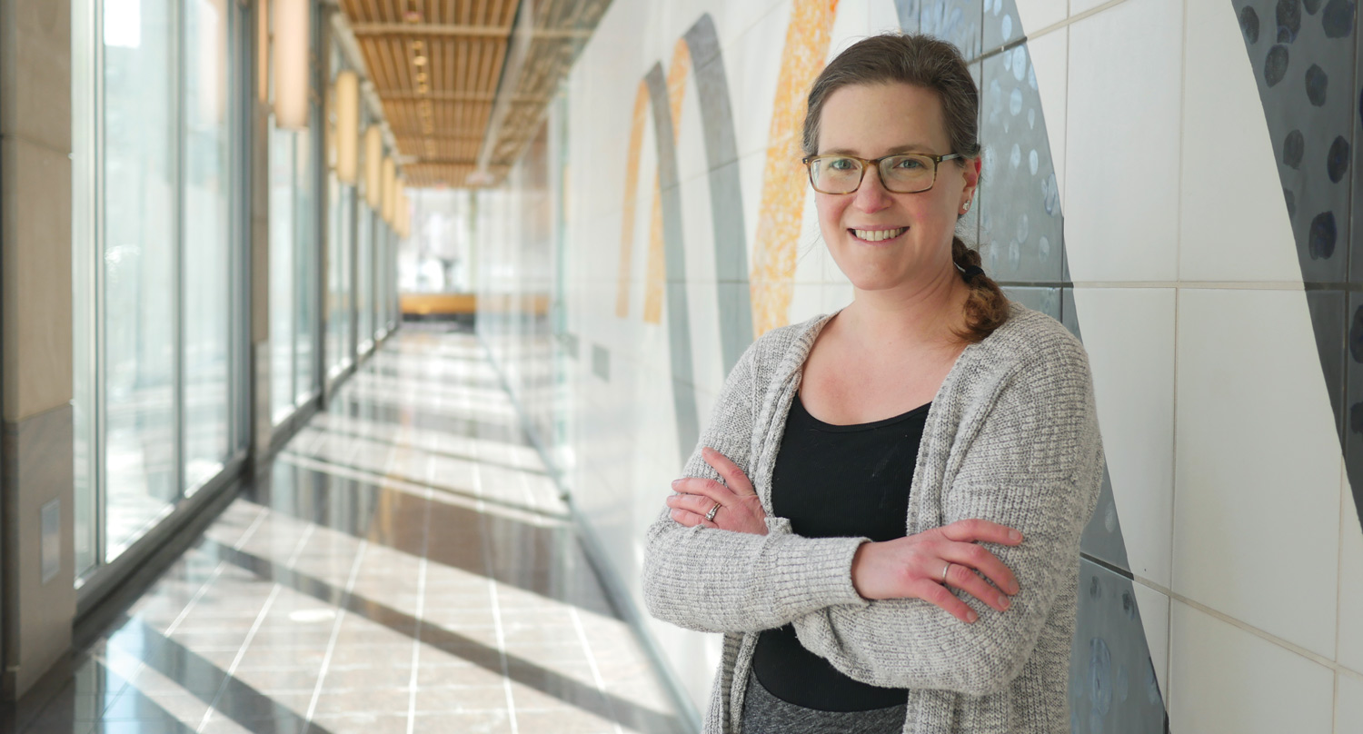
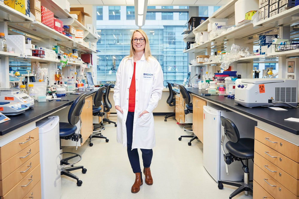
Immune cells communicate across scales in time and space, forming circuits that control their destructive capacity. Harikesh Wong employs a variety of quantitative approaches, including advanced fluorescence microscopy and computational modeling, to study these circuits within intact tissue environments. Ultimately, he seeks to understand how imbalanced immune cell communication — due to genetic or environmental variation — results in detrimental outcomes, including chronic infection, autoimmunity, and the formation of tumors.
Wong received a bachelor’s degree from McMaster University followed by a PhD in cell biology from the University of Toronto. Next, he pursued a postdoc at the National Institutes of Health in immunology and systems biology. Wong joins MIT as an assistant professor in the Department of Biology and a core member of the Ragon Institute of MGH, MIT and Harvard.