Research Area: Human Disease
PhD students Maureen Buckley and Krista Pullen discuss the role of sex hormones in infectious diseases, with a focus on HIV/AIDS and COVID-19.
June 8, 2022
Education
- Graduate: PhD, 2011, MIT; MD, 2013, Harvard Medical School
- Undergraduate: BA, 2006, Biology, University of Chicago
Research Summary
Diverse commensal microbes colonize every surface of our bodies. We study the constant communication between these microbes and our immune system. We focus on our largest organ: the skin. By employing microbial genetics, immunologic approaches, and mouse models, we can dissect (1) the molecular signals used by microbes to educate our immune system and (2) how different microbial communities alter immune responses. Ultimately, we aim to harness these microbe-host interactions to engineer novel vaccines and therapeutics for human disease.
Awards
- Howard Hughes Medical Institute Hanna H. Gray Fellow, 2018-2026
- A.P. Giannini Postdoctoral Research Fellowship, 2018
- Dermatology Foundation Research Fellowship, 2017
Eva Frederick | Whitehead Institute
April 28, 2022
Whitehead Institute Member Sebastian Lourido and his lab members study the parasite Toxoplasma gondii. The parasite causes the disease toxoplasmosis, which can be dangerous for pregnant or immunocompromised patients.
As the parasite evolved over millennia, its phylum (the Apicomplexan parasites) split off from other branches of life, which poses a challenge to researchers hoping to understand its genetics. “Toxoplasma is very highly diverged from the organisms that we typically study, like mice, yeast and [nematodes],” said Lourido lab researcher and Massachusetts Institute of Technology (MIT) graduate student Tyler Smith. “Our lab focuses a lot on developing toolkits to probe and study the genomes of these parasites.”
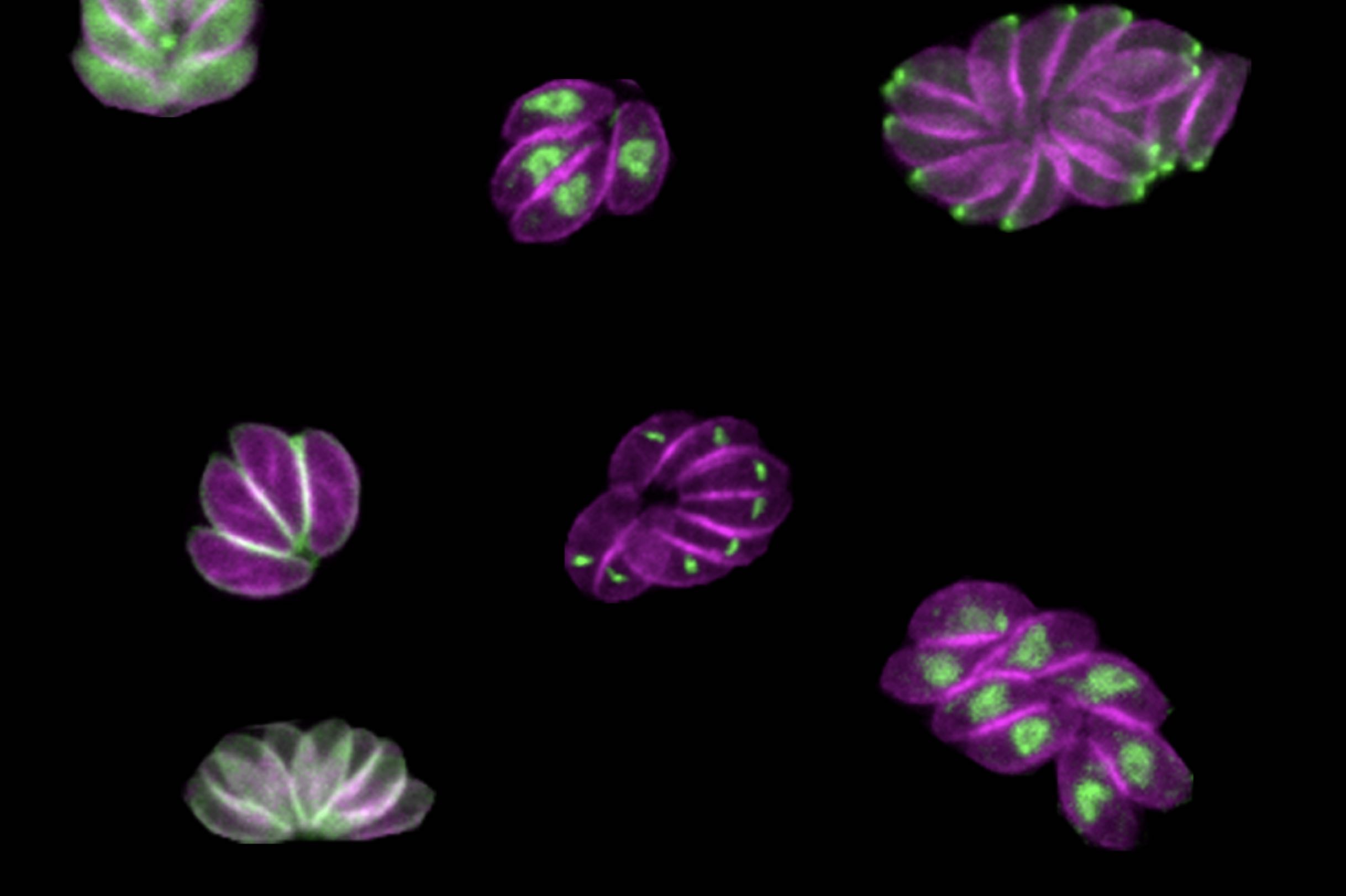
Now, in a paper published in the journal Nature Microbiology on April 28, Smith and colleagues describe a new method for determining the role of genes within the genome of the parasite. The method can be conducted by a single investigator, and goes a step beyond simply assessing whether or not a given gene is essential for survival. By inserting specific sequences — such as those encoding fluorescent markers or sequences that can turn a gene on and off — throughout the Toxoplasma genome, the method allows the researchers to visualize where an individual gene’s product resides within the parasites and identify when in the life cycle important genes became essential, providing more detailed information than a traditional CRISPR screen.
Although the method could theoretically be used with any gene family, Smith and Lourido decided to first focus on a family of proteins called kinases, the genetic code for which comprises around 150 of Toxoplasma’s 8,000 total genes.
“Kinases are interesting from a basic biology perspective because they are signaling hubs of basic biological processes,” said Smith, who is first author of the study. “From a more translational perspective, kinases are really common drug targets. We have a lot of inhibitors that work with kinases. For some cancers that are linked to specific kinases, the inhibitors can be chemotherapies.”
Using the method, researchers discovered a gene encoding a previously unstudied kinase which they named SPARK. They were able to show that the SPARK kinase is involved in the process of the parasites entering and leaving host cells, and future research on inhibitors of SPARK could lead to new treatments for toxoplasmosis. “Identifying these kinases that are really vital for these critical decision points in a parasite’s life cycle could be really fruitful for developing new therapeutics,” said Lourido, who is also an associate professor of biology at MIT.
New dimensions of screening
Many CRISPR screens use gene editing technology to knock out genes throughout the genomes of a sample of cells, creating a population where every gene in the genome is mutated in at least one of the cells. Then, by looking at which mutations have detrimental effects on the cells, researchers can extrapolate which genes are essential for survival.
But the workings of a whole organism are infinitely more complicated than just survival or death, and researchers are often faced with a challenge when it comes to figuring out exactly what different gene products are doing in the cells. That’s why Smith and Lourido decided to design a method of screening for Toxoplasma genes that could provide more information about what the products of those genes do. “CRISPR screens can tell you which genes are important, but it doesn’t give you much information about why they’re important,” Smith said. “We were seeking to make a kind of platform that could look at other dimensions.”
Smith and Lourido used CRISPR technology to introduce small amounts of new DNA into the parasites’ genes that code for kinases. The new DNA included sequences encoding a fluorescent marker protein and sequences that could be used to manipulate gene expression levels.
After creating a population of parasites modified this way, the researchers then used imaging to determine where the fluorescently tagged proteins had ended up in the cells, and to observe what happened in the cells when the proteins were turned off. “Being able to see different cell division phenotypes — for instance parasites that either failed to replicate at all, or tried to replicate but would have some abnormalities — that gets us closer and allows us to generate hypotheses as to actually why these kinases are important, not just whether or not they are important,” Smith said.
The depletion of some proteins caused the parasites to die instantly, while others affected the parasites at a later point in their life cycles, so they would drop out of the population more slowly. “Cells with mutations in these kinases replicate fine, but a problem might arise when they need to leave their host cell and enter a new host cell later on down the line,” Smith said.
A “SPARK” of inspiration
After the screen, the researchers followed up on one of these kinases in particular, which they called SPARK (short for Store Potentiating/Activating Regulatory Kinase). Mutants depleted of SPARK died, but not until a later phase of the life cycle. Smith and Lourido conducted further experiments to understand SPARK’s role, and found evidence that the protein was involved in the release of calcium in the cell that is required for a parasite to enter or leave a host cell.
“The thing I found very interesting about SPARK is that it’s a kinase that’s very different from the analogous kinase in other model organisms, but is conserved throughout all of the apicomplexan phylum,” Smith said. “That’s the phylum that includes Toxoplasma and a bunch of other single-celled parasites like Plasmodium, which is the malaria parasite.”
Because SPARK is far different from its human analog and essential to the parasite’s life cycle, a SPARK-specific kinase inhibitor could be used to treat toxoplasmosis by killing the parasite without affecting the patient. “The hope would be that you can target SPARK and inhibit it without hitting mammalian kinases,” Smith said. “It’s easy enough to design something that kills a cell, but the trick is only killing parasites and not your own cells.”
In the future, the researchers hope to turn their new screening method to other families of genes, such as transcription factors, to understand their function in the parasites. “Our results have been quite encouraging in that we think this method will be scalable, and we can target larger gene sets in the future,” Smith said. “I think the ultimate end goal would be to do the whole genome.”
“There’s this whole universe of parasite proteins that we know so little about, where this type of analysis will be incredibly insightful.” Lourido said. “We’re really very excited about scaling it up further.
Greta Friar | Whitehead Institute
April 18, 2022
Drug overdose, mostly from opioid use, is the leading cause of accidental death in the United States. Prior studies of twins have revealed that genetics play a key role in opioid use disorder. Researchers know that a mixture of genetic and environmental risk factors contribute to heritability of the disorder, but identifying the specific risk factors is challenging. Opioid use disorder is complex, so instead of one or a few genes causing the disorder, there may be many contributing factors that can combine in different ways. Researchers want to understand which genes contribute to opioid use disorder because this will lead to a better understanding of its underlying biology and could help identify people who will be most at risk if exposed to opioids, enabling researchers, health care providers, and social services to develop strategies for prevention, treatment, and support.
The usual approach for finding genes associated with disease risk is to do a genome wide association study, which compares the genetics of many people to identify patterns in different gene versions occurring in association with a disease. This approach is being used to look at opioid use disorder, but requires many more patient samples than are currently available to reach clear conclusions. Researchers from multiple research universities and institutes, including Whitehead Institute Member Olivia Corradin and her former PhD advisor, Case Western Reserve University Professor Peter Scacheri; as well as Icahn School of Medicine Professor Schahram Akbarian; Eric O. Johnson, a distinguished fellow at RTI International; Dr. Kiran C. Patel College of Allopathic Medicine at Nova South Eastern University Professor Deborah C. Mash; and Richard Sallari of Axiotl, Inc., developed a shortcut for identifying genes that are associated with opioid use disorder and may contribute to it using only a small number of patient samples. Genome wide studies may require hundreds of thousands of samples, but this new method, described in their research published in the journal Molecular Psychiatry on March 17, uses only around 100 samples—51 cases and 51 controls—to narrow in on five candidate genes.
“With this work, we think we’re only seeing the tip of the iceberg of the complex, diverse factors contributing to opioid overdose,” says Corradin, who is also an assistant professor of biology at the Massachusetts Institute of Technology. “However, we hope our findings can help prioritize genes for further study, to speed up the identification of risk markers and possible therapeutic targets.”
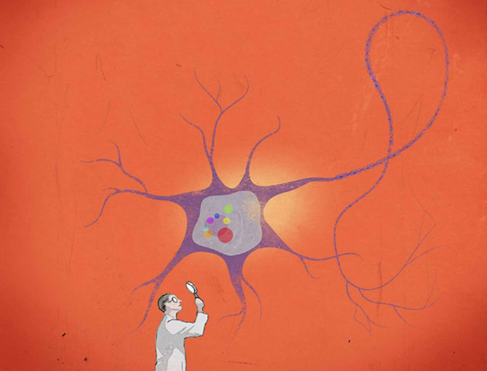
In order to learn more about the underlying biology of opioid use disorder, the researchers analyzed brain tissue samples from people who had died of opioid overdoses and compared them with samples from people with no known opioid use history who died of other accidental causes. They specifically looked at neurons from the dorsolateral prefrontal cortex, an area of the brain known to play important roles in addiction. Instead of analyzing the genes in these cells directly, the researchers instead looked at the regulators of the genes’ activity, and searched for changes in these regulators that could point them to genes of interest.
To identify a gene, first map its community
Genes have DNA regions, often close to the gene, that can ratchet up and down the gene’s expression, or the strength of its activity in certain cells. Researchers have only recently been able to map the three-dimensional organization of DNA in a cell well enough to identify all of the regulators that are close to and acting upon target genes. Corradin and her collaborators call a gene’s collection of close regulatory elements its “plexus.” Their approach finds genes of interest by searching for patterns of variation across each gene’s entire plexus, which can be easier to spot with a small sample size.
The patterns that the researchers look for in a plexus are epigenetic changes: differences in the chemical tags that affect regulatory DNA and in turn, modify the expression of the regulators’ target gene. In this case, the researchers looked at a type of epigenetic tag called H3K27 acetylation, which is linked to increases in the activity of regulatory regions. They found nearly 400 locations in the DNA that consistently had less H3K27 acetylation in the brains of people who died of opioid overdose, which would lower activity of target genes. They also identified under-acetylated DNA locations that were often specific to individuals rather than uniform across all opioid overdose cases. The researchers then looked at how many of those locations belonged to regulatory elements in the same plexus. Surprisingly, these individual-specific changes often occurred within the same gene’s plexus. A gene whose plexus had been heavily affected as a collective was flagged as a possible contributor to opioid use disorder.
“We know that the factors that contribute to opioid use disorder are numerous, and that it’s an extremely complex disease that by definition is going to be extremely heterogeneous,” Scacheri says. “The idea was to figure out an approach that embraces that heterogeneity, and then try to spot the themes within it.”
Using this approach, the researchers identified five candidate genes, ASTN2, KCNMA1, DUSP4, GABBR2, and ENOX1. One of the genes, ASTN2, is related to pain tolerance, while KCNMA1, DUSP4, and GABBR2 are active in signaling pathways that have been linked more broadly to addiction. Follow up experiments can confirm whether these genes contribute to opioid use disorder.
The five genes and their plexi are also involved in the heritability of generalized anxiety disorder, metrics of tolerance for risk-taking, and educational attainment. Heritability of these traits and opioid use disorder have previously been found to coincide, and people with opioid use disorder often also have generalized anxiety. Furthermore, heritability of these traits and opioid use disorder all have been associated with early childhood adversity. These connections suggest the possibility that early childhood adversity could be contributing to the epigenetic changes observed by the researchers in the brains of people who died of opioid overdose—a useful hypothesis for further research.
The researchers hope that these results will provide some insights into the genetics and neurobiology of opioid use disorder. They are interested in moving their research forward in several ways: they would like to see if they can identify more candidate genes by increasing their sample number, examine different parts of the brain and different cell types, and further analyze the genes already identified. They also hope that their results demonstrate the potency of their approach, which was able to discern useful patterns and identify candidate genes from the neurons of only 51 cases.
“We’re trying a different approach here that relies on this idea of convergence and leverages our understanding of the three-dimensional architecture of DNA, and I hope this approach will be applied to further our understanding of all sorts of complex diseases,” Scacheri says.

Greta Friar | Whitehead Institute
April 20, 2022
Stem cells are the versatile building blocks from which every cell type in the body, from neurons, to skin cells, to blood cells, is ultimately descended. Researchers have also figured out how to turn stem cells into different cell types in the lab, which has been helpful for studying health and disease in their normal cellular contexts, and could be used to generate cells for medical transplants. Whitehead Institute Founding Member Rudolf Jaenisch not only uses these cells in his research, but has spent much of his career discovering and improving the methods for making accurate laboratory models out of stem cell-derived cells.
One challenge that Jaenisch’s lab is focusing on is how to eliminate the differences between cell types as they are found in the body and their stem cell-derived equivalents. In particular, they have found that stem-cell derived cells are often immature, more closely resembling the cells found in fetuses rather than in adults. These differences can make the cells less accurate research models and prevent them from being medically useful as functional transplant cells. Stem cells in the body receive complex cocktails of molecular signals as they transform into different cell types. The challenge for researchers lies in figuring out which of the many molecular signals in the body are relevant and then get the recipe exactly right in their recreations.
Postdoc Haiting Ma in the Jaenisch lab decided to tackle this problem for hepatocytes, the main type of cell in the liver. In work published in Cell Stem Cell on April 21, Jaenisch and Ma share their findings on why stem cell-derived liver cells resemble fetal liver cells, and what’s needed to make them mature—including an important role for a thyroid hormone.
The liver filters everything that enters the body through the digestive system. It helps to store and modify nutrients, safely break down toxins and waste, process medications, and more. There is still a lot to learn about how the liver functions, and what goes wrong in a number of liver-associated diseases, and accurate stem cell-derived models will help with that research. Liver cells are also needed to treat end-stage liver disease, and if researchers could mass produce stem cell-derived liver cells that can function safely in an adult liver, this could help to meet the demand for liver cell transfusions.
For this study, Jaenisch and Ma grew liver cells from stem cells in two setups: a typical 2D culture, in which the cells were grown in a dish, and a 3D spheroid, in which cells that started out in the normal culture were then allowed to grow into three-dimensional balls of cells. The spheroids can be designed to mimic some aspects of the cells’ natural environment in ways that a 2D culture cannot. In each case, the researchers exposed the cells to a carefully timed mixture of signals to prompt them to develop into liver cells. The researchers then analyzed cells from both the 2D and 3D cultures and compared them to primary liver cells, or cells from a body, using a variety of techniques to look for differences related to DNA and gene expression. They found that the cells cultivated in the 3D system were closer to cells from the adult body than those in the 2D system.
“The 3D culture not only contributes to maturation of the liver cells, but it can also be used to scale up production of the cells, which could be very useful for cell therapies in the future,” Ma says.
However, both sets of lab-derived cells lacked important features of adult liver cells. The analyses pointed to one important missing factor in particular: in the adult liver cells, a hormone receptor called Thyroid Hormone Receptor Beta (THRB) binds to a number of places in the DNA. THRB then senses the presence or absence of thyroid hormones, and regulates a variety of gene expression processes accordingly. However, the researchers found that while the stem cell-derived liver cells made the right amount of THRB, something was preventing it from binding where it should and performing its function.
Normally, THRB has a partner that helps it bind to DNA, the thyroid hormone T3. When the researchers added T3 to their 2D and 3D cultures, this led to more typical binding of THRB, which in turn made the cells—especially the cells from the 3D culture—more closely resemble adult liver cells in a number of ways. Improved THRB binding increased the expression of key liver genes, restored the activity of regulatory elements in the DNA that modify gene expression, and reduced the expression of a fetal liver gene. The researchers also gained insights into the molecules that THRB interacts with and the mechanisms by which it affects liver maturation, painting a more complete picture of its key roles in liver cells.
Altogether, this work led to a better recipe for making adult liver cells from stem cells in the lab–using the 3D spheroid culture and adding T3. When cells developed with this approach were incorporated into the livers of mice, the cells integrated successfully and the liver maintained normal function long term.
The new and improved stem cell-derived liver cells are still not a perfect match for adult liver cells—the researchers have ideas about which missing characteristics they could tackle next—but the current cells’ ability to seamlessly integrate into the liver, as well as indicators from the analyses that they would be good models for liver-associated diseases, suggest that they will be useful in a variety of projects.
“As we improve the authenticity of our stem cell-derived cell types, we open up new opportunities for research,” Jaenisch says. “We can build more accurate models in which to study high-impact diseases, such as liver diseases, diabetes, and chronic viral infections, and using those models we can develop strategies for treatment and prevention.”
Greta Friar | Whitehead Institute
April 11, 2022
Cancer is at its most deadly when it spreads and forms tumors in new tissues. This process, called metastasis, is responsible for the vast majority of cancer deaths, and yet there is still a lot that researchers do not know about how and when it happens. Whitehead Institute Founding Member Robert Weinberg, also the Daniel K. Ludwig Professor for Cancer Research at the Massachusetts Institute of Technology, studies the mechanisms behind metastasis. One such mechanism is a process called the epithelial-mesenchymal transition (EMT), which causes epithelial cells, which normally stick tightly together, to lose their cohesion, enabling them to move around and even invade nearby tissue. This EMT program also operates during embryonic development. Cancer cells can co-opt this process and use it travel from their original tumor site to distant tissues throughout the body. Some of the cancer cells that spread are able, on rare occasions, to form new tumors in these tissues—metastases—while the great majority of these cells remain dormant after entering the distant tissues.
New research from Weinberg and postdoc Yun Zhang shows that cells change in diverse ways through the actions of the EMT, which can influence whether cells are able to form new tumors after they spread. The work, published in Nature Cell Biology on April 11, 2022, also identifies two regulators of the EMT and shows that loss of each regulator leads to a different metastatic risk profile.
“Using triple negative breast cancer as a model, we are trying to go a bit deeper into understanding the molecular mechanisms that regulate the EMT, how cells enter into different EMT intermediate states, and which of these states contribute to metastasis,” Zhang says.
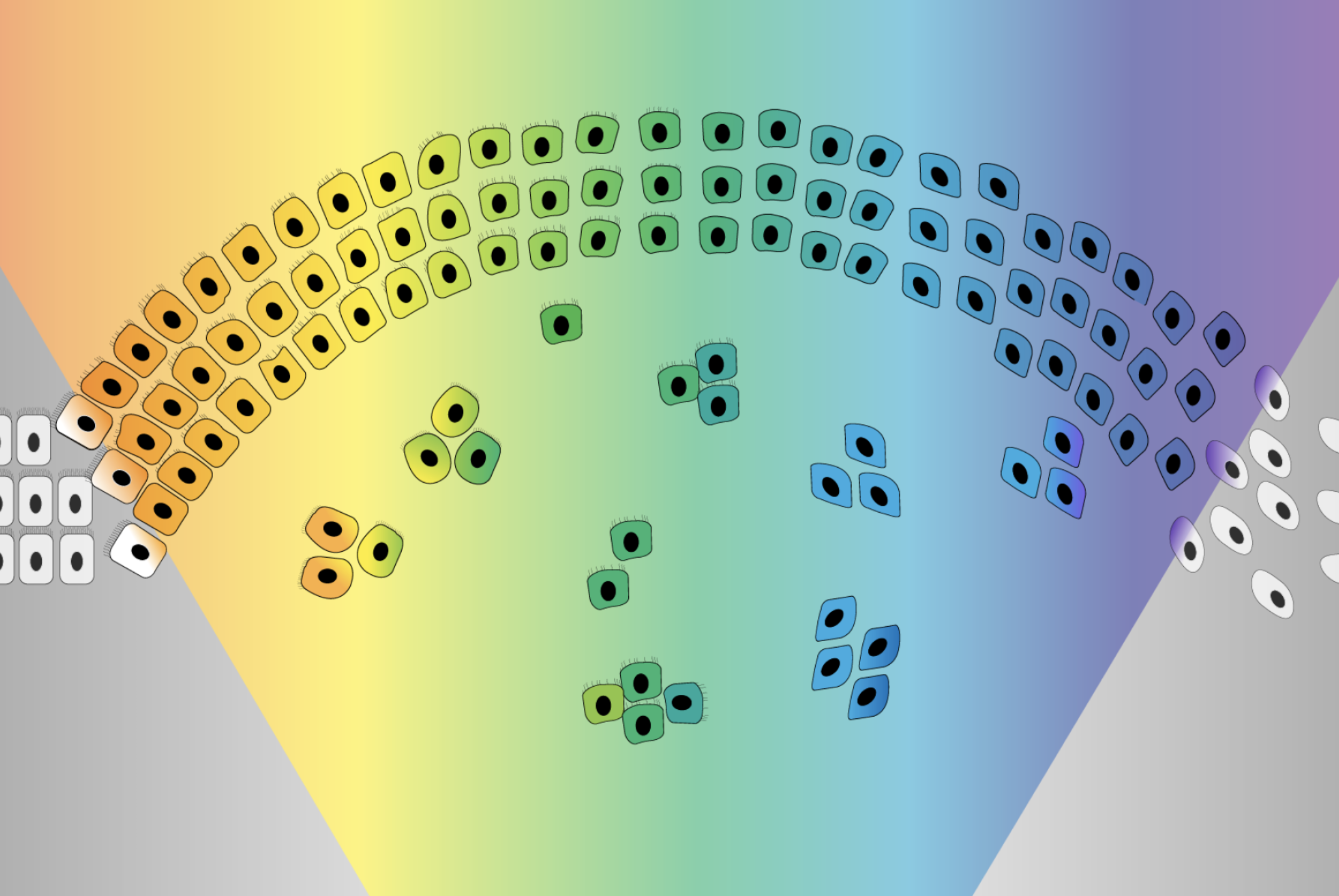
The EMT was originally imagined as a sort of binary switch, in which cells start out epithelial and become mesenchymal, much like a light switch being flicked from off to on. However, researchers are learning that the EMT works more like a dimmer switch that can be shifted along a spectrum of brightness. Cells that undergo the EMT usually end up in hybrid states between the epithelial and mesenchymal extremes. These cells in the middle of the spectrum, which have some characteristics of each extreme, are called “quasi-mesenchymal” cells, and it turns out that they–rather than cells that become fully mesenchymal–are the most capable of metastasizing and forming new tumors throughout the body.
Protected versus plastic cells
Weinberg and Zhang set out to better understand the EMT spectrum and what controls cells’ movement along it. First, they compared epithelial cells to each other and found that some were more plastic or prone to transitioning along the EMT spectrum than others. They also used the CRISPR gene editing tool to screen for genes that might be regulating the cells’ plasticity. If researchers can learn what makes a cell become quasi-mesenchymal—posing a high risk for metastasis—they might be able use this information, at some time in the future, to develop strategies to prevent cells from entering this high-risk state.
The CRISPR gene screen turned up a number of molecules that seemed to influence cells’ epithelial-mesenchymal plasticity. Two groups of these molecules had especially strong effects: PRC2, a complex that operates in chromosomes to silence or inactivate genes, and KMT2D-COMPASS, a complex that helps activate genes. Both complexes help to keep cells in a stable epithelial state. Loss of either complex makes cells more prone to moving along the EMT spectrum.
The researchers then determined how the loss of either complex enables the EMT. PRC2 normally silences several key EMT-related genes. When PRC2 is lost, those genes activate, which in turn sensitizes the cell to a signal that can trigger the EMT. The loss of KMT2D-COMPASS affects how well PRC2 can bind its targets, leading to the same signal sensitivity. In spite of the similar mechanisms at play, the loss of PRC2 versus KMT2D-COMPASS leads cells to transition to end up in different EMT states, an exciting finding for the researchers. Cells without KMT2D-COMPASS became fully mesenchymal, while cells without PRC2 became hybrid or quasi-mesenchymal. Consequently, cells without PRC2 were much more capable of metastasis than cells without KMT2D-COMPASS (or cells in which both complexes were active) in mouse models. When the researchers looked at historical data from breast cancer patients, they observed the same pattern: people with faulty PRC2 component genes had worse outcomes. These findings provide further evidence that cells in the middle of the EMT spectrum are most likely to metastasize.
This work supports the understanding of the EMT as a spectrum rather than a simple switch, and shows that different EMT regulators can program cells to transition to different parts of the EMT spectrum. Additionally, the finding that loss of PRC2 is linked to metastasis has implications for cancer drugs currently in development that work by inactivating PRC2. Benefits of the drugs may outweigh risks for patients with certain types of cancer for which PRC2 is an effective target. However, Weinberg and Zhang caution that researchers leading clinical trials of PRC2-targeting drugs should be careful about selecting patients and monitoring outcomes. In the types of cancer cells that the researchers looked at, even temporary PRC2 inactivation, such as from a therapy trial, was sufficient to trigger cells to become EMT hybrids with increased metastatic capacity.
Weinberg and Zhang intend to continue exploring the genes identified in their CRISPR screen to see if they can identify other hybrid states along the EMT spectrum, in which cells have different combinations of epithelial and mesenchymal features. They hope that by deepening their understanding of the gene expression profiles of cancer cells associated with different EMT trajectories, they can contribute to the development of therapies for people with potentially metastatic cancers.
“Understanding when and how cancer cells become able to form life-threatening metastases is crucial in order to help the many patients for whom this is a risk,” Weinberg says. “This work provides new insights into the mechanisms that enable cells to metastasize and the roles that different EMT programs can play.”

Whitehead Institute
March 25, 2022
For the hundreds of thousands of people diagnosed with breast cancer each year, surgery to remove the cancerous tissue is often the best option — but this relatively simple procedure comes with some drawbacks. In more than a few cases, the surgical removal of a tumor can lead to an increased risk of the cancer reemerging in other locations in the body.
In a 2018 study, a postdoc in the lab of Whitehead Institute Member Bob Weinberg discovered that, at least in mice, this phenomenon was due to a bodily butterfly effect: the creation of a wound site in one place in the body, which necessitated subsequent wound healing, caused immune system changes affecting distant parts of the body.
These changes occurred as bone marrow cells responded to the wounding with a flood of inflammatory cells that entered into the wound site and, at the same time, scattered throughout the body. These dispersed inflammatory cells weakened the ability of the immune system to control the outgrowth of a distantly located metastatic tumor. Without this immune control, which otherwise could keep the metastasis at a very small size, the metastasis would grow out aggressively.
Hence, wounding in one part of the body provoked metastasis outgrowth at a distant site. This suggested, among other things, that the outgrowth of metastatic tumors, which is often seen in women who have recently undergone a mastectomy, might be actively provoked by the post-surgical wound-healing process.
Weinberg’s work also presented a way to potentially avoid this effect, using a preventative measure that’s probably sitting in your bathroom cabinet right now: the cheap and common class of drugs known as NSAIDS, which includes ibuprofen and aspirin. When mice were given NSAIDS before and after tumor removal surgeries, they experienced a fivefold lower rate of cancer recurrence at the site of metastasis than a control group given opioids. These NSAIDs could therefore be used in place of the opioids, which are often used to treat post-surgical pain.
The human body is full of undiscovered connections like this one and adding in foreign substances further complicates matters. While a treatment might work well in a Petri dish, researchers describe whole -body metabolism as “a whole different kettle of fish.”
The way drugs move through the body and interact with internal systems is called pharmacokinetics. When a person is given a medicine — either orally, through a chemotherapy method, or via injection — that drug must be able to find its way to its target in a high enough concentration to have an effect, and then when its purpose is served, it must be able to leave the body safely and not build up to a harmful amount.
Much like Weinberg’s work on NSAIDS in breast cancer, Whitehead Institute’s basic research has led to other surprising discoveries about drug activities in the human body. Read on to learn about research that is changing the way new drugs are designed, making existing treatments less toxic, and more.
Concentration is key
When it comes to the action of drugs in the human body, concentration is key. Just ask Rick Young, a Whitehead Institute Member and professor of biology at MIT. In 2018, Young’s lab, which had previously studied the regulatory circuitry involved in transcription (the copying of DNA into RNA), shifted its focus after discovering tiny droplets within cells that concentrate the molecular materials needed to transcribe the DNA.
The droplets, called transcriptional condensates, were the newest in a slew of recent discoveries of other such groupings of cellular components. Some of these aggregations facilitate RNA splicing while others help to form ribosomes.
For Young, the discovery of transcription-related condensates sparked an interest in how these droplets were affecting the action of drugs. Previous theories held that transcription was able to take place in cells because there was a sufficient concentration of necessary proteins, such as RNA polymerase and other accessory proteins. As the Young lab showed, these collaborating cellular players were actually being concentrated in the condensates,
In 2020, Young and Ann Boija and Isaac Klein, two postdocs in his lab, took their investigation a step further, analyzing the mechanism by which several cancer drugs are concentrated in cellular condensates, and how that concentration could affect their action in individual cells and thus in the body. They found that cancer drugs sort themselves into specific types of condensates, independently of their targets, which can allow them to build up into high concentrations in these localized areas within cells.
“This could have enormous implications for the way we discover and develop drugs,” said Rick Young. “If drugs had properties that had them partitioning into a condensate where their target lives, then they would enjoy two properties of condensates: they would be compartmentalized, and they would be at much higher concentrations than if they diffuse through the cell.”
Young’s work on condensates led him to co-create a pharmaceutical company called Dewpoint Therapeutics, with the goal of reformulating treatments for cancer or neurological conditions such as amyotrophic lateral sclerosis by targeting biomolecular condensates. Whitehead Institute Founding Member Rudolf Jaenisch serves as a scientific advisor.
Trouble in parasites
While researchers in Young’s lab investigate how drugs could be more efficiently targeted, Sebastian Lourido’s lab is taking a different tack — why do some drugs stop working as time progresses?
The malaria drug artemisinin was developed in China in the 1970s, and completely changed the way the world treated malaria. In the following decades, however, the parasites that cause malaria, several species within the genus Plasmodium, have slowly grown less susceptible to the drug.
In a paper published in September of 2020, Whitehead Institute Member Lourido and collaborators identified two parasite genes that were negatively impacting the actions of the drug in the parasite’s cells.
Researchers liken artemisinin to a “ticking time bomb,” which needs another molecule, called heme, to light its fuse. Heme, a small molecule that is one component of hemoglobin, helps transport electrons and deliver oxygen to tissues. When heme encounters artemisinin, it activates the drug, allowing the creation of small, toxic chemical radicals. These proceed to react with the parasites proteins, fats, and metabolites, eventually leading to its death.
In order to understand how some parasites were becoming less vulnerable to the drug, Lourido, along with researchers Clare Harding, Boryana Petrova and Saima Sidik, ran a genetic screen on a related parasite, Toxoplasma gondii. The screen allowed them to assess which mutations in the parasites’ genomes were beneficial for their survival and which ones were harmful.
The screen revealed two genes that affected how susceptible the parasites were to treatment with artemisinin. One, called Tmem14c, seemed to be protecting the parasites. The gene is analogous to a gene that transports heme out of mitochondria where it is generated. Lourido hypothesized that when the Tmem14c protein is working properly, it helps the cells shuttle heme and its building blocks and get them where they need to go in the cell. When this gene is knocked out or mutated, heme can build up in the parasite cells, making them more likely to activate the artemisinin “bomb.”
Another gene, when mutated, made the parasites less sensitive to artemisinin. The gene, called DegP2, encodes a protein that plays a role in heme metabolism, so when it was mutated, less heme was available in the cells to activate the drug.
This knowledge provides useful insights for treatment methods, said Lourido. For example, healthcare providers should take into consideration the fact that heme is key in artemisinin’s action, and avoid combining the drug with other treatments that might lower the amount of heme in parasite cells. “Understanding how different pathways within the cell participate to render parasites susceptible to these antiparasitic drugs helps us better pair them with other compounds that are going to be synergistic and not work against our own goal of defeating parasites,” Lourido said.
Taking the edge off toxic treatments
Another application of fundamental pharmacokinetics research involves mitigating the harmful effects of drugs. Consider the chemotherapy drug methotrexate. Methotrexate was the very first targeted drug ever made. Developed more than 60 years ago by Dr. Sidney Farber, the drug acts by inhibiting a key molecule in the metabolic process that builds DNA and RNA, thereby interfering with basic functions of the cell and with DNA synthesis, repair and replication, helping to destroy cancerous cells in the body.
Methotrexate is still a widely used component of chemotherapy cocktails, especially for pediatric leukemia. In the human body, though, methotrexate is like a bull in a china shop. It is very effective at knocking back cancer, but the drug’s life-threatening side effects, including gut, liver, kidney and brain damage, often lead doctors to terminate their patients’ treatment early, or seriously compromise the survivors’ quality of life.
The drug was much-studied in the 70s, but research trailed off in the subsequent decades due to limits on the existing technologies. Nearly fifty years later, Naama Kanarek, then a postdoctoral researcher in the lab of former Whitehead Institute Member David Sabatini, decided to take a fundamental research approach to studying the effects of methotrexate, in hopes that she might discover some way to make the drug less toxic.
“We now have access to genetic tools that allow us to address long standing questions in a way that was not possible before,” said Kanarek, who now runs her own lab at Boston Children’s Hospital. “We can use a CRISPR screen, and instead of focusing on what is known, we can ask what is unknown about the drug. We can find new genes that are involved in the response of cells to the drug that were not found before simply because the tools were not there.”
The screen revealed one gene in particular that seemed to be playing a role in how sensitive cancer cells were to methotrexate, the researchers reported in Nature in July of 2018. The cells’ sensitivity is important, Kanarek said, because if the cells can be made more vulnerable to methotrexate, the duration of treatment or required dose could be reduced. “If we can reduce dose because we can improve efficacy, then we can reduce toxicity without compromising on the cure rates and that is good news to the patients,” Kanarek said.
The gene in question, called FTCD, encodes an enzyme involved in the breakdown of the amino acid histidine. When the gene was knocked out, cancer cells were less sensitive to methotrexate. When the pathway was boosted with the addition of extra histidine, cells became more sensitive.
Former Whitehead Institute Director Susan Lindquist, who passed away 2016, performed similar work on the natural product amphotericin B, a drug which is used to treat some fungal infections. The drug is especially useful because fungi have not yet developed a resistance to it, as they have with so many other treatments. But amphotericin B also has some serious drawbacks; it can cause kidney damage, heart failure, and other serious and even fatal side effects.
These side effects mostly happened because amphotericin B works by binding to a chemical group called a sterol. In fungi, it binds to molecules called ergosterols in the cell wall, destabilizing the cells. Unfortunately humans also have a prevalent sterol: cholesterol. When amphotericin B binds to cholesterol in human cell membranes, it can damage human cells.
Using chemical synthesis methods, Lindquist and colleagues at Whitehead Institute and elsewhere were able to tweak the structure of the drug to bind only to ergosterol molecules and not cholesterol, bypassing most of the harmful side effects.
Why fundamental research
Drug development is often an extremely targeted pursuit, but for Whitehead Institute scientists, their advances have mostly come from a simple curiosity about the cellular mechanisms. For example, Rick Young didn’t set out to study condensates, but an inquiry into the fundamentals of transcription led him in this entirely new direction.
Such fundamental research has the potential to branch in any number of different ways. “Fundamental science can lead in directions that you would not foresee,” said the Institute’s Associate Director of Intellectual Property Shoji Takahashi. Basic research into drug behavior is essential and can contribute to life-changing therapies down the line.

Merrill Meadow | Whitehead Institute
March 23, 2022
Metabolism is the sum of life-sustaining chemical reactions occurring in cells and across whole organisms. The human genome codes for thousands of metabolic enzymes, and specific metabolic pathways play significant roles in many biological processes—from breaking down food to release energy, to normal proliferation and differentiation of cells, to pathologies underlying diabetes, cancer, and other diseases.
For decades, Whitehead Institute researchers have helped both to clarify how metabolism works in healthy states and to identify how metabolic processes gone awry contribute to diseases. Among Whitehead Founding Member Harvey Lodish’s wide-ranging accomplishments, for example, are the identification of genes and proteins involved in development of insulin resistance and stress responses in fat cells. His lab explored the hormones controlling fatty acid and glucose metabolism, broadening understanding of obesity and type 2 diabetes. In 1995, the lab cloned adiponectin, a hormone made exclusively by fat cells. A long series of studies has shown that adiponectin causes muscle to burn fatty acids faster – so they are not stored as fat – and increases the metabolism of the sugar glucose. More recently the lab identified and characterized types of RNAs that are specifically expressed in fat cells – including a microRNA unique to brown fat, which burns rather than stores fatty acids. In addition, former Member David Sabatini’s discovery of the mTOR protein and his subsequent work elaborating many ways in which the mTOR pathway affects cells function has proven to be fundamental to understanding the relationship between metabolism and an array of diseases.
Today, Institute researchers continue to pioneer a deeper understanding of how metabolic processes contribute to health and disease – with long-term implications that could range from new treatments for obesity and type 2 diabetes to methods for slowing the aging process. Here are a few examples of Whitehead Institute scientists’ creative and pioneering work in the field of metabolism.
Understanding hibernation and torpor
Research inspiration comes in many forms. For example, Whitehead Institute Member Siniša Hrvatin – who joined the faculty in January 2022 from Harvard Medical School (HMS) – was inspired to pursue his current research by science-fiction tales about suspended animation for long-term space travel. And during graduate school, he realized that the ability of some mammals to enter a state of greatly reduced metabolism – such as occurs in hibernation – was a mild but real-world form of suspended animation.
Hrvatin’s doctoral research in Doug Melton’s lab at Harvard University focused primarily on stem cell biology. But his subsequent postdoctoral research positions at Massachusetts Institute of Technology (MIT) and HMS enabled him to begin exploring the mechanisms and impact of reduced metabolic states in mammals. The timing was serendipitous, too, because he was able to use the growing array of genetic tools that were becoming available – and create some new tools of his own as well.
“To survive extreme environments, many animals have evolved the ability to profoundly decrease metabolic rate and body temperature and enter states of dormancy, such as hibernation and torpor,” Hrvatin says. Hibernating animals enter repeated states of significantly reduced metabolic activity, each lasting days to weeks. By comparison, daily torpor is shorter, with animals entering repeated periods of lower-than-normal metabolic activity lasting several hours.
Hrvatin’s lab studies the mysteries of how animals and their cells initiate, regulate, and survive these adaptations. “Our long-term goal is to determine if these adaptations can be harnessed to create therapeutic applications for humans.” He and his team are focusing on three broad questions regarding the mechanisms underlying torpor in mice and hibernation in hamsters.
First: How do the animals’ brains initiate and regulate the metabolic processes involved in this process? During his postdoctoral research, Hrvatin published details of his discovery of neurons involved in the regulation of mouse torpor. “Now we are investigating how these torpor-regulating neurons receive information about the body’s energy-state,” he explains, “and studying how these neurons then drive a decrease of metabolic rate and body temperature throughout the body.”
Second: How do individual cells – and their genomes – adapt to extreme or changing environments; and how do these adaptations differ between types of organisms?
“Cells from hibernating organisms ranging from rodents to bears have evolved the ability to survive extreme cold temperatures for many weeks to months,” Hrvatin notes. “We are using genetic screens to identify species-specific mechanisms of tolerance to extreme cold. Then we will explore whether these mechanisms can be induced in non-hibernating organisms – potentially to provide health benefits.”
Third: Can we deliberately and specifically slow down tissue damage, disease progression, and or aging in cells and whole organisms by inducing torpor or hibernation – or facets of those states? It has long been known that hibernating animals live longer than closely related non-hibernators; that cancer cells do not replicate during hibernation; and that cold can help protect neurons from the effects of loss of oxygen. However, the cellular mechanisms underlying these phenomena remain largely unknown. Hrvatin’s lab will induce a long-term hibernation-like state in mice and natural hibernation in hamsters, and study how those states affect processes such as tissue repair, cancer progression, and aging.
“In the lab, if you take many human cell types and put them in a cold environment they die, but cells from hibernators survive,” Hrvatin notes. “We’re fascinated by the cellular processes underlying those survival capacities. As a starting place, we are using novel CRISPR screening approaches to help us identify the genomic mechanisms involved.”
And then? “Ultimately, we hope to take on the biggest question: Is it possible to transfer some of those survival abilities to humans?
Solving a mitochondrial conundrum
When Whitehead Institute postdoctoral researcher Jessica Spinelli was studying cancer metabolism in graduate school, she became interested in what seemed to be a scientific paradox regarding mitochondria, the cell’s energy-producing organelles: Mitochondria are believed to be important for tumor growth; but they generally need oxygen to function, and substantial portions of tumors have very low oxygen levels. Pursuing research in the lab of former Whitehead Institute Member David Sabatini, Spinelli sought to understand how those facts fit together and whether mitochondria could somehow adapt to function with limited oxygen levels.
Recently, Spinelli and colleagues published an answer to the conundrum – one that could inform research into medical conditions including ischemia, diabetes and cancer. In a Science paper for which Spinelli was first author, the team demonstrated that when cells are deprived of oxygen, a molecule called fumarate can serve as a substitute and enable mitochondria to continue functioning.
As Spinelli explains, humans need oxygen molecules for the cellular respiration process that takes place in our cells’ mitochondria. In this process – called the electron transport chain – electrons are passed along in a sort of cellular relay race that, ultimately, allows the cell to create the energy needed to perform its vital functions. Usually, oxygen is necessary to keep that process operating.
Using mass spectrometry to measure the quantities of molecules produced through cellular respiration in varied conditions, Spinelli and the team found that cells deprived of oxygen had a high level of succinate molecules, which form when electrons are added to a molecule called fumarate. “From this, we hypothesized that the accumulation of succinate in low-oxygen environments is caused by mitochondria using fumarate as a substitute for oxygen’s role in the electron transport chain,” Spinelli explains. “That could explain how mitochondria function with relatively little oxygen.” The next step was to test that hypothesis in mice, and those studies provided several interesting findings: Only mitochondria in kidney, liver, and brain tissues could use fumarate in the electron transport chain. And even in normal conditions, mitochondria in these tissues used both fumarate and oxygen to function – shifting to rely more heavily on fumarate when oxygen was reduced. In contrast, heart and skeletal muscle mitochondria made minimal use of fumarate and did not function well with limited oxygen.
“We foresee some exciting work ahead, learning exactly how this process is regulated in different tissues,” Spinelli says, “and, especially, in solid tumor cancers, where oxygen levels vary between regions.”
Seeking a more accurate model of diabetes
Max Friesen, a postdoctoral researcher in the lab of Whitehead Institute Founding Member Rudolf Jaenisch, studies the role of cell metabolism in type 2 diabetes (T2D). An increasingly prevalent disease that affects millions of people around the world, T2D is hard to study in the lab. This has made it very challenging for scientists to detail the cellular mechanisms through which it develops – and therefore to create effective therapeutics.
“It has always been very hard to model T2D, because metabolism differs greatly between species,” Friesen says. “That fact leads to complications when we use animal models to study this disease. Mice, for example, have much higher metabolism and faster heart rates than humans. As a result, researchers have developed many approaches that cure diabetes in mice but that fail in humans.” Nor do most in vitro culture systems—cells in a dish—effectively recapitulate the disease.
But, building on Jaenisch’s pioneering success in developing disease models derived from human stem cells, Friesen is working to create a much more accurate in vitro system for studying diabetes. His goal is to make human stem cell-derived tissues that function as they would in the human body, closely recapitulating what happens when an individual develops diabetes. Currently, Friesen is differentiating human stem cells into metabolic tissues such as liver and adipocytes (fat). He has improved current differentiation protocols by adapting these cells to a culture medium that is much closer to the environment they see in the human body. Serendipitously, the process also makes the cells responsive to insulin at levels that are present in the human bloodstream. “This serves as a great model of a healthy cell that we can then turn into a disease model by exposing the cell to diabetic hyperinsulinemia,” Friesen says.
These advances should enable him to gain a better understanding of how metabolic pathways – such as the insulin signaling pathway – function in a diabetic model versus a healthy control model. “My hope is that our new models will enable us to figure out how dietary insulin resistance develops, and then identify a therapeutic intervention that blocks that disease-causing process,” he explains. “It would be fantastic to help alleviate this growing global health burden.”
Alumna, Margaret ‘Mo’ Okobi ’16, prepares for a career combining medicine and public health research to improve mental health care services in vulnerable communities
Leah Campbell | School of Science
March 22, 2022
With only one year left of medical school at Harvard, Mo Okobi ’16, took a leave of absence. She didn’t want to take a break as much as a “step back” — a moment to reassess and reframe what she was learning in school within the bigger picture of the U.S. healthcare system. During that year, Okobi earned her master’s in public health from the Harvard School of Public Health.
“It’s so important to keep a broader view of the trends in medicine,” she says, “to look at what medications and therapies we’re using and what is actually working for patients.”
When she applied to the MPH program, Okobi couldn’t have known that her year-long leave to study public health would overlap with a pandemic. Needless to say, it was good timing. Okobi’s joint training in public health and medicine has also uniquely positioned her for a career combining clinical practice and research around chronic mental illness.
Though she says she never expected to find herself on this path, Okobi’s experiences at MIT shaped her approach to medicine and her commitment to providing quality mental healthcare to underserved communities.
Okobi was interested in many aspects of medicine when she enrolled at MIT. She majored in biology, with a minor in chemistry, to explore her broad interests in STEM. She joined MIT Emergency Medical Services, a student-run, volunteer ambulance service, for the same reason.
Being a certified EMT, though, she says, was one of the most formative aspects of her MIT education. She describes it as an “intensive introduction to medicine,” cementing her excitement about her future as a doctor. It was also one of her first exposures to mental health care, and Okobi believes that mental health emergencies made up a large proportion, if not a majority, of their calls.
“MIT EMS was really my first opportunity to actually work with patients very closely and see them in their moments of need,” she says.
While deciding to become a doctor was easy, Okobi’s journey to research wasn’t so smooth. She participated in one official undergraduate research experience at MIT, admitting, “I kind of hated it.” Working in a basic science biology lab, Okobi realized that the bench wasn’t for her.
Fortunately, her mentor, Hazel Sive, a former MIT professor of biology and now Dean of the College of Science at Northeastern University, took the time to talk with Okobi and figure out where she’d thrive. Sive, a South African, connected Okobi with the MIT Africa Program, through which she spent a summer in Johannesburg with the South African National Health Laboratory Services.
That experience, which she describes as a “pivotal framing moment,” helped Okobi understand how a career combining clinical practice and research might look. She worked with scientists studying HIV transmission from mother to child, assessing the quality of testing and resource gaps across different provinces. Near the end of her internship, Okobi was able to go into an HIV clinic and do antibody testing.
“I’m looking at these numbers. I’m making all these graphs and writing this paper, but I’m also seeing the people,” she says. “I loved the clinical work. I loved meeting people and knowing their stories.”
At graduation, Okobi was recognized as a Ronald E. McNair Scholar, an award that goes every year to Black undergraduates who have excelled academically and contributed to the experience of students on campus from underrepresented groups. The award was established in honor of McNair, PhD ’77, an accomplished astronaut who received his doctorate in physics from MIT and tragically perished onboard the Challenger space shuttle in 1986.
Having found a passion for applied, public health research, Okobi spent a year before medical school at a healthcare data analytics startup called Aetion. Aetion uses data from hospitals and insurance companies to analyze healthcare trends like medication usage and clinical outcomes. For a self-described “numbers nerd” like Okobi, it was a great way to learn how healthcare studies are designed and see the big picture behind clinical decisions.
It was her second year of medical school, though, that focused Okobi’s health interests around psychiatry for marginalized populations. During her clinical rotations, she worked at Cambridge Health Alliance (CHA), a public, community-centered hospital. At CHA, she served many non-English speakers and MassHealth recipients and was able to do rotations in outpatient, inpatient, and emergency psychiatric settings.
“I really got to see it from all angles,” Okobi says of her psychiatry rotations. “I loved the practice of creating space for people to talk about their lives…. it’s about the medicine and mental illness, but it’s also about seeing the whole person.”
As for what’s next, Okobi will be heading west for her residency in psychiatry at the University of California San Francisco.
Her work with CHA convinced Okobi to apply for residencies in psychiatry. She’s primarily interested in acute care settings, particularly for those with severe, chronic mental illnesses like schizophrenia. For her, one of the joys of inpatient care is working closely with patients on a daily basis, to, as she describes it, “try to leave a positive, lasting impression on those first encountering psychiatric care.”
Yet, armed with her degree in public health, she’s also committing to combining her clinical practice with ongoing research. At Harvard, she organized a mental health survey for medical and dental students in her class to improve access to mental health resources. Since 2020, she’s been participating in research on psychiatric emergency care utilization with the Boston Emergency Services Team. In 2020, she returned to Aetion as a consultant with their new FDA-backed COVID-19 research group, to lead epidemiological studies examining COVID-19 risk factors.
Looking back, Okobi knows that it may seem like she had a clear professional plan from the get-go. But she stresses that that wasn’t her experience at all, and current students should understand that things have a way of working out if you’re open to trying things.
“My path was very much like ping pong, never knowing where I’m going next,” she says. “The uncertainty never really ends, but I take refuge in those moments of serendipity, when I find something or someone that excites me and challenges me to be a better version of myself.”