Revealing an imperfect actor in plant biotechnology

Whitehead Institute researchers detect the chemical mistakes of a common herbicide-resistance enzyme, then successfully re-engineer it for enhanced precision.

Nicole Davis | Whitehead Institute
November 29, 2017

A research team led by MIT’s Whitehead Institute for Biomedical Research has harnessed metabolomic technologies to unravel the molecular activities of a key protein that enables plants to withstand a common herbicide.

Their findings reveal how the protein — a kind of catalyst or enzyme first isolated in bacteria and introduced into plants such as corn and soybeans in the 1990s — can sometimes act imprecisely, and how it can be successfully re-engineered to be more precise. The new study, which appears online in the journal Nature Plants, raises the standards for bioengineering in the 21st century.

“Our work underscores a critical aspect of bioengineering that we are now becoming technically able to address,” says senior author Jing-Ke Weng, a member of the Whitehead Institute and an assistant professor of biology at MIT. “We know that enzymes can behave indiscriminately. Now, we have the scientific capabilities to detect their molecular side effects, and we can leverage those insights to design smarter enzymes with enhanced specificity.”

Plants provide an extraordinary model for scientists to study how metabolism changes over time. Because they cannot escape from predators or search for new food sources when supplies run low, plants must often grapple with an array of environmental insults using what is readily available — their own internal biochemistry.

“Although they appear to be stationary, plants have rapidly evolving metabolic systems,” Weng explains. “Now, we can gain an unprecedented view of these changes because of cutting-edge techniques like metabolomics, allowing us to analyze metabolites and other biochemicals on a broad scale.”

Key players in this evolutionary process, and a major focus of research in Weng’s laboratory, are enzymes. Traditionally, these naturally occurring catalysts have been viewed as mini-machines, taking the proper starting material (or substrate) and flawlessly converting it to the correct product. But Weng and other scientists now recognize that they make mistakes, often by latching on to an unintended substrate.

“This concept, known as enzyme promiscuity, has a variety of implications, both in enzyme evolution and more broadly, in human disease,” Weng says.

It also has implications for bioengineering, as Bastien Christ, a postdoctoral fellow in Weng’s laboratory, and his colleagues recently discovered.

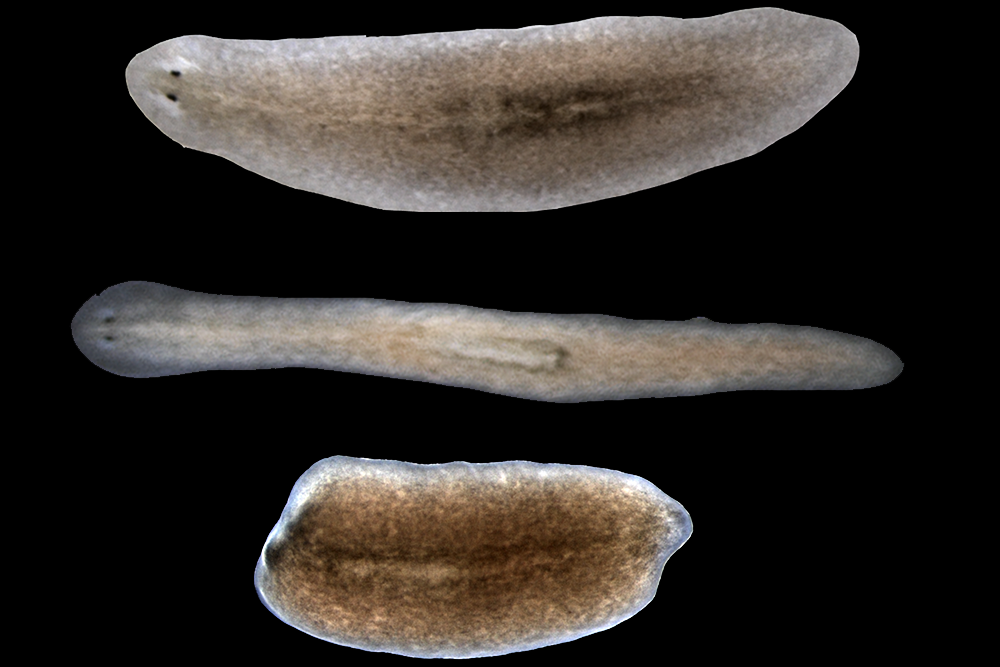
Christ, then a graduate student in Stefan Hörtensteiner’s lab at the University of Zurich in Switzerland, was studying a particular strain of the flowering plant Arabidopsis thaliana as part of a separate project when he made a puzzling observation. He found that two biochemical compounds were present at unusually high levels in the plant’s leaves.

Strangely, these compounds (called acetyl-aminoadipate and acetyl-tryptophan) weren’t present in any of the normal, so-called wild type plants. As he and his colleagues searched for an explanation, they narrowed in on the source: an enzyme, called BAR, that was engineered into the plants as a kind of chemical beacon, enabling scientists to more readily study them.

But BAR is more than just a tool for scientists. It is also one of the most commonly deployed traits in genetically modified crops such as soybeans, corn, and cotton, enabling them to withstand a widely-used herbicide (known as phosphinothricin or glufosinate).

For decades, scientists have known that BAR, originally isolated from bacteria, can render the herbicide inactive by tacking on a short string of chemicals, made of two carbons and one oxygen (also called an acetyl group). As the researchers describe in their Nature Plants paper, BAR has a promiscuous side, and can work on other substrates, too, such as the amino acids tryptophan and aminoadipate (a lysine derivative).

That explains why they can detect the unintended products (acetyl-tryptophan and acetyl-aminoadipate) in crops genetically engineered to carry BAR, such as soybeans and canola.

Their research included detailed studies of the BAR protein, including crystal structures of the protein bound to its substrates. This provided them with a blueprint for how to strategically modify BAR to make it less promiscuous, and favor only the herbicide as a substrate and not the amino acids. Christ and his colleagues created several versions that lack the non-specific activity of the original BAR protein.

“These are natural catalysts, so when we borrow them from an organism and put them into another, they may not necessarily be perfect for our purposes,” Christ says. “Gathering this kind of fundamental knowledge about how enzymes work and how their structure influences function can teach us how to select the best tools for bioengineering.”

There are other important lessons, too. When the BAR trait was first evaluated by the U.S. Food and Drug Administration (FDA) in 1995 for use in canola, and in subsequent years for other crops, metabolomics was largely non-existent as a technology for biomedical research. Therefore, it could not be applied toward the characterization of genetically engineered plants and foods, as part of their regulatory review. Nevertheless, acetyl-aminoadipate and acetyl-tryptophan, which are normally present in humans, have been reviewed by the FDA and are safe for human and animal consumption.

Weng and his colleagues believe their study makes a strong case for considering metabolomics analyses as part of the review process for future genetically engineered crops.

“This is a cautionary tale,” Weng says.

The work was supported by the Swiss National Science Foundation, the EU-funded Plant Fellows program, the Pew Scholar Program in the Biomedical Sciences, and the Searle Scholars Program.

Muscle plays surprising role in tissue regeneration

Whitehead Institute researchers have pinpointed distinct muscle subsets that orchestrate and pattern regrowth.

Nicole Davis | Whitehead Institute
November 22, 2017

Researchers at the Whitehead Institute have illuminated an important role for different subtypes of muscle cells in orchestrating the process of tissue regeneration.

In a paper appearing online today in Nature, they reveal that a subtype of muscle fibers in flatworms is required for triggering the activity of genes that initiate the regeneration program. Notably, in the absence of these muscles, regeneration fails to proceed. Another type of muscle, they report, is required for giving regenerated tissue the proper pattern — for example, forming one head instead of two.

“One of the central mysteries in organ and tissue regeneration is: How do animals initiate all of the cellular and molecular steps that lead to regeneration?” says senior author Peter Reddien, a member of Whitehead Institute, professor of biology at MIT, and investigator with the Howard Hughes Medical Institute. “We’ve helped answer this question by revealing a surprising molecular program that operates within a subgroup of muscle cells that helps establish the molecular information required for proper tissue regeneration after injury.”

For more than a decade, Reddien and the researchers in his laboratory have studied the biological mechanisms that underlie regeneration in a tiny flatworm called planarians. These worms possess some impressive regenerative capabilities: When sliced in two, each piece of the worm can regrow the body parts needed to form two complete organisms. In previous studies, Reddien’s team identified a set of always-on genes, known as position control genes (PCGs), that provide cells with region-specific instructions, like a set of GPS coordinates, that tell cells where they are in the body, and thus what body part to regenerate. Interestingly, PGCs are active in planarian muscle cells, suggesting muscle may play a major role in the regeneration process.

“This discovery raised a lot of questions about how muscle participates in this process,” Reddien says.

In planarians, there are a handful of muscle cell types. For example, if you imagine the worms as simple cylindrical tubes, there are longitudinal muscle fibers, which run head-to-tail along the tubes’ long axis. There are also circular fibers, which are perpendicular to the longitudinal fibers and hug the tubes’ outer circumference.

To assess the roles of these different muscle cell types in regeneration, first author Lucila Scimone and her colleagues needed a method to selectively remove them. When myoD, a gene found specifically in the longitudinal fibers, is inhibited, those fibers fail to form. Similarly, the nkx1-1 gene marks the circular fibers, and when its function is reduced, they do not develop. Using these genes as molecular scalpels, Scimone and her co-authors could test the effects of ablating these distinct muscle groups on regeneration.

Surprisingly, when the longitudinal fibers were removed, the results were dramatic. The worms live quite normally, but when they are injured (the head removed, for example) they cannot regenerate the missing parts.

“This is an amazing result; it tells us that these longitudinal fibers are essential for orchestrating the regeneration program from the very beginning,” says Scimone, a scientist in Reddien’s lab.

As the researchers dug deeper into the finding, they learned that the functions of two critical genes are disrupted when longitudinal fibers are missing. These genes, called notum and follistatin, are known for their fundamental roles in regeneration, controlling head-versus-tail decisions and sustained cell proliferation, respectively, following tissue injury.

In addition to this essential role for longitudinal fibers, the research team also uncovered a key role for circular fibers. When these muscles are missing, planarians are able to regenerate missing body parts, but what regrows is abnormally patterned. For example, two heads may be regenerated within a single outgrowth, instead of one.

These results underscore an important and previously unappreciated role for muscle, widely known for its contractile properties, in instructing the tissue regeneration program. The Whitehead researchers will continue to probe the role of different muscle cell types in planarian regeneration and also explore whether other animals with regenerative capabilities possess a similar muscle-localized program for conferring positional information.

“It’s hard to understand what limits humans’ abilities to regenerate and repair wounds without first knowing what mechanisms are enabling some animals, like planarians, to do it so amazingly well,” Reddien says.

This work was supported by the National Institutes of Health, Howard Hughes Medical Institute, and the Eleanor Schwartz Charitable Foundation.

Eric S. Lander

Education

  • PhD, 1981, Oxford University
  • AB, 1978, Mathematics, Princeton University

Research Summary

Following the successful completion of the Human Genome Project, the challenge now is to decipher the information encoded within the human genetic code — including genes, regulatory controls and cellular circuitry. Such understanding is fundamental to the study of physiology in both health and disease. At the Broad Institute, my lab collaborates with other to discover and understand the genes responsible for rare genetic diseases, common diseases, and cancer; the genetic variation and evolution of the human genome; the basis of gene regulation via enhancers, long non-coding RNAs, and three-dimensional folding of the genome; the developmental trajectories of cellular differentiation; and the history of the human population.

Awards

  • William Allan Award, American Society of Human Genetics, 2018
  • James R. Killian Jr. Faculty Achievement Award, MIT, 2016
  • Block Memorial Award for Distinguished Achievement in Cancer Research, Ohio State University, 2013
  • AAAS Philip Hauge Abelson Prize, 2015
  • Breakthrough Prize in Life Sciences, 2013
  • Harvey Prize for Human Health, Technion University, Israel, 2012
  • Dan David Prize, 2012
  • Albany Prize in Medicine and Biomedical Research, Albany Medical College, 2010
  • Gairdner Foundation International Award, Canada, 2002
  • Max Delbruck Medal, Berlin, 2001
  • MacArthur Foundation, MacArthur Fellowship, 1987
Iain M. Cheeseman

Education

  • PhD, 2002, University of California, Berkeley
  • BS, 1997, Biology, Duke University

Research Summary 

Our lab is fascinated by the molecular machinery that directs core cellular processes, and in particular how these processes are modulated and rewired across different physiological contexts. Our work has focused on the proteins that direct chromosome segregation and cell division, including the macromolecular kinetochore structure that mediates chromosome-microtubule interactions. Although cell division is an essential cellular process, this machinery is remarkably flexible in its composition and properties, which can vary dramatically between species and are even modulated within the same organism — over the cell cycle, during development, and across diverse physiological situations. To define the basis by which the kinetochore and other core cellular structures are rewired to adapt to diverse situations and functional requirements, we are currently investigating diverse transcriptional, translational, and post-translational mechanisms that act to generate proteomic variability both within individual cells and across tissues, cell state, development, and disease.

Awards

  • Global Consortium for Reproductive Longevity and Equality (GCRLE) Scholar Award, 2020
  • MIT Undergraduate Research Opportunities Program (UROP) Outstanding Mentor – Faculty, 2019
  • American Society for Cell Biology (ASCB) Early Career Life Scientist Award, 2012
  • Searle Scholar Award, 2009-2012

Follow on Twitter

Mary Gehring

Education

  • PhD, 2005, University of California, Berkeley
  • BA, 1998, Biology, Williams College

Research Summary

We focus on plant epigenetics — that is, the heritable information that influences cellular function but is not encoded in the DNA sequence itself. We use genetic, genomic and molecular biology approaches to study the fidelity of epigenetic inheritance and the dynamics of epigenomic reprogramming during reproduction, primarily in the model plant Arabidopsis thaliana. More specifically, we investigate the interplay among repetitive sequences, DNA methylation and chromatin structure in these dynamic processes.

Awards

  • Rosalind Franklin Young Investigator Award, 2013
  • Pew Scholar in the Biomedical Sciences, 2011
Troy Littleton

Education

  • PhD, 1994, Baylor College of Medicine; MD, 1997, Baylor College of Medicine
  • BS, 1989, Biochemistry, Louisiana State University

Research Summary

Using Drosophila, we study how neurons form synaptic connections, as well as how synapses transmit information and change during learning and memory. We also investigate how alterations in neuronal signaling underlie several neurological diseases, including epilepsy, autism, and Huntington’s Disease. We hope to bridge the gap between the molecular components of the synapse and the physiological responses they mediate.

Alan D. Grossman

Education

  • PhD, 1984, University of Wisconsin, Madison
  • BS, 1979, Biochemistry, Brown University

Research Summary

We use a variety of approaches to investigate several of the fundamental and conserved processes used by bacteria for propagation and growth, adaptation to stresses, and acquisition of new genes and traits via horizontal gene transfer. Our long term goals are to understand many of the molecular mechanisms and regulation underlying basic cellular processes in bacteria. Our organism of choice for these studies is usually the Gram positive bacterium Bacillus subtilis.

Our current efforts are focused in two important areas of biology: 1) The control of horizontal gene transfer, specifically the lifecycle, function, and control of integrative and conjugative elements (ICEs). These elements are widespread in bacteria and contribute greatly to the spread of antibiotic resistances between organisms. 2) Regulation of the initiation of DNA replication and the connections between replication and gene expression, with particular focus on the conserved replication initiator and transcription factor DnaA. This work is directly related to mechanisms controlling bacterial growth, survival, and stress responses.

Awards

  • National Academy of Sciences, 2014
  • American Academy of Arts and Sciences, 2008
  • American Academy of Microbiology 1998
  • Eli Lilly Company Research Award, 1997
Retinoic acid regulates transitions in mouse sperm production
November 7, 2017

CAMBRIDGE, MA – Sperm production requires progression through a well-orchestrated series of transitions in the testes that move diploid spermatogonia cells, with two complete sets of chromosomes, through a series of transitions to produce haploid sperm, with one copy of each chromosome, poised to swim and fertilize an available egg. There are four major transitions in sperm production, or spermatogenesis. The first is spermatogonial differentiation, during which spermatogonia differentiate, losing their stem-cell like qualities. The resulting spermatocytes then initiate meiosis and undergo two rounds of cell division to generate haploid spermatids. The spermatids undergo elongation and then the resulting sperm are released.

The signals that control progression through these transitions were poorly understood until 2015, when David Page, Member and Director of Whitehead Institute, professor of biology at Massachusetts Institute of Technology, and investigator with Howard Hughes Medical Institute and colleagues determined that retinoic acid (RA), a derivative of vitamin A that has been shown to play a key role in a number of developmental processes, was responsible for coordinating the first two stages of spermatogenesis-differentiation and meiosis. Now, in a paper published this week in the journal Proceedings of the National Academy of Sciences, Page, first author Tsutomu Endo, and colleagues extend those findings to show that RA signaling in mice coordinates the second two transitions as well.

Diagram of model by which retinoic acid coordinates spermatogenesisThe researchers used chemical manipulation of RA levels to determine that RA controlled the second two transitions, spermatid elongation and sperm release, in addition to the first two. With this knowledge in hand, the researchers were then able to drill down and get a better picture of how RA regulates male gamete production. One outstanding question has been how males are able to continually produce sperm throughout their lifetime, in contrast with females whose egg production and maturation is limited. Page and colleagues measured RA levels in the testes and discovered that it is cyclically produced, driving production of sperm during the male lifetime. In addition to the timing of RA production, the researchers also examined its source. From which cells was the RA signal coming? During the first two transitions, they determined that the RA was coming from the somatic Sertoli cells, the support cells of the testes, and in the second two transitions they determined that it was being released by the germ cells themselves-the meiotic (pachytene-stage) spermatocytes were found to be secreting RA to other germ cells in the testes.

These findings not only contribute to our fundamental understanding of male gamete formation, they also provide important clues for the field of reproductive technology. For years, scientists have been working on making gametes in the laboratory, but have had difficulty making functional sperm. This discovery of the role of RA in spermatogenesis adds important tools to the toolbox of assisted reproduction. The work shows that RA is required in both the early and late transitions of spermatogenesis and sheds light on an important component of laboratory efforts for sperm production.

Other researchers involved include Elizaveta Freinkman and Dirk G. de Rooij.

This research was supported by Howard Hughes Medical Institute (HHMI) and the United States Department of Defense (DoD W81XWH-15-1-0337)

Written by Lisa Girard
****
David Page’s primary affiliation is with Whitehead Institute for Biomedical Research, where his laboratory is located and all his research is conducted. He is also a Howard Hughes Medical Institute Investigator and a Professor of Biology at the Massachusetts Institute of Technology.
 ****
Paper cited: Endo, T et al.  Periodic production of retinoic acid by meiotic and somatic cells coordinates four transitions in mouse spermatogenesis. Proc Natl Acad Sci. DOI: 10.1073/pnas.1710837114. Epub 2017 Nov 6.
Other work cited: Endo T et al. Periodic retinoic acid-STRA8 signaling intersects with periodic germ-cell competencies to regulate spermatogenesis. Proc Natl Acad Sci. DOI: 10.1073/pnas.1505683112. Epub 2015 Apr 20.
A new workflow for natural product characterization comes ashore with red algae
October 30, 2017

CAMBRIDGE, Mass. – A few years ago while paddling off the coast near La Jolla, California, avid surfer Roland Kersten noticed a piece of red algae (Laurenica pacifica) bobbing alongside his surfboard. Kersten, whose background is in natural product chemistry, was intrigued.

Natural products—chemicals from living organisms such as plants and algae—represent a rich source of potential therapeutics. A majority of anti-cancer drugs are natural product-based or inspired. One such well-known natural product—the potent anti-cancer drug Taxol—was identified in the bark of a yew tree.

Marine algae, like the red algae Kersten found, are often rich in compounds called sesquiterpenes, some of which have been shown to have potential medicinal attributes. Since the 1970s, scientists identified many sesquiterpenes produced by Laurencia species with anti-cancer properties. The identification techniques usually required about tens of milligrams of purified compounds, which were obtained from more than a kilogram of algae. Because Laurencia and the reef ecosystems in which it thrives are protected, and such large-scale harvesting for scientific or medicinal purposes is no longer tenable, Kersten had to devise a different approach to analyze its sesquiterpenes.

Kersten received a collection permit to clamber over the rocky shore at deep low tide to collect a hand-sized sample of the red algae. Now a postdoctoral fellow in the lab of Whitehead Member and Massachusetts Institute of Technology assistant professor of biology Jing-Ke Weng, Kersten’s first task was to search the RNA sequences of all genes expressed in his red algae sample to find those whose product seemed likely to be enzymes that make sesquiterpenes.  In order to determine the product generated by these enzymes, he engineered them in yeast and isolated its sesquiterpene products.

In order to define the first step in the biogenesis of sesquiterpenes in red algae, Kersten wanted to see the precise 3-D structure of the isolated sesquiterpene. But the small handful of algae he had obtained produced only a fraction of the amount required for x-ray crystallography, the established method for determining a compound’s absolute structure. So Kersten tried a method recently developed by collaborator Makoto Fujita at the University of Tokyo that requires only a few nanograms of material: soaking extracted compounds into a special crystalline sponge, which supports the sample’s molecular shape while it is bombarded with x-rays to accurately determine the 3-D conformation of a molecule. A new combination of the crystalline sponge method and nuclear magnetic resonance spectroscopy by the Fujita group revealed the structure of prespatane.

With the compound’s structure in hand, Kersten is closer to understanding how Laurenciabiosynthesizes its sesquiterpenes and how to engineer yeast to produce the same molecules for medicinal research at scale—without touching the red algae flourishing on protected reefs. And the novel workflow—spanning genomics, metabolomics, synthetic biology, and x-ray crystallography with crystalline sponges—established by Weng, Kersten, and their collaborators may expedite the identification of other promising compounds produced by organisms from both land and sea.

Other contributors to this work include Shoukou Lee of Tokyo University, Daishi Fujita of Tokyo University and Whitehead Institute, Tomáš Pluskal of Whitehead Institute.  The team also collaborated with researchers from Scripps Institution of Oceanography and Salk Institute of Biological Sciences.

This work was supported by Howard Hughes Medical Institute, the Simons Foundation, the Helen Hay Whitney Foundation, the Pew Scholars Program in the Biomedical Sciences, the Searle Scholars Program, and the Japan Science and Technology Agency.

 Written by Nicole Giese Rura
* * *
Jing-Ke Weng’s primary affiliation is with Whitehead Institute for Biomedical Research, where his laboratory is located and all his research is conducted. He is also an assistant professor of biology at Massachusetts Institute of Technology.
* * *
Full Citation:
“A Red Algal Bourbonane Sesquiterpene Synthase Defined by Microgram-scale NMR-coupled Crystalline Sponge XRD Analysis”
Journal of the American Chemical Society, online October 30, 2017.
Roland D. Kersten (1,6), Shoukou Lee (2,6) , Daishi Fujita (1,2) , Tomáš Pluskal (1) , Susan Kram (3), Jennifer E. Smith (3) , Takahiro Iwai (2) , Joseph P. Noel (4) , Makoto Fujita (2), Jing-Ke Weng (1,5).
1. Whitehead Institute for Biomedical Research, 455 Main Street, Cambridge, MA, United States
2. Graduate School of Engineering, The University of Tokyo, JST-ACCEL, Tokyo, Japan
3. Scripps Institution of Oceanography, University of California San Diego, La Jolla, CA, United States
4. Howard Hughes Medical Institute, Jack H. Skirball Center for Chemical Biology and Proteomics, The Salk Institute for Biological Studies, La Jolla, CA, United States
5. Department of Biology, Massachusetts Institute of Technology, Cambridge, MA, United States
6. These authors contributed equally
Department of Biology hosts its first Science Slam

Eight biology trainees had just three minutes to explain their research and earn favor with the judges and audience in new yearly event.

Raleigh McElvery | Department of Biology
October 5, 2017

Nearly 300 spectators crowded into a lecture hall at the Ray and Maria Stata Center on a recent Tuesday to witness the first annual Science Slam, hosted by MIT’s Department of Biology.

A science slam features a series of short presentations where researchers explain their work in a compelling manner and — as the name suggests — make an impact. The presentations aren’t just talks, they’re performances geared towards a science-literate but non-specialized public audience. In this case, competitors were each given one slide and three minutes to tell their scientific tales and earn votes from audience members and judges.

The jury included Ellen Clegg, editorial page editor of The Boston Globe and co-author of two award-winning books, “ChemoBrain” and “The Alzheimer’s Solution;” Emilie Marcus, CEO of Cell Press and editor-in-chief of the flagship journal, Cell; and Ari Daniel, an independent science reporter who produces digital videos for PBS NOVA and co-produces the Boston branch of Story Collider.

Among the competitors were five graduate students and three postdocs who hailed from labs scattered throughout Building 68, the Whitehead Institute, the Broad Institute, the Koch Institute for Integrative Cancer Research, and the Picower Institute for Learning and Memory. The storytellers were:

  • Sahin Naqvi, from David Page’s lab, who spoke about the evolution of genetic sex differences in mammals, as well as how these differences impact the likelihood of developing certain diseases based on gender;
  • Sudha Kumari, from Darrell Irvine’s lab, who spoke about her work investigating immune cell interactions — specifically how T cells communicate using physical contact;
  • Deniz Atabay, from Peter Reddien’s lab, who spoke about the ways cells in flatworms self-organize during regeneration to re-form organs, tissues, and even neural circuits;
  • Emma Kowal, from Christopher Burge’s lab, who spoke about her goals to demystify the ways in which certain noncoding regions of genetic sequence, known as introns, contribute to protein production;
  • Xin Tang, from Rudolf Jaenisch’s lab, who spoke about a technique to illuminate the seemingly invisible changes in brain cells that trigger disease, using a glowing enzyme from a firefly;
  • Nicole Aponte, from Troy Littleton’s lab, who spoke about her ability to manipulate brain cell activity in the fruit fly, and study how defects in neuronal connections contribute to developmental disorders;
  • Karthik Shekhar, from Aviv Regev’s lab, who spoke about his efforts to identify and manipulate different types of brain cells, understanding how they assemble into complex networks to facilitate learning, memory, and — in some cases — disease; and
  • Monika Avello, from Alan Grossman’s lab, who spoke about “bacterial sexology,” that is, how and why these organisms choose to block unwanted sexual advances from fellow bacteria.

Vivian Siegel, who oversees the department’s communications efforts, moderated the event. Siegel and the Building 68 communications team joined forces with three members of the Building 68 MIT Postdoctoral Association — Ana Fiszbein, Isabel Nocedal, and Peter Sudmant — to publicize the event and to host two pre-slam workshops, as well as one-on-one training sessions with individual participants.

“Participating in a Science Slam seemed like a great way for our trainees to learn how to communicate to a nonspecialized audience, which is something they will need to be able to do throughout their careers,” Siegel said. “We really wanted to develop a camaraderie among the participants, and bring trainees together from across the department to help each other tell compelling stories about their science.”

Kowal — whose talk was titled “Gone but Not Forgotten: How Do Introns Enhance Gene Expression?”  — ultimately took home both the audience and jury cash prizes. Kowal completed her undergraduate degree in chemical and physical biology at Harvard before coming to MIT for graduate school. Her dream is to write science fiction, so she decided she’d better study science so she’d know what to write about.

“I really enjoyed seeing people get stoked about introns, and the fact that they enhance gene expression,” she said. “It’s a great way to get comfortable explaining your project in a compelling way to a broad audience. Since you’ll probably be telling people about your work for a while, I think it’s a very good use of time to practice doing that.”