Signaling factor seeking gene
Greta Friar | Whitehead Institute
September 25, 2019

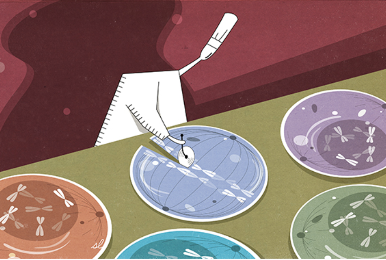
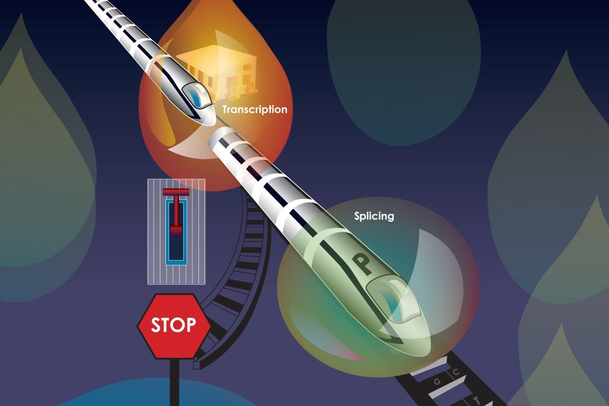
Cambridge, MA — During embryonic development, stem cells begin to take on specific identities, becoming distinct cell types with specialized characteristics and functions, in order to form the diverse organs and systems in our bodies. Cells rely on two main classes of regulators to define and maintain their identities; the first of these are master transcription factors, keystone proteins in each cell’s regulatory network, which keep the DNA sequences associated with crucial cell identity genes accessible for transcription — the process by which DNA is “read” into RNA. The other main regulators are signaling factors, which transmit information from the environment to the nucleus through a chain of proteins like a game of cellular telephone. Signaling factors can prompt changes in gene transcription as the cells react to that information.

One long-standing conundrum of how cell identity is determined is that many species, including humans, use the same core signaling pathways, with the same signaling factors, in all of their cells, yet this uniform machinery can cue a diverse array of cell-type specific gene activity, like an identical line of code being entered in many computers and causing each to start running a completely different program. New research from Whitehead Institute Member Richard Young, who is also a professor of biology at the Massachusetts Institute of Technology, published online in Molecular Cell on September 25, sheds light on how the same signaling factor can lead to so many distinct responses — with the help of a mechanism called phase separation.

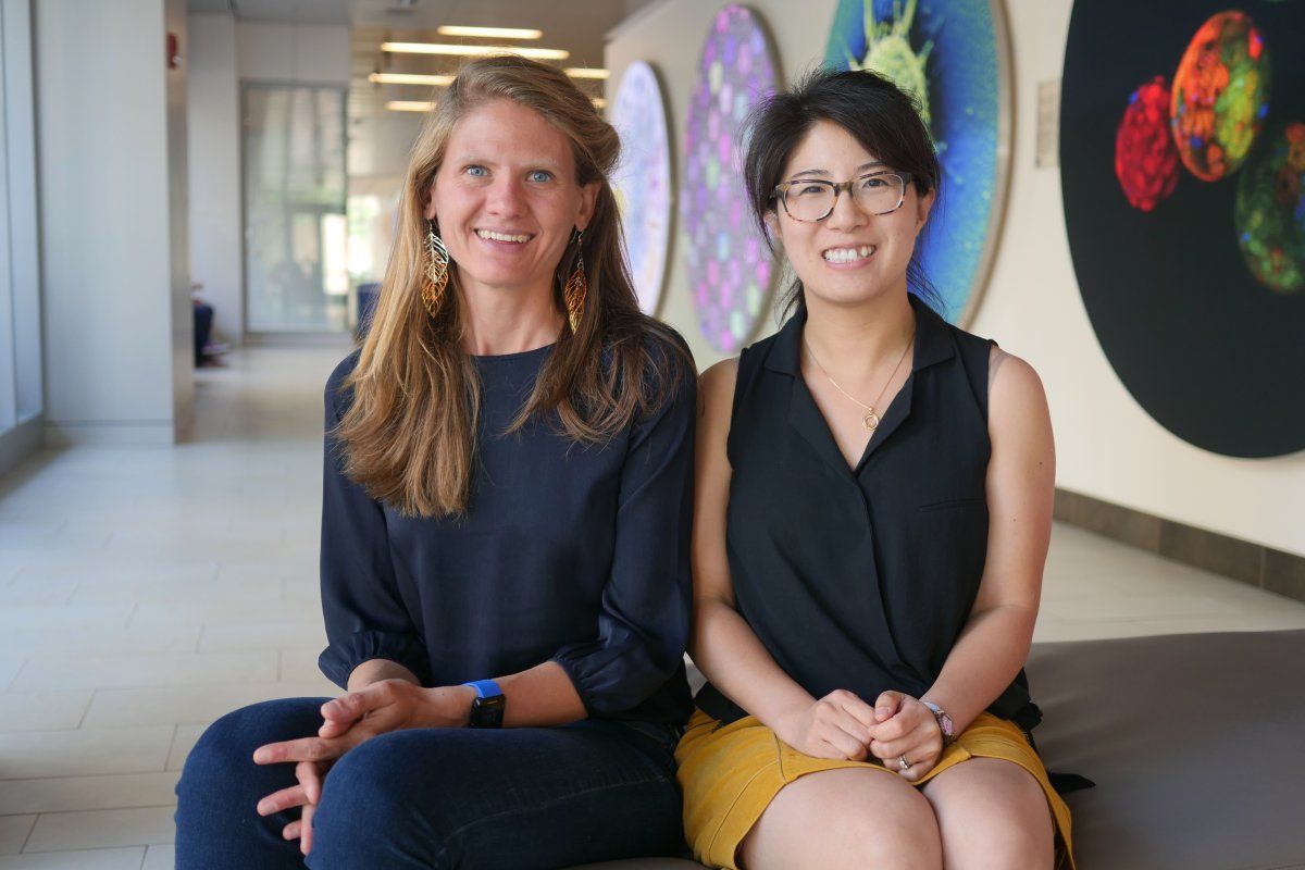
Co-senior author on the paper Jurian Schuijers, previously a postdoctoral researcher in Young’s lab and now a professor at the Center for Molecular Medicine at the University Medical Center Utrecht, was drawn to this puzzle after previously working in a signaling lab: “Two cells of different types that are right next to each other in the body can receive the exact same signal and have different reactions, and there was not a satisfying explanation for how that happens,” Schuijers says.

Young’s lab had previously found that signaling factors in pathways important for development tend to concentrate at super enhancers, clusters of DNA sequences that increase transcription of crucial cell identity genes. Because super enhancers are established at the genes important to identity in each cell, their activity is cell-type specific, so this co-localization provided a partial explanation of the puzzle, but it raised the question of how signaling factors are recruited to super enhancers. Young found the vague explanations that had been put forward, such as super enhancers being the most accessible DNA to signaling factors and their co-factors, unconvincing.

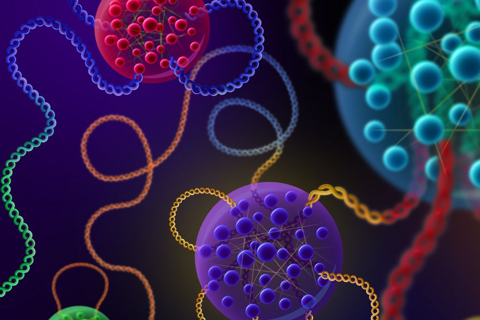
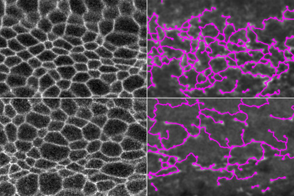
Young and his team suspected that an explanation for signaling factor recruitment might lie in their research on transcriptional condensates — droplets that form at super enhancers and concentrate transcriptional machinery there using phase separation, meaning the molecules separate out of their surroundings to form a distinct liquid compartment, like a drop of vinegar in a pool of oil. The proteins in condensates can do this because they contain intrinsically disordered regions (IDRs), stretches of amino acids that remain flexible, like wet spaghetti, and do not become fixed into a single shape the way most protein structures do. This property allows them to mesh together to form a condensate. The researchers reasoned that if signaling factors were joining transcriptional condensates, that could explain their concentration at super enhancers.

Young’s team confirmed that signaling factors in several of the most important pathways for embryonic development in mammals — WNT, TGF-β and JAK/STAT — contained IDRs. They further found that these factors were able to use their IDRs to form and join condensates, and that, in mouse cells, they appeared to join the condensates at super enhancers upon activation of their respective pathways.

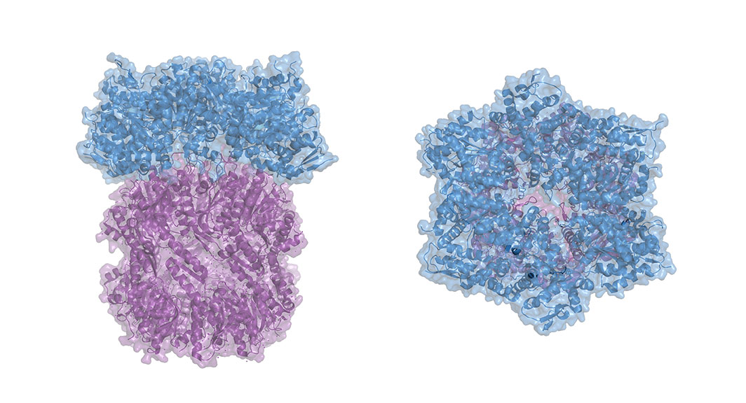
The researchers then decided to focus on beta catenin, the signaling factor at the end of the Wnt signaling pathway, a pathway essential for development; it helps to coordinate things like body axis patterning and cell fate specification, proliferation and migration. When Wnt signaling goes awry in embryos, they fail to develop, and when it goes awry in adults it is implicated in diseases including cancer. The beta catenin protein has IDRs on both of its ends and a structured middle section, called the Armadillo repeat domain, where it binds to other transcription factors. Typically, beta catenin binds to transcription factors in the TCF/LEF family, which in turn bind to DNA—beta catenin cannot bind to DNA on its own — anchoring the signaling factor at the right site and prompting gene transcription. However, the researchers found that beta catenin could concentrate at super enhancers even when it could not bind to its usual partners, suggesting that transcriptional condensates were a sufficient recruitment mechanism. The researchers then created two abridged beta catenin molecules: one version that only contained the IDRs and one that only contained the Armadillo repeat domain. Both partial factors were able to concentrate at super enhancers, but neither was as effective as the combined whole.

“If you ask most people how these factors find their target locations in the genome, they would say it’s through their DNA binding domains,” says first author Alicia Zamudio, a graduate student in Young’s lab. “This research suggests that factors use both their structured DNA binding domains and their unstructured domains to find the right locations to bind in the genome and to activate target genes.”

One advantage for cells of using IDRs, versus DNA binding alone, might be reducing the time it takes for signaling factors to concentrate near the right genes, the researchers say. Speed is of the essence for some signaling pathways in order for cells to be able to respond quickly to environmental stimuli. Transcriptional condensates are larger in size and much fewer in number than DNA binding sites or DNA-binding co-factors, and so they shrink the space that a signaling factor entering the nucleus must search.

This research could provide new opportunities for drug discovery. Signaling pathways and super enhancers are both co-opted by oncogenes to drive the spread of cancer, so transcriptional condensates could be a promising target to disrupt both oncogenic signaling and oncogene transcription. Young also hopes that this research, which adds to his lab’s growing body of work on transcriptional condensates, will lead to a new appreciation of the disordered regions of proteins.

“For a long time, researchers have mostly ignored the intrinsically disordered regions of proteins — we literally cut them off when identifying the crystal structures — much in the same way that researchers used to study genes and ignore ‘junk DNA,’” Young says. “But, just as with junk DNA, we are discovering that the overlooked, less obviously functional regions of these molecules are very important after all.”


This work is supported by NIH grant GM123511 and NSF grant PHY1743900 (R.A.Y.), NIH grant GM117370 (D.J.T.), NSF Graduate Research Fellowship (A.V.Z.), NIH grant T32CA009172 (I.A.K.), and DFG Research Fellowship DE 3069/1-1 (T.M.D.).


Written by Greta Friar

***

Richard Young’s primary affiliation is with Whitehead Institute for Biomedical Research, where his laboratory is located and all his research is conducted. He is also a professor of biology at the Massachusetts Institute of Technology.

***

Full citation:

“Mediator condensates localize signaling factors to key cell identity genes”

Molecular Cell, published online September 25, 2019. DOI: 10.1016/j.molcel.2019.08.016

Alicia V. Zamudio (1, 2), Alessandra Dall’Agnese (1), Jonathan E. Henninger (1), John C. Manteiga(1, 2), Lena K. Afeyan (1, 2), Nancy M. Hannett (1), Eliot L. Coffey (1, 2), Charles H. Li (1, 2), Ozgur Oksuz (1), Benjamin R. Sabari (1), Ann Boija (1), Isaac A. Klein (1,3), Susana W. Hawken (4), Jan-Hendrik Spille (5), Tim-Michael Decker (6), Ibrahim I. Cisse (5), Brian J. Abraham (1,7), Tong I. Lee (1), Dylan J. Taatjes (6), Jurian Schuijers (1,8,9), and Richard A. Young (1, 2, 9).

1. Whitehead Institute for Biomedical Research, Cambridge, MA 02142, USA

2. Department of Biology, Massachusetts Institute of Technology, Cambridge, MA, 02139, USA 3. Department of Medical Oncology, Dana-Farber Cancer Institute, Harvard Medical School, Boston, MA, 02215, USA

4. Program in Computational and Systems Biology, Massachusetts Institute of Technology, Cambridge, MA, 02139 USA

5. Department of Physics, Massachusetts Institute of Technology, Cambridge, MA, 02139, USA 6. Department of Biochemistry, University of Colorado, Boulder, CO 80303, USA

7. St. Jude Children’s Research Hospital, Memphis, TN, 038105, USA

8. Present address: Center for Molecular Medicine, University Medical Center Utrecht, Utrecht, 3584 CX, The Netherlands.

9. Equal contribution

Understanding genetic circuits and genome organization

Assistant professors Pulin Li and Seychelle Vos are investigating how cells become tissues and the proteins that organize DNA.

Raleigh McElvery | Department of Biology
September 12, 2019

MIT’s Department of Biology welcomed two new assistant professors in recent months: Pulin Li began at the Whitehead Institute in May, and Seychelle Vos arrived at Building 68 in September. Their respective expertise in genetic circuits and genome organization will augment the department’s efforts to explore cell biology at all levels — from intricate molecular structures to the basis for human disease.

“Pulin and Seychelle bring new perspectives and exciting ideas to our research community,” says Alan Grossman, department head. “I’m excited to see them start their independent research programs and look forward to the impact that they will have.”

From cells to tissues

Growing up in Yingkou, China, Li was exposed to science at a young age. Her dad worked for a pharmaceutical company researching traditional Chinese medicine, and Li would spend hours playing with his lab tools and beakers. “I can still vividly remember the smell of his Chinese herbs,” she says. “Maybe that’s part of the reason why I’ve always been interested in biology as it relates to medical sciences.”

She earned her BS in life sciences from Peking University, and went on to pursue a PhD in chemical biology at Harvard University studying hematopoietic stem cells. Li performed chemical screens to find drugs that would make stem cell transplantation in animal models more efficient, and eventually help patients with leukemia. In doing so, she became captivated by the molecular mechanisms that control cell-to-cell communication.

“I would like to eventually go back to developing new therapies and medicines,” she says, “but that translational research requires a basic understanding of how things work at a molecular level.”

As a result, her postdoc at Caltech was firmly rooted in basic biology. She investigated the genetic circuits that underlie cell-cell communication in developing and regenerating tissues, and now aims to develop new methods to study these same processes here at MIT.

Traditional genetic approaches involve breaking components of a system one at a time to investigate the role they play. However, Li’s lab will adopt a “bottom-up” approach that involves building these systems from the ground up, adding the components back into the cell one by one to pinpoint which genetic circuits are sufficient for programming tissue function. “Building up a system, rather than tearing it down, allows you to test different circuit designs, tune important parameters, and understand why a circuit has evolved to perform a specific function,” she explains.

She is most interested in determining which aspects of cellular communication are critical for tissue formation, in hopes of understanding the diversity of life forms in nature, as well as inspiring new methods to engineer or regenerate different tissues.

“My dream would be to put a bunch of genetic circuits into cells in such a way that they could enable the cells to self-organize into certain patterns and shapes, and replace damaged tissues in a patient,” she says.

Proteins that organize DNA

Although Vos was born in South Africa, her family moved so frequently for her father’s job that she doesn’t call any one place home. “If I had to pick, I’d say it would be the middle of the Atlantic Ocean,” she says.

Both of her grandparents on her mother’s side were researchers, and encouraged various scientific escapades, like bringing wolf spiders to kindergarten for show-and-tell. Her grandmother on her father’s side found her early passions “mildly disturbing,” but dutifully fulfilled her requests for high-resolution insect microscopy books nonetheless.

“I really wanted to know how plants and animals worked starting from a young age, thanks to my grandparents,” Vos says.

In high school she was already conducting research on the side at Clemson University, South Carolina, and went on to earn her BS in genetics from the University of Georgia. She began her PhD in molecular cell biology at the University of California at Berkeley intending to study immunology, but surprised herself by becoming taken with structural biology instead.

Purifying proteins and solving structures required a much different skill set than performing screens and manipulating genomes, but she very much enjoyed her work on topoisomerase, the enzyme that modifies DNA so it doesn’t become too coiled.

She continued conducting biochemical and structural research during her postdoc at the Max Planck Institute for Biophysical Chemistry in Germany. There, she used cryogenic electron microscopy to probe how different RNA polymerase II complexes are regulated during transcription in eukaryotes.

Today, she’s a molecular biologist at her core, but she’s prepared to use “whatever technique gets the answer.” As she explains: “You need biochemistry to solve structures and genetics to understand how they’re working within the whole organism, so it’s all related.”

In her new lab in Building 68, she will continue investigating gene expression, but this time in the context of genome organization. DNA must be compacted in order to fit into a cell, and Vos will study the proteins that organize DNA so it can be compressed without interfering with gene expression. She also wants to know how those same proteins are affected by gene expression.

“How gene regulation impacts compaction is a really critical question to address because different cell types are organized in different ways, and that impacts which genes are ultimately expressed,” she says. “We still don’t really understand how these processes work at an atomic level, so that’s where my expertise in biochemistry and structural biology can be useful.”

When asked what they are most excited about as the school year begins, both Li and Vos say the same thing: the diverse skills and expertise of the students and faculty.

“It’s not just about solving one structure, people here want to understand the entire process,” Vos says. “Biology is a conglomeration of many different fields, and if we can have engineers, mathematicians, physicists, chemists, biologists, and others work together, we can begin to tackle pressing questions.”

To divide or not to divide?
Nicole Davis, Whitehead Institute
August 15, 2019
An emerging view of RNA transcription and splicing

Whitehead Institute scientists find chemical modification contributes to trafficking between non-membrane-bound compartments that control gene expression.

Nicole Davis | Whitehead Institute
August 9, 2019

Cells often create compartments to control important biological functions. The nucleus is a prime example; surrounded by a membrane, it houses the genome. Yet cells also harbor enclosures that are not membrane-bound and more transient, like oil droplets in water. Over the past two years, these droplets (called “condensates”) have become increasingly recognized as major players in controlling genes. Now, a team led by Whitehead Institute scientists helps expand this emerging picture with the discovery that condensates play a role in splicing, an essential activity that ensures the genetic code is prepared to be translated into protein. The researchers also reveal how a critical piece of cellular machinery moves between different condensates. The team’s findings appear in the Aug. 7 online issue of Nature.

“Condensates represent a real paradigm shift in the way molecular biologists think about gene control,” says senior author Richard Young, a member of the Whitehead Institute and professor of biology at MIT. “Now, we’ve added a critical new layer to this thinking that enhances our understanding of splicing as well as the major transcriptional apparatus RNA polymerase II.”

Young’s lab has been at the forefront of studying how and when condensates form as well as their functions in gene regulation. In the current study, Young and his colleagues, including first authors Eric Guo and John Manteiga, focused their efforts on a key transition that happens when genes undergo transcription — an early step in gene activation whereby an RNA copy is created from the genes’ DNA template. First, all of the molecular machinery needed to make RNA, including a large protein complex known as RNA polymerase II, assembles at a given gene. Then, specific chemical modifications to RNA polymerase II allow it to begin transcribing DNA into RNA. This shift from so-called transcription initiation to active transcription also involves another important molecular transition: As RNA molecules begin to grow, the splicing apparatus must also move in and carry out its job.

“We wanted to step back and ask, ‘Do condensates play an important role in this switch, and if so, what mechanism might be responsible?’” explains Young.

For roughly three decades, it has been recognized that the factors required for splicing are stored in compartments called speckles. Yet whether these speckles play an active role in splicing, or are simply storage vessels, has remained unclear.

Using confocal microscopy, the Whitehead team discovered condensates filled with components of the splicing machinery in the vicinity of highly active genes. Notably, these structures exhibited similar liquid-like characteristics to those condensates described in prior studies from Young’s lab that are involved in transcription initiation.

“These findings signaled to us that there are two types of condensates at work here: one involved in transcription initiation and the other in splicing and transcriptional elongation,” said Manteiga, a graduate student in Young’s lab.

With two different condensates at play, the researchers wondered: How does the critical transcriptional machinery, specifically RNA polymerase II, move from one condensate to the other?

Guo, Manteiga, and their colleagues found that chemical modification, specifically the addition of phosphate groups, serves as a kind of molecular switch that alters the protein complex’s affinity for a particular condensate. With fewer phosphate groups, it associates with the condensates for transcription initiation; when more phosphates are added, it enters the splicing condensates. Such phosphorylation occurs on one end of the protein complex, which contains a specialized region known as the C-terminal domain (CTD). Importantly, the CTD lacks a specific three-dimensional structure, and previous work has shown that such intrinsically disordered regions can influence how and when certain proteins are incorporated into condensates.

“It is well-documented that phosphorylation acts as a signal to help regulate the activity of RNA polymerase II,” says Guo, a postdoc in Young’s lab. “Now, we’ve shown that it also acts as a switch to alter the protein’s preference for different condensates.”

In light of their discoveries, the researchers propose a new view of splicing compartments, where speckles serve primarily as warehouses, storing the thousands of molecules required to support the splicing apparatus when they are not needed. But when splicing is active, the phosphorylated CTD of RNA Pol II serves as an attractant, drawing the necessary splicing materials toward the gene where they are needed and into the splicing condensate.

According to Young, this new outlook on gene control has emerged in part through a multidisciplinary approach, bringing together perspectives from biology and physics to learn how properties of matter predict some of the molecular behaviors he and his team have observed experimentally. “Working at the interface of these two fields is incredibly exciting,” says Young. “It is giving us a whole new way of looking at the world of regulatory biology.”

Support for this work was provided by the U.S. National Institutes of Health, National Science Foundation, Cancer Research Institute, Damon Runyon Cancer Research Foundation, Hope Funds for Cancer Research, Swedish Research Council, and German Research Foundation DFG.

Study furthers radically new view of gene control

Along the genome, proteins form liquid-like droplets that appear to boost the expression of particular genes.

Anne Trafton | MIT News Office
August 8, 2019

In recent years, MIT scientists have developed a new model for how key genes are controlled that suggests the cellular machinery that transcribes DNA into RNA forms specialized droplets called condensates. These droplets occur only at certain sites on the genome, helping to determine which genes are expressed in different types of cells.

In a new study that supports that model, researchers at MIT and the Whitehead Institute for Biomedical Research have discovered physical interactions between proteins and with DNA that help explain why these droplets, which stimulate the transcription of nearby genes, tend to cluster along specific stretches of DNA known as super enhancers. These enhancer regions do not encode proteins but instead regulate other genes.

“This study provides a fundamentally important new approach to deciphering how the ‘dark matter’ in our genome functions in gene control,” says Richard Young, an MIT professor of biology and member of the Whitehead Institute.

Young is one of the senior authors of the paper, along with Phillip Sharp, an MIT Institute Professor and member of MIT’s Koch Institute for Integrative Cancer Research; and Arup K. Chakraborty, the Robert T. Haslam Professor in Chemical Engineering, a professor of physics and chemistry, and a member of MIT’s Institute for Medical Engineering and Science and the Ragon Institute of MGH, MIT, and Harvard.

Graduate student Krishna Shrinivas and postdoc Benjamin Sabari are the lead authors of the paper, which appears in Molecular Cell on Aug. 8.

“A biochemical factory”

Every cell in an organism has an identical genome, but cells such as neurons or heart cells express different subsets of those genes, allowing them to carry out their specialized functions. Previous research has shown that many of these genes are located near super enhancers, which bind to proteins called transcription factors that stimulate the copying of nearby genes into RNA.

About three years ago, Sharp, Young, and Chakraborty joined forces to try to model the interactions that occur at enhancers. In a 2017 Cell paper, based on computational studies, they hypothesized that in these regions, transcription factors form droplets called phase-separated condensates. Similar to droplets of oil suspended in salad dressing, these condensates are collections of molecules that form distinct cellular compartments but have no membrane separating them from the rest of the cell.

In a 2018 Science paper, the researchers showed that these dynamic droplets do form at super enhancer locations. Made of clusters of transcription factors and other molecules, these droplets attract enzymes such as RNA polymerases that are needed to copy DNA into messenger RNA, keeping gene transcription active at specific sites.

“We had demonstrated that the transcription machinery forms liquid-like droplets at certain regulatory regions on our genome, however we didn’t fully understand how or why these dewdrops of biological molecules only seemed to condense around specific points on our genome,” Shrinivas says.

As one possible explanation for that site specificity, the research team hypothesized that weak interactions between intrinsically disordered regions of transcription factors and other transcriptional molecules, along with specific interactions between transcription factors and particular DNA elements, might determine whether a condensate forms at a particular stretch of DNA. Biologists have traditionally focused on “lock-and-key” style interactions between rigidly structured protein segments to explain most cellular processes, but more recent evidence suggests that weak interactions between floppy protein regions also play an important role in cell activities.

In this study, computational modeling and experimentation revealed that the cumulative force of these weak interactions conspire together with transcription factor-DNA interactions to determine whether a condensate of transcription factors will form at a particular site on the genome. Different cell types produce different transcription factors, which bind to different enhancers. When many transcription factors cluster around the same enhancers, weak interactions between the proteins are more likely to occur. Once a critical threshold concentration is reached, condensates form.

“Creating these local high concentrations within the crowded environment of the cell enables the right material to be in the right place at the right time to carry out the multiple steps required to activate a gene,” Sabari says. “Our current study begins to tease apart how certain regions of the genome are capable of pulling off this trick.”

These droplets form on a timescale of seconds to minutes, and they blink in and out of existence depending on a cell’s needs.

“It’s an on-demand biochemical factory that cells can form and dissolve, as and when they need it,” Chakraborty says. “When certain signals happen at the right locus on a gene, the condensates form, which concentrates all of the transcription molecules. Transcription happens, and when the cells are done with that task, they get rid of them.”

A new view

Weak cooperative interactions between proteins may also play an important role in evolution, the researchers proposed in a 2018 Proceedings of the National Academy of Sciences paper. The sequences of intrinsically disordered regions of transcription factors need to change only a little to evolve new types of specific functionality. In contrast, evolving new specific functions via “lock-and-key” interactions requires much more significant changes.

“If you think about how biological systems have evolved, they have been able to respond to different conditions without creating new genes. We don’t have any more genes that a fruit fly, yet we’re much more complex in many of our functions,” Sharp says. “The incremental expanding and contracting of these intrinsically disordered domains could explain a large part of how that evolution happens.”

Similar condensates appear to play a variety of other roles in biological systems, offering a new way to look at how the interior of a cell is organized. Instead of floating through the cytoplasm and randomly bumping into other molecules, proteins involved in processes such as relaying molecular signals may transiently form droplets that help them interact with the right partners.

“This is a very exciting turn in the field of cell biology,” Sharp says. “It is a whole new way of looking at biological systems that is richer and more meaningful.”

Some of the MIT researchers, led by Young, have helped form a company called Dewpoint Therapeutics to develop potential treatments for a wide variety of diseases by exploiting cellular condensates. There is emerging evidence that cancer cells use condensates to control sets of genes that promote cancer, and condensates have also been linked to neurodegenerative disorders such as amyotrophic lateral sclerosis (ALS) and Huntington’s disease.

The research was funded by the National Science Foundation, the National Institutes of Health, and the Koch Institute Support (core) Grant from the National Cancer Institute.

Biologists and mathematicians team up to explore tissue folding

An algorithm developed to study the structure of galaxies helps explain a key feature of embryonic development.

Anne Trafton | MIT News Office
July 25, 2019

As embryos develop, they follow predetermined patterns of tissue folding, so that individuals of the same species end up with nearly identically shaped organs and very similar body shapes.

MIT scientists have now discovered a key feature of embryonic tissue that helps explain how this process is carried out so faithfully each time. In a study of fruit flies, they found that the reproducibility of tissue folding is generated by a network of proteins that connect like a fishing net, creating many alternative pathways that tissues can use to fold the right way.

“What we found is that there’s a lot of redundancy in the network,” says Adam Martin, an MIT associate professor of biology and the senior author of the study. “The cells are interacting and connecting with each other mechanically, but you don’t see individual cells taking on an all-important role. This means that if one cell gets damaged, other cells can still connect to disparate parts of the tissue.”

To uncover these network features, Martin worked with Jörn Dunkel, an MIT associate professor of physical applied mathematics and an author of the paper, to apply an algorithm normally used by astronomers to study the structure of galaxies.

Hannah Yevick, an MIT postdoc, is the lead author of the study, which appears today in Developmental Cell. Graduate student Pearson Miller is also an author of the paper.

A safety net

During embryonic development, tissues change their shape through a process known as morphogenesis. One important way tissues change shape is to fold, which allows flat sheets of embryonic cells to become tubes and other important shapes for organs and other body parts. Previous studies in fruit flies have shown that even when some of these embryonic cells are damaged, sheets can still fold into their correct shapes.

“This is a process that’s fairly reproducible, and so we wanted to know what makes it so robust,” Martin says.

In this study, the researchers focused on the process of gastrulation, during which the embryo is reorganized from a single-layered sphere to a more complex structure with multiple layers. This process, and other morphogenetic processes similar to fruit fly tissue folding, also occur in human embryos. The embryonic cells involved in gastrulation contain in their cytoplasm proteins called myosin and actin, which form cables and connect at junctions between cells to form a network across the tissue. Martin and Yevick had hypothesized that the network of cell connectivity might play a role in the robustness of the tissue folding, but until now, there was no good way to trace the connections of the network.

To achieve that, Martin’s lab joined forces with Dunkel, who studies the physics of soft surfaces and flowing matter — for example, wrinkle formation and patterns of bacterial streaming. For this study, Dunkel had the idea to apply a mathematical procedure that can identify topological features of a three-dimensional structure, analogous to ridges and valleys in a landscape. Astronomers use this algorithm to identify galaxies, and in this case, the researchers used it to trace the actomyosin networks across and between the cells in a sheet of tissue.

“Once you have the network, you can apply standard methods from network analysis — the same kind of analysis that you would apply to streets or other transport networks, or the blood circulation network, or any other form of network,” Dunkel says.

Among other things, this kind of analysis can reveal the structure of the network and how efficiently information flows along it. One important question is how well a network adapts if part of it gets damaged or blocked. The MIT team found that the actomyosin network contains a great deal of redundancy — that is, most of the “nodes” of the network are connected to many other nodes.

This built-in redundancy is analogous to a good public transit system, where if one bus or train line goes down, you can still get to your destination. Because cells can generate mechanical tension along many different pathways, they can fold the right way even if many of the cells in the network are damaged.

“If you and I are holding a single rope, and then we cut it in the middle, it would come apart. But if you have a net, and cut it in some places, it still stays globally connected and can transmit forces, as long as you don’t cut all of it,” Dunkel says.

Folding framework

The researchers also found that the connections between cells preferentially organize themselves to run in the same direction as the furrow that forms in the early stages of folding.

“We think this is setting up a frame around which the tissue will adopt its shape,” Martin says. “If you prevent the directionality of the connections, then what happens is you can still get folding but it will fold along the wrong axis.”

Although this study was done in fruit flies, similar folding occurs in vertebrates (including humans) during the formation of the neural tube, which is the precursor to the brain and spinal cord. Martin now plans to apply the techniques he used in fruit flies to see if the actomyosin network is organized the same way in the neural tube of mice. Defects in the closure of the neural tube can lead to birth defects such as spina bifida.

“We would like to understand how it goes wrong,” Martin says. “It’s still not clear whether it’s the sealing up of the tube that’s problematic or whether there are defects in the folding process.”

The research was funded by the National Institute of General Medical Sciences and the James S. McDonnell Foundation.

Researchers determine what makes some proteins “slippery” enough to evade destruction
Raleigh McElvery
July 10, 2019

All cells must balance generating new proteins with eliminating excess or damaged ones by way of powerful degradation machines — which, much like wood chippers, chew up proteins and spit them out. But, these proteins are often folded into intricate structures, and must be unfurled before they can be fed into these degradation machines, broken into tiny bits, and ultimately recycled. In bacteria, a molecular motor known as ClpX must grip the end of the ill-fated protein and apply force to straighten it. However, until now, researchers weren’t sure precisely how ClpX gripped its target tightly enough to accomplish this task.

There had been evidence to suggest that some amino acids — the chemical building blocks that comprise proteins — are “slippery” and thus more difficult to grip. In a new study published in eLife, researchers at the MIT Department of Biology examined each amino acid’s individual contribution to grip. By parsing the physical basis for this molecular interaction, they hope to better understand how some proteins evade destruction.

“Previous studies had shown that small amino acids were notoriously hard to grip, but no one really understood why,” says Tristan Bell, graduate student and first author on the paper. “It’s like watching a game of tug-of-war and knowing that a person’s hands are important for pulling on the rope, but having no idea what allows the hands to get a good grip on the rope.”

ClpX, he explains, is roughly shaped like donut with loops protruding into the center hole. These loops grip the target protein, jamming it against the surface of ClpX, and unfolding it so it can be threaded through the hole and shredded.

The researchers engineered proteins with tails comprised of various amino acid combinations, and measured how well ClpX could grip them, both in bacteria and in test tubes. They determined that ClpX can only grip between six and eight amino acids at a time, and that only a handful of the 20 possible amino acids could actually be “well-gripped.” When ClpX was able to grasp multiple amino acids simultaneously, its grip strength increased.

“We think that somehow the charge is preventing ClpX from making strong contacts with the target protein, preventing it from achieving a stable grip state,” Bell says.Just like in previous experiments, large amino acids appeared easier to grip than small ones, “similar to the way a knotted rope is easier to grasp than a smooth, slippery one,” Bell says. But, regardless of size, amino acids that carried electric charge seemed to be more slippery.

The team thinks that proteins with slippery tails might have an evolutionary advantage, because they are harder to grip and therefore less likely to be degraded.

Invaders like viruses have been known to insert a slippery sequence into certain proteins to prevent the host cell from destroying them and thus promoting replication. Even healthy cells produce proteins with strategically placed slippery sequences, which allow a portion of the protein to break away from the degradation machinery unscathed. In the bacteria Caulobacter crescentus, this planned breakage actually produces a version of one protein that’s needed for DNA replication.

“Next,” Bell says, “we’re hoping to look across entire proteomes in different organisms to find more proteins that escape destruction.”

“Tristan’s experiments and results reveal some of the molecular determinants of grip in the bacterial degradation machines we study,” says Bob Sauer, the Salvador E. Luria Professor of Biology and senior author on the study. “Many of the rules he discovered apply to related machines that function in all biological organisms, including humans, emphasizing the common evolution of these machines.”

Citation:
“Interactions between a subset of substrate side chains and AAA+ motor pore loops determine grip during protein unfolding”
eLife, online June 28, 2019, DOI: 10.7554/eLife.46808
Tristan A. Bell, Tania A. Baker, and Robert T. Sauer.

Researchers identify important proteins hijacked by pathogens during cell-to-cell spread
Raleigh McElvery
July 9, 2019

Listeria monocytogenes, the food-borne bacterium responsible for listeriosis, can creep from one cell to the next, stealthily evading the immune system. This strategy of cell-to-cell spread allows them to infect many different cell types, and can spur complications like meningitis. Yet the molecular details of this spread remain a mystery.

In a paper recently published in Molecular Biology of the Cell, researchers from the MIT Department of Biology, University of California, Berkeley, and Chan Zuckerberg Biohub are beginning to piece together the elusive means by which Listeria moves from one cell to the next. This mode of transport, the scientists suggest, looks a lot like trans-endocytosis, a process that healthy, uninfected cells use to exchange organelles and various cytoplasmic components. In fact, the two processes are so similar that Listeria may be co-opting the host cell’s trans-endocytosis machinery for its own devices.

Although the particulars of trans-endocytosis are poorly understood, the process permits neighboring cells to exchange materials via membrane-bound compartments called vacuoles, which release their cargo upon reaching their final destination.

Much like trans-endocytosis, cell-to-cell spread relies on vacuoles to ferry Listeria. First, the pathogen commandeers the host cell’s own machinery to assemble a tail of proteins that allows it to rocket around inside the cell and ram against both the membrane of the host and that of the adjacent cell. The resulting protrusion is then somehow engulfed into a double-membrane vacuole, and the bacteria burst through their containment to begin the process anew in the recipient cell.

“There’s been a lot of work looking at Listeria cell-to-cell spread,” says Rebecca Lamason, the Robert A. Swanson (1969) Career Development Assistant Professor in the MIT Department of Biology and senior author on the study. “But we still don’t really understand the molecular mechanisms that allow the bacteria to manipulate the membrane to promote engulfment. Depending on what we uncover, we might also be able to apply that information to better grasp how an uninfected cell regulates trans-endocytosis.”

Lamason and her team anticipated that the same proteins implicated in trans-endocytosis would also be involved in Listeria cell-to-cell spread, which would indicate that the pathogen was appropriating these proteins for its own purposes. The researchers made a list of 115 host genes of interest, and then used an RNAi screen to identify just 22 that are critical for cell-to-cell spread.

They were excited to find that, of those 22 genes, several are also implicated in endocytosis, which suggests Listeria is using a similar strategy. These include genes encoding caveolin proteins that control membrane trafficking and remodeling, as well as another protein called PACSIN2 that interacts with caveolins to regulate protrusion engulfment.

Now that the researchers have pinpointed these key proteins, the next step is to determine how they work together in order to promote cell-to-cell spread — especially since the protrusions created by Listeria are much larger than those required for trans-endocytosis.

“As we drill down even deeper into the molecular mechanisms, it will be interesting to see where trans-endocytosis and cell-to-cell spread differ, and where they are similar,” Lamason says. “Our hope is that investigating the mechanisms of bacterial spread will reveal fundamental insights into host intercellular communication.”

Citation:
“RNAi screen reveals a role for PACSIN2 and caveolins during bacterial cell-to-cell spread”
Molecular Biology of the Cell, online June 26, 2019, DOI: 10.1091/mbc.E19-04-0197
Allen G. Sanderlin, Cassandra Vondrak, Arianna J. Scricco, Indro Fedrigo, Vida Ahyong, and Rebecca L. Lamason

Measuring chromosome imbalance could clarify cancer prognosis

A study of prostate cancer finds “aneuploid” tumors are more likely to be lethal than tumors with normal chromosome numbers.

Anne Trafton | MIT News Office
May 13, 2019

Most human cells have 23 pairs of chromosomes. Any deviation from this number can be fatal for cells, and several genetic disorders, such as Down syndrome, are caused by abnormal numbers of chromosomes.

For decades, biologists have also known that cancer cells often have too few or too many copies of some chromosomes, a state known as aneuploidy. In a new study of prostate cancer, researchers have found that higher levels of aneuploidy lead to much greater lethality risk among patients.

The findings suggest a possible way to more accurately predict patients’ prognosis, and could be used to alert doctors which patients might need to be treated more aggressively, says Angelika Amon, the Kathleen and Curtis Marble Professor in Cancer Research in the Department of Biology and a member of the Koch Institute for Integrative Cancer Research.

“To me, the exciting opportunity here is the ability to inform treatment, because prostate cancer is such a prevalent cancer,” says Amon, who co-led this study with Lorelei Mucci, an associate professor of epidemiology at the Harvard T.H. Chan School of Public Health.

Konrad Stopsack, a research associate at Memorial Sloan Kettering Cancer Center, is the lead author of the paper, which appears in the Proceedings of the National Academy of Sciences the week of May 13. Charles Whittaker, a Koch Institute research scientist; Travis Gerke, a member of the Moffitt Cancer Center; Massimo Loda, chair of pathology and laboratory medicine at New York Presbyterian/Weill Cornell Medicine; and Philip Kantoff, chair of medicine at Memorial Sloan Kettering; are also authors of the study.

Better predictions

Aneuploidy occurs when cells make errors sorting their chromosomes during cell division. When aneuploidy occurs in embryonic cells, it is almost always fatal to the organism. For human embryos, extra copies of any chromosome are lethal, with the exceptions of chromosome 21, which produces Down syndrome; chromosomes 13 and 18, which lead to developmental disorders known as Patau and Edwards syndromes; and the X and Y sex chromosomes. Extra copies of the sex chromosomes can cause various disorders but are not usually lethal.

Most cancers also show very high prevalence of aneuploidy, which poses a paradox: Why does aneuploidy impair normal cells’ ability to survive, while aneuploid tumor cells are able to grow uncontrollably? There is evidence that aneuploidy makes cancer cells more aggressive, but it has been difficult to definitively demonstrate that link because in most types of cancer nearly all tumors are aneuploid, making it difficult to perform comparisons.

Prostate cancer is an ideal model to explore the link between aneuploidy and cancer aggressiveness, Amon says, because, unlike most other solid tumors, many prostate cancers (25 percent) are not aneuploid or have only a few altered chromosomes. This allows researchers to more easily assess the impact of aneuploidy on cancer progression.

What made the study possible was a collection of prostate tumor samples from the Health Professionals Follow-up Study and Physicians’ Health Study, run by the Harvard T.H. Chan School of Public Health over the course of more than 30 years. The researchers had genetic sequencing information for these samples, as well as data on whether and when their prostate cancer had spread to other organs and whether they had died from the disease.

Led by Stopsack, the researchers came up with a way to calculate the degree of aneuploidy of each sample, by comparing the genetic sequences of those samples with aneuploidy data from prostate genomes in The Cancer Genome Atlas. They could then correlate aneuploidy with patient outcomes, and they found that patients with a higher degree of aneuploidy were five times more likely to die from the disease. This was true even after accounting for differences in Gleason score, a measure of how much the patient’s cells resemble cancer cells or normal cells under a microscope, which is currently used by doctors to determine severity of disease.

The findings suggest that measuring aneuploidy could offer additional information for doctors who are deciding how to treat patients with prostate cancer, Amon says.

“Prostate cancer is terribly overdiagnosed and terribly overtreated,” she says. “So many people have radical prostatectomies, which has significant impact on people’s lives. On the other hand, thousands of men die from prostate cancer every year. Assessing aneuploidy could be an additional way of helping to inform risk stratification and treatment, especially among people who have tumors with high Gleason scores and are therefore at higher risk of dying from their cancer.”

“When you’re looking for prognostic factors, you want to find something that goes beyond known factors like Gleason score and PSA [prostate-specific antigen],” says Bruce Trock, a professor of urology at Johns Hopkins School of Medicine, who was not involved in the research. “If this kind of test could be done right after a prostatectomy, it could give physicians information to help them decide what might be the best treatment course.”

Amon is now working with researchers from the Harvard T.H. Chan School of Public Health to explore whether aneuploidy can be reliably measured from small biopsy samples.

Aneuploidy and cancer aggressiveness

The researchers found that the chromosomes that are most commonly aneuploid in prostate tumors are chromosomes 7 and 8. They are now trying to identify specific genes located on those chromosomes that might help cancer cells to survive and spread, and they are also studying why some prostate cancers have higher levels of aneuploidy than others.

“This research highlights the strengths of interdisciplinary, team science approaches to tackle outstanding questions in prostate cancer,” Mucci says. “We plan to translate these findings clinically in prostate biopsy specimens and experimentally to understand why aneuploidy occurs in prostate tumors.”

Another type of cancer where most patients have low levels of aneuploidy is thyroid cancer, so Amon now hopes to study whether thyroid cancer patients with higher levels of aneuploidy also have higher death rates.

“A very small proportion of thyroid tumors is highly aggressive and lethal, and I’m starting to wonder whether those are the ones that have some aneuploidy,” she says.

The research was funded by the Koch Institute Dana Farber/Harvard Cancer Center Bridge Project and by the National Institutes of Health, including the Koch Institute Support (core) Grant.