Three innovative research projects in literature, plant epigenetics, and chemical engineering will be supported by Professor Amar G. Bose Research Grants.
MIT Resource Development
Now in its seventh year, the Professor Amar G. Bose Research Grants support visionary projects that represent intellectual curiosity and a pioneering spirit. Three MIT faculty members have each been awarded one of these prestigious awards for 2019 to pursue diverse questions in the humanities, biology, and engineering.
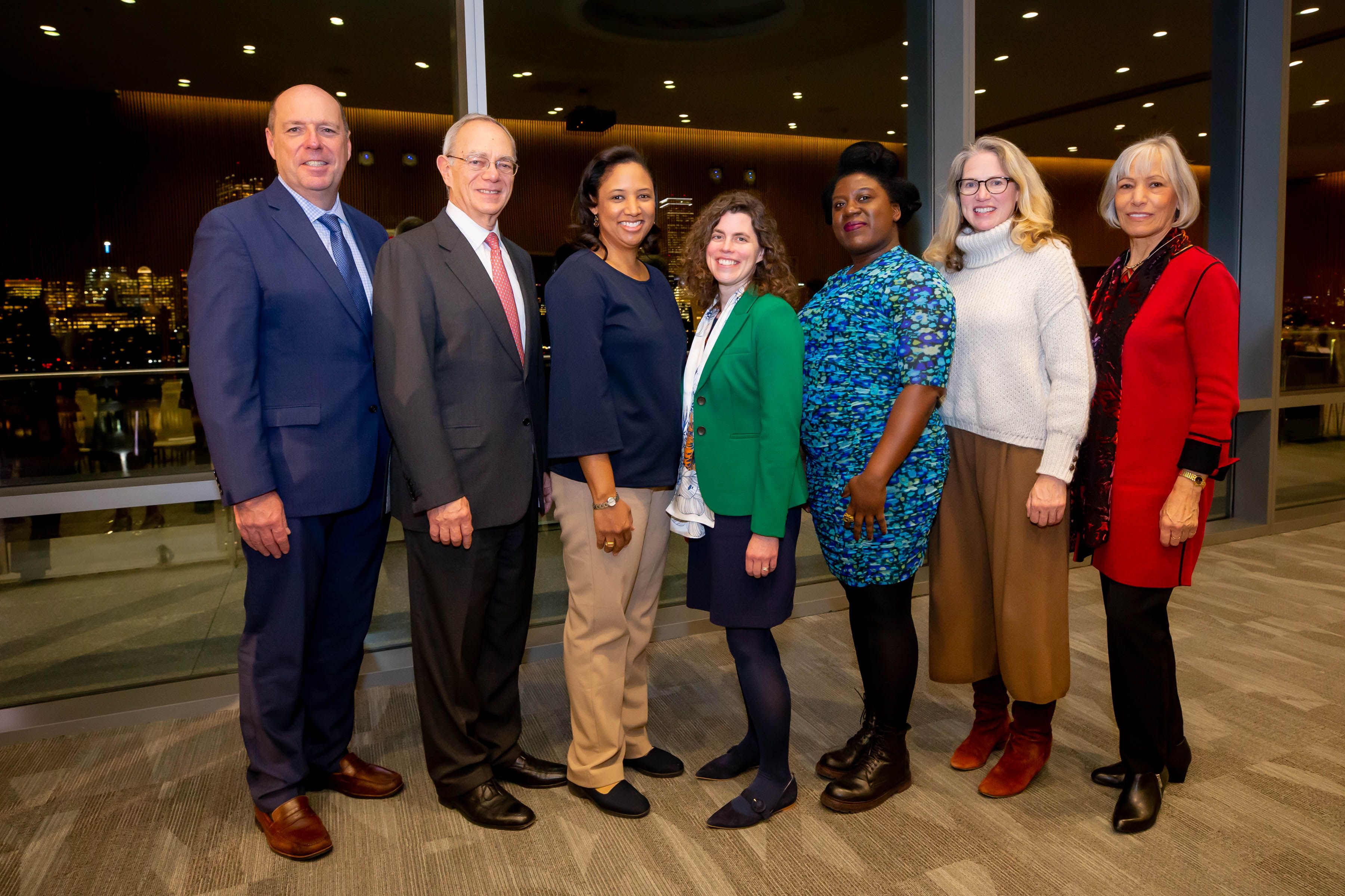
At a ceremony hosted by MIT President L. Rafael Reif on Nov. 25 and attended by past awardees, Provost Martin Schmidt, the Ray and Maria Stata Professor of Electrical Engineering and Computer Science, formally announced this year’s Amar G. Bose Research Fellows: Sandy Alexandre, Mary Gehring, and Kristala L.J. Prather.
The fellowships are named for the late Amar G. Bose ’51, SM ’52, ScD ’56, a longtime MIT faculty member and the founder of the Bose Corporation. Speaking at the event, President Reif expressed appreciation for the Bose Fellowships, which enable highly creative and unusual research in areas that can be hard to fund through traditional means. “We are tremendously grateful to the Bose family for providing the support that allows bold and curious thinkers at MIT to dream big, challenge themselves, and explore.”
Judith Bose, widow of Amar’s son, Vanu ’87, SM ’94, PhD ’99, congratulated the fellows on behalf of the Bose family. “We talk a lot at this event about the power of a great innovative idea, but I think it was a personal mission of Dr. Bose to nurture the ability, in each individual that he met along the way, to follow through — not just to have the great idea but the agency that comes with being able to pursue your idea, follow it through, and actually see where it leads,” Bose said. “And Vanu was the same way. That care that was epitomized by Dr. Bose not just in the idea itself, but in the personal investment, agency, and nurturing necessary to bring the idea to life — that care is a large part of what makes true change in the world.”
The relationship between literature and engineering
Many technological innovations have resulted from the influence of literature, one of the most notable being the World Wide Web. According to many sources, Sir Tim Berners-Lee, the web’s inventor, found inspiration from a short story by Arthur C. Clarke titled “Dial F for Frankenstein.” Science fiction has presaged a number of real-life technological innovations, including the defibrillator, noted in Mary Shelley’s “Frankenstein;” the submarine, described in Jules Verne’s “20,000 Leagues Under the Sea;” and earbuds, described in Ray Bradbury’s “Fahrenheit 451.” But the data about literature’s influence on STEM innovations are spotty, and these one-to-one relationships are not always clear-cut.
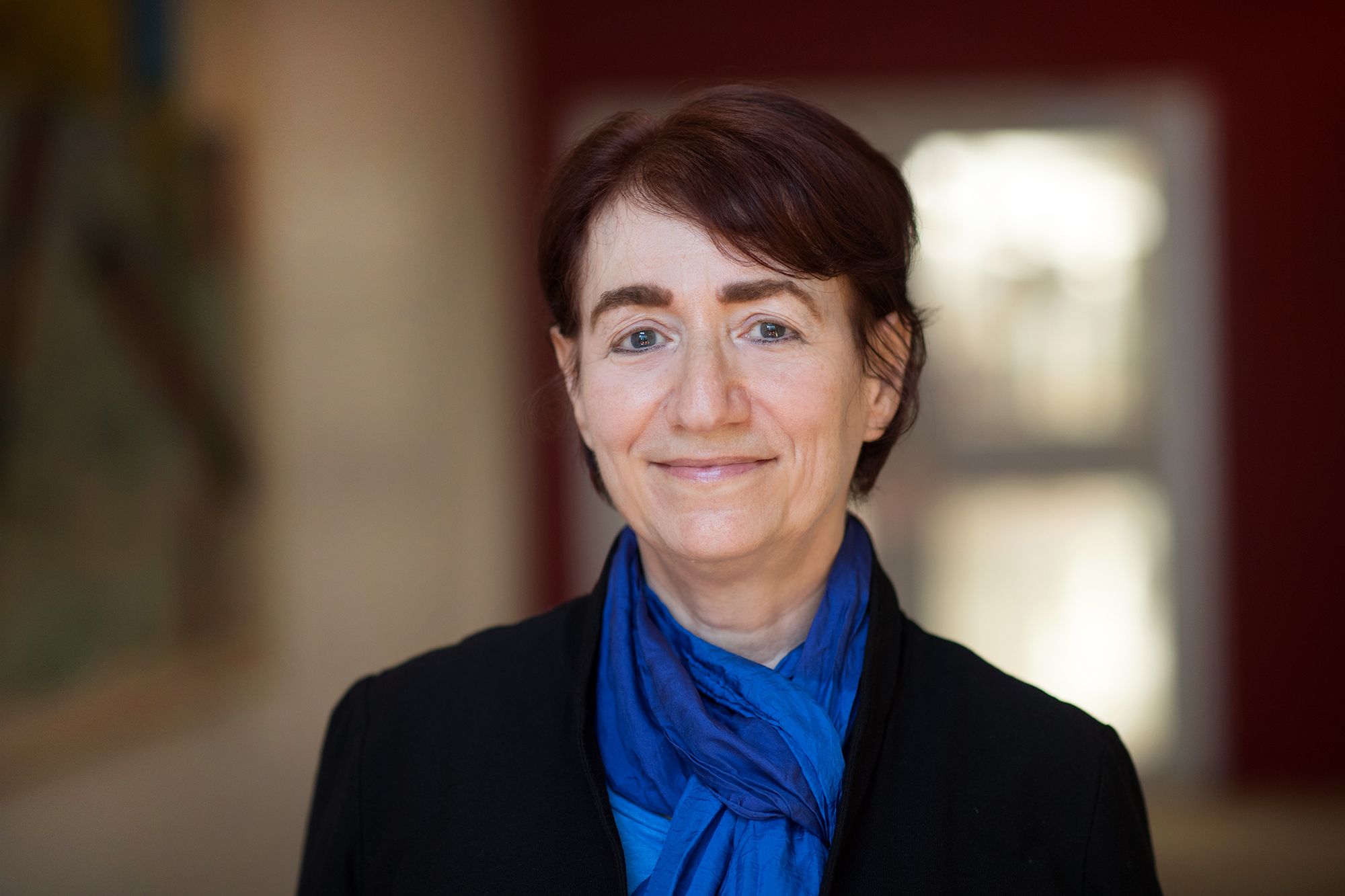
Sandy Alexandre, associate professor of literature, intends to change that by creating a large-scale database of the imaginary inventions found in literature. Alexandre’s project will enact the step-by-step mechanics of STEM innovation via one of its oft-unsung sources: literature. “To deny or sever the ties that bind STEM and literature is to suggest — rather disingenuously — that the ideas for many of the STEM devices that we know and love miraculously just came out of nowhere or from an elsewhere where literature isn’t considered relevant or at all,” she says.
During the first phase of her work, Alexandre will collaborate with students to enter into the database the imaginary inventions as they are described verbatim in a selection of books and other texts that fall under the category of speculative fiction—a category that includes but is not limited to the subgenres of fantasy, Afrofuturism, and science fiction. This first phase will, of course, require that students carefully read these texts in general, but also read for these imaginary inventions more specifically. Additionally, students with drawing skills will be tasked with interpreting the descriptions by illustrating them as two-dimensional images.
From this vast inventory of innovations, Alexandre, in consultation with students involved in the project, will decide on a short list of inventions that meet five criteria: they must be feasible, ethical, worthwhile, useful, and necessary. This vetting process, which constitutes the second phase of the project, is guided by a very important question: what can creating and thinking with a vast database of speculative fiction’s imaginary inventions teach us about what kinds of ideas we should (and shouldn’t) attempt to make into a reality? For the third and final phase, Alexandre will convene a team to build a real-life prototype of one of the imaginary inventions. She envisions this prototype being placed on exhibit at the MIT Museum.
The Bose research grant, Alexandre says, will allow her to take this project from a thought experiment to lab experiment. “This project aims to ensure that literature no longer play an overlooked role in STEM innovations. Therefore, the STEM innovation, which will be the culminating prototype of this research project, will cite a work of literature as the main source of information used in its invention.”
Nature’s role in chemical production
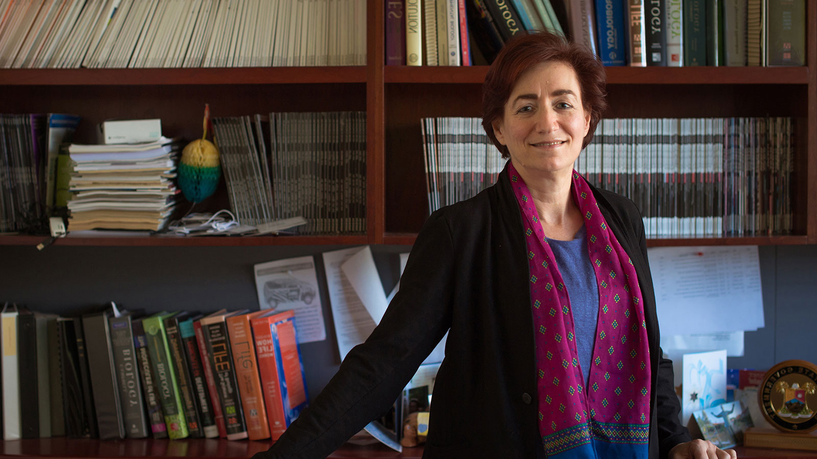
Kristala L.J. Prather ’94, the Arthur D. Little Professor of Chemical Engineering, has been focused on using biological systems for chemical production during the 15 years she’s been at the Institute. Biology as a medium for chemical synthesis has been successfully exploited to commercially produce molecules for uses that range from food to pharmaceuticals — ethanol is a good example. However, there is a range of other molecules with which scientists have been trying to work, but they have faced challenges around an insufficient amount of material being produced and a lack of defined steps needed to make a specific compound.
Prather’s research is rooted in the fact that there are a number of naturally (and unnaturally) occurring chemical compounds in the environment, and cells have evolved to be able to consume them. These cells have evolved or developed a protein that will sense a compound’s presence — a biosensor — and in response will make other proteins that help the cells utilize that compound for its benefit.
“We know biology can do this,” Prather says, “so if we can put together a sufficiently diverse set of microorganisms, can we just let nature make these regulatory molecules for anything that we want to be able to sense or detect?” Her hypothesis is that if her team exposes cells to a new compound for a long enough period of time, the cells will evolve the ability to either utilize that carbon source or develop an ability to respond to it. If Prather and her team can then identify the protein that’s now recognizing what that new compound is, they can isolate it and use it to improve the production of that compound in other systems. “The idea is to let nature evolve specificity for particular molecules that we’re interested in,” she adds.
Prather’s lab has been working with biosensors for some time, but her team has been limited to sensors that are already well characterized and that were readily available. She’s interested in how they can get access to a wider range of what she knows nature has available through the incremental exposure of new compounds to a more comprehensive subset of microorganisms.
“To accelerate the transformation of the chemical industry, we must find a way to create better biological catalysts and to create new tools when the existing ones are insufficient,” Prather says. “I am grateful to the Bose Fellowship Committee for allowing me to explore this novel idea.”
Prather’s findings as a result of this project hold the possibility of broad impacts in the field of metabolic engineering, including the development of microbial systems that can be engineered to enhance degradation of both toxic and nontoxic waste.
Adopting orphan crops to adapt to climate change
In the context of increased environmental pressure and competing land uses, meeting global food security needs is a pressing challenge. Although yield gains in staple grains such as rice, wheat, and corn have been high over the last 50 years, these have been accompanied by a homogenization of the global food supply; only 50 crops provide 90% of global food needs.
However, there are at least 3,000 plants that can be grown and consumed by humans, and many of these species thrive in marginal soils, at high temperatures, and with little rainfall. These “orphan” crops are important food sources for farmers in less developed countries but have been the subject of little research.
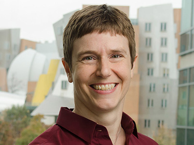
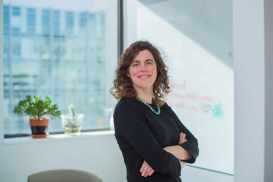
Mary Gehring, associate professor of biology at MIT, seeks to bring orphan crops into the molecular age through epigenetic engineering. She is working to promote hybridization, increase genetic diversity, and reveal desired traits for two orphan seed crops: an oilseed crop, Camelina sativa (false flax), and a high-protein legume, Cajanus cajan (pigeon pea).
C. sativa, which produces seeds with potential for uses in food and biofuel applications, can grow on land with low rainfall, requires minimal fertilizer inputs, and is resistant to several common plant pathogens. Until the mid-20th century, C. sativa was widely grown in Europe but was supplanted by canola, with a resulting loss of genetic diversity. Gehring proposes to recover this genetic diversity by creating and characterizing hybrids between C. sativa and wild relatives that have increased genetic diversity.
“To find the best cultivars of orphan crops that will withstand ever increasing environmental insults requires a deeper understanding of the diversity present within these species. We need to expand the plants we rely on for our food supply if we want to continue to thrive in the future,” says Gehring. “Studying orphan crops represents a significant step in that direction. The Bose grant will allow my lab to focus on this historically neglected but vitally important field.”
 Cornish remembers the “intellectually vibrant environment” at MIT and the cross-fertilization between labs in the department, and learned as much from other postdocs as from faculty. Petra Levin, a postdoc in professor