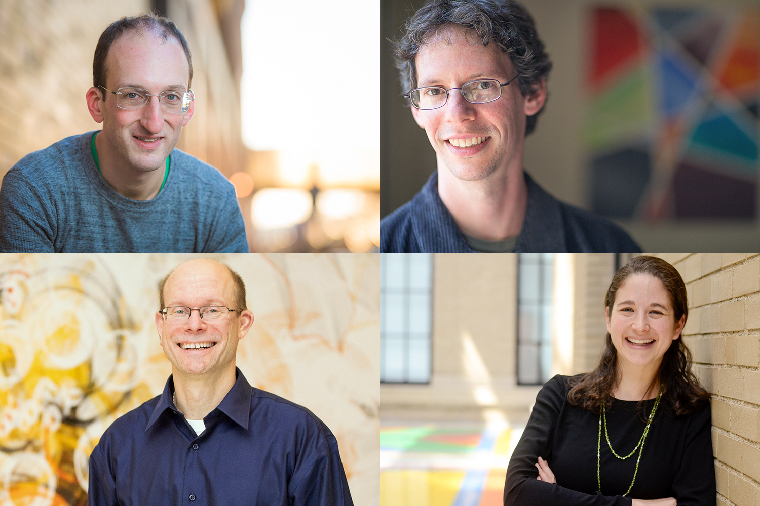
A research tool of a different color
Greta Friar | Whitehead Institute
November 18, 2020

Melanosomes are the organelles, or structures, inside our cells, that produce melanin, the molecule that gives our skin, hair and eyes their color. Melanosomes produce several different forms of melanin, including black/brown coloration and yellow/red coloration, and the many variations in levels at which each coloration can be produced in an individual generate the wide variety of skin, hair, and eye colors in the world.

Many genes that have been associated with skin color encode proteins that are active in melanosomes, but their specific functions are unknown, leaving gaps in researchers’ understanding of the underlying biology of skin color. In order to help researchers get a more detailed understanding of melanosome biology, Whitehead Institute Member David Sabatini’s lab has developed a tool, called MelanoIP, with which researchers can rapidly and specifically isolate melanosomes from the cell and analyze their contents. Using this tool, researchers can uncover the identity of the proteins at work there and explain mechanistically how genetic variation contributes to differences in skin color. In research published in Nature on November 18, Sabatini and graduate student Charles Hank Adelmann unveil MelanoIP and explain how they used it to crack the identity of melanosome protein MFSD12.

MelanoIP is the latest in a series of tools based on a method that Sabatini, who is also a professor of biology at Massachusetts Institute of Technology and an investigator with the Howard Hughes Medical Institute, and collaborators developed to rapidly extract specific organelles from the cell for investigation. Sabatini and former graduate student Walter Chen first developed the method to isolate mitochondria. The process starts with researchers creating a tag that localizes to the organelle type of interest. Then they expose the contents of the whole cell to beads covered in antibodies that latch onto the tags, which pull the organelles with them when they are collected. The lab has since adapted this process to use on lysosomes, the recycling centers of the cell, and peroxisomes, organelles important in several metabolic processes—and now, melanosomes.

The first melanosome protein that Sabatini and Adelmann turned their attention to, MFSD12, was known to be linked to the production of red coloration or pheomelanin. When MFSD12 is suppressed, this leads to darker skin color in humans and mice, because the melanosomes are generating brown/black melanin but not any of the lighter red melanin. However, MFSD12’s exact role was unknown. Using MelanoIP, Adelmann discovered that MFSD12 is required for the import of the amino acid cysteine into melanosomes, which is a necessary component in red melanin synthesis. Adelmann’s research suggests that MFSD12 is itself the transporter, but further work is needed to confirm whether it works alone or in conjunction with other molecules.

One reason that the Sabatini lab picked the melanosome as the next organelle to apply their IP toolkit to is because of its close relation to the lysosome, one of the organelles for which the lab had already built such a tool. This close relation proved relevant in Adelmann’s research on MFSD12, when he discovered that the protein is also required for the transport of cysteine into lysosomes. People with the rare genetic disorder cystinosis are affected by the buildup of cystine, another form of cysteine, in lysosomes. Adelmann found that by inhibiting MFSD12, and preventing cysteine from entering lysosomes, he could reverse the buildup of cystine in cells with the genetic mutation linked to cystinosis, suggesting a potential therapeutic use for MFSD12 inhibitors.

Adelmann is now turning his attention to cracking the identity of more of the proteins active in melanosomes and uncovering more of the biology underlying variation in skin color.

***

Written by Greta Friar

***

Adelmann, Charles H. et al. “MFSD12 mediates the import of cysteine into melanosomes and lysosomes.” Nature, Nov. 18, 2020. DOI: 10.1038/s41586-020-2937-x

Vaccine Booster

Biologist Jianzhu Chen works to enhance immune response

Mark Wolverton | Spectrum
November 16, 2020

Jianzhu Chen, professor of biology and a member of the Koch Institute for Integrative Cancer Research at MIT, is pursuing a different strategy from most of his colleagues working on SARS-CoV-2, the virus that causes Covid-19. “We focus on the immune system and fundamental mechanisms as well as their application in cancer immunotherapy, vaccine development, and metabolic diseases,” he explains. Rather than trying to develop a specific vaccine, Chen is pursuing vaccine platform technologies that can be used to enhance any vaccine.

This effort is built on Chen’s previous work on dengue fever, a severe tropical disease transmitted by mosquitos. “We have been working to improve a vaccine against dengue virus infection,” he says, “which has this phenomenon called antibody-dependent enhancement,” in which “non-neutralizing” antibodies bind to the virus but do not destroy it. The immune system’s pathogen-eating macrophages then consume these virus-antibody complexes and become infected themselves, making a subsequent infection worse.

Chen’s team has identified vaccine adjuvants, or enhancing agents, that can increase neutralizing (that is, effective) antibodies while reducing non-neutralizing antibody response in mice and nonhuman primates. The team is confident that using a similar strategy against Covid-19 would improve any vaccine’s effectiveness.

Addressing cytokine storm

Chen is also focusing on the dangerous hyperinflammatory response seen in Covid-19: the cytokine storm that can result when the immune system overreacts to infection.

“We have been working on macrophage biology for quite some time,” Chen says. “SARS-CoV-2 infection is a hyperinflammatory response, and macrophages probably play a critical role in that response.”

“We have identified many compounds, including FDA-approved drugs, bioactive compounds, and natural products that can modulate macrophage activity to become anti-inflammatory,” he says. Such macrophage modulation would likely be used in conjunction with other treatments as a therapeutic strategy for already-infected patients.

A promising result from either research project could be used along with a Covid-19 vaccine to enhance immune response while preventing or reducing the severity of any possible reinfection. But it’s too early to tell what might happen. “We don’t have a vaccine yet,” Chen notes. “It’s not clear when we’ll have one. Even when we have one, it’s not clear how well it will work. It could be 95% protection; it could be 50%. Some of them may not confer much protection at all. But even 50% or 60% is a significant number of people.”

Another challenge, Chen acknowledges, is that medical research must move from theory to lab and ultimately into the real world. Vaccines can be designed and modeled on computers but eventually “we have to test them to see if they work as we expect,” he says. “You have to immunize mice or some other animals and then challenge them with SARS-CoV-2 to see whether the vaccine protects the animals from infection or dramatically minimize disease symptoms. These kinds of studies can’t be modeled computationally.”

Chen also hopes that his particular contributions will have benefits beyond the pandemic. “We’re aiming to develop a vaccine platform prototyped on SARS-CoV-2 that can be used for the development of many other vaccines as well, using the most appropriate technologies.” If that happens, science will have dug at least one substantial jewel out of the depths of an unprecedented public health crisis.

Regulating the regulators
Whitehead Institute
November 12, 2020

MicroRNAs are short RNA sequences that maintain a tight control on which genes are expressed and when. They do this by regulating which messenger RNA (mRNA) transcripts — the single-stranded templates for proteins — are actually read by the cell. But what controls these cellular controllers?

In a new study published Nov. 12 in Science, researchers in David Bartel’s lab at Whitehead Institute show that mRNAs and other RNAs often turn the tables on their microRNA regulators — and show that the path to microRNA degradation is not what scientists expected it to be.

 “A lot of people know that microRNAs repress mRNAs — that’s textbook,” said Charlie Shi, a graduate student in Bartel’s lab and first author on the paper. “But in certain cases, this logic is reversed. And I think that’s really interesting and weird, this idea that often the tables are turned.”

When transcripts attack

MicroRNAs typically control gene expression by binding to mRNA transcripts, and then working together with a protein called Argonaute to “silence” those transcripts by causing them to be more rapidly degraded. Because microRNAs are held cozily inside of the Argonaute protein, they are shielded from destructive enzymes in the cell, and are thus fairly long-lived by cellular standards. They can persist for up to a week, causing the destruction of many mRNA molecules over that time.

Sometimes, however, a microRNA binds to a special target site on an mRNA transcript that leads to premature destruction of the microRNA. This phenomenon — called target-directed microRNA degradation, or TDMD — happens naturally in cells, and is a way to control how much of certain microRNAs are allowed to persist at any given time.

Bartel’s lab began studying this form of degradation after researchers in the lab discovered that an RNA called CYRANO, which doesn’t code for any proteins, leads to the degradation of a specific microRNA called miR-7. This interaction was interesting to the researchers because the mechanism did not seem to line up with the current theories about TDMD.

Previous models of TDMD suggested that special target sites, like the one in CYRANO, cause one end of the microRNA to stick out of Argonaute and become vulnerable to the addition and subtraction of nucleotides by cytoplasmic enzymes.  This process, called tailing and trimming, was thought to be a key step in the path to degradation of the microRNA.

“But when you knock out the enzyme that causes tailing of miR-7, it has no impact on the degradation,” Shi said. “So that’s curious, right? So how can we really perturb this supposedly responsible system and have no impact?”

A new model

In order to further probe the mechanism of TDMD, the researchers focused in on this interaction between the CYRANO noncoding RNA and miR-7. Shi designed a CRISPR screen to identify genes essential for the microRNA’s degradation when it encountered a CYRANO transcript.

The screen yielded one gene that was essential to the microRNA’s degradation, called ZSWIM8. When they looked up the gene’s function, the researchers found that it codes for a component of a ubiquitin ligase. Ubiquitin — so named because it is found in virtually all types of cells — serves as a flag to mark proteins for degradation in a cellular garbage disposal called the proteasome.

The finding of the ZSWIM8 ubiquitin ligase implied that CYRANO-mediated microRNA degradation involves destruction of the Argonaute protein. In this new molecular model for TDMD, the regulating RNA, CYRANO, binds to the microRNA, mir-7, encased in its protective Argonaute protein, and then recruits the ZSWIM8 ubiquitin ligase.  This ligase then sticks a few ubiquitin molecules onto the microRNA’s Argonaute, leading Argonaute to be degraded, and thereby exposing its microRNA cargo to be destroyed by enzymes in the cell.  Importantly, this process does not require any trimming and tailing of the microRNA.

“The discovery of this unanticipated pathway for TDMD illustrates the power of CRISPR screens, which can simultaneously query essentially every protein in the cell, including those that you never dreamed would be involved,” said Bartel, who is also an investigator of the Howard Hughes Medical Institute and a professor of biology at Massachusetts Institute of Technology.

A multitude of microRNAs

When the researchers looked at other known examples of TDMD, they found the ZSWIM8 was essential in all of them. Having identified this key part of the degradation pathway allowed them to seek out more microRNAs that are subject to this regulation.

“When we started this project, there were only around four examples in nature of endogenous RNAs that are encoded by the cell that can perform TDMD,” Shi said. “We had a feeling that there would be many more, and so by finding a factor that was required for TDMD in a general way — ZSWIM8 –we were then able to ask and answer the question, ‘how widespread this phenomenon?’”

As it turns out, TDMD is fairly common in multicellular organisms. The researchers looked for evidence of the microRNA degradation mechanism in different cell types — two from mice, and one from fruit flies — and found that in any given cell, up to 20 different microRNAs were regulated by TDMD out of a couple hundred total microRNAs in the cell.

The researchers also observed this mechanism in human cells and nematodes, suggesting that TDMD as a method for regulating microRNAs dates back to the common ancestor of these disparate species. That definitely creates a lot of questions for us,” Shi said. “Each one of these microRNAs is a story.”

Two treatment methods enhance chemotherapy by the same means: cellular senescence
Raleigh McElvery
November 10, 2020

In 2019, cancer researchers from MIT found a drug that targeted an elusive molecular interaction previously considered “undruggable.” In so doing, they opened up a new realm of chemotherapy. This drug, called JH-RE-06, sensitized tumors to treatment, but the scientists didn’t fully understand how it exerted its effects. Now, in a pair of studies published in PNAS, the same team is closer to determining which cellular processes this drug alters to enhance cancer therapy.

Many widely-used chemotherapies, like cisplatin, kill tumors by damaging their DNA and inducing programmed cell death. But cells are resilient, and many can continue to function with the help of repair enzymes that simply bypass the damage and continue to replicate the DNA. As a result, some tumors not only defy death, but gain mutations that render them more resistant to treatment or spur new malignancies elsewhere.

“If you’re not making a patient better, it’s very likely you’re making them worse,” says Michael Hemann, associate professor of biology and co-author on both studies. Rather than fully replacing conventional DNA-damaging treatments — which could take decades — Hemann suggests an effective “medium-term” solution: augmenting low doses of cisplatin with safer agents that strengthen the chemotherapy’s tumor-killing capacity.

The team had tested this approach back in 2019, when they first identified JH-RE-06 and saw it enhanced chemotherapy treatment. These experiments revealed that JH-RE-06 bound to an especially shallow (and infamously undruggable) pocket of one DNA repair enzyme called REV1. This barred REV1 from interacting with another key enzyme, and prevented the cancer cells from recovering after cisplatin treatment. But what happened next to cause the tumors to shrink was unclear.

As they began their next round of experiments, the researchers expected to find that the drug would simply enhance the way cisplatin kills tumors via programmed cell death.

Nimrat Chatterjee, Walker’s former postdoc and lead author of the first study, treated mice and individual cells with a combination of cisplatin and JH-RE-06. She expected to see signs of programmed cell death, but for months, she saw no such markers.“We thought that if we blocked the DNA repair process with the JH compound, we’d see more programmed cell death,” says co-author Graham Walker, American Cancer Society Professor and Howard Hughes Medical Institute Professor. “As it turns out, we did see more cell death — just not the kind we were expecting.”

One evening, just as she was about to head home for the day, she peeked through her microscope at the cancer cells treated with JH-RE-06 and cisplatin. She noticed they were fluorescing a strange green color.

“At first, I didn’t know what I was seeing,” she recalls. But after some follow-up, it became clear that the mysterious green color was coming from lipid-containing residues that usually appear as cells age and stop dividing. The cells appeared to be in a permanently dormant state known as senescence — not yet dead but unable to proliferate. JH-RE-06 was altering cisplatin function by triggering a second molecular pathway independent of programmed cell death.

“That was one of the best ‘aha’ moments of my scientific career so far,” Chatterjee says. “REV1, the DNA repair enzyme that JH-RE-06 binds, may have other novel biological functions and a larger role in cancer cell etiology than we originally thought. We’re now grappling with more questions about REV1 than ever before.”

Around the same time, Faye-Marie Vassel PhD ’20, Walker and Hemann’s former joint graduate student and lead author of the second study, witnessed a similar phenomenon in her own experiments. She was investigating a different way of inhibiting the two key DNA repair enzymes that enable cancer cells to survive chemotherapy. Instead of probing JH-RE-06, which latches onto REV1, she tried deleting REV1’s binding partner, called REV7. This protein is particularly influential because it serves an important role in fixing double-stranded breaks in addition to interacting with REV1.

When Vassel deleted REV7 from mice with non-small cell lung cancer, the tumors became more sensitive to cisplatin, as expected. But, like Chatterjee, she saw signs of senescence rather than programmed cell death. The two studies had converged on a common biology: adding JH-RE-06 or deleting REV7 strengthened the effects of cisplatin by inducing this dormant state.

Cancer detection and treatment methods have improved dramatically in the last two decades, but drug-resistant cancers like non-small cell lung cancer remain difficult to combat, Vassel says. “Our experiments are the first to show that senescence induction is likely a consequence of REV7 inhibition,” she adds. “Inhibiting REV7 in tandem with cisplatin therapy may prove to be an effective strategy for enhancing a chemotherapeutic response.”

Chemotherapies that trigger programmed cell death have been the mainstay of cancer treatment for decades. But studies like these show that triggering senescence may be a promising complementary strategy. Most senescent cells are eventually cleared by the immune system, and the researchers suspect this is how cancer cells treated with JH-RE-06 or REV7 inhibitors would be eliminated from the body.

Walker and Hemann agree that, at the moment, their sister studies raise more questions than answers. As Walker explained, “We’ve pried open a new discovery, and hopefully set the stage for many exciting experiments to come.”

Top image: Genetically-engineered mouse model for lung cancer. Credit: Credit: National Cancer Institute, National Institutes of HealthNIH Image Gallery/Flickr (CC BY-NC)
The REV7 image was originally published in: “Structural basis of Rev1-mediated assembly of a quaternary vertebrate translesion polymerase complex consisting of Rev1, heterodimeric polymerase (Pol) ζ, and Pol κ.”
Journal of Biological Chemistry, online August 2, 2012, DOI: 10.1074/jbc.M112.394841
Jessica Wojtaszek, Chul-Jin Lee, Sanjay D’Souza, Brenda Minesinger, Hyungjin Kim, Alan D. D’Andrea, Graham C. Walker, and Pei Zhou.

Citations:
“REV1 inhibitor JH-RE-06 enhances tumor cell response to chemotherapy by triggering senescence hallmarks”
Proceedings of the National Academy of Sciences, online November 9, 2020, DOI: 10.1073/pnas.2016064117
Nimrat Chatterjee , Matthew A Whitman, A Harris , Sophia M Min , Oliver Jonas , Evan C Lien , Alba Luengo , Matthew G Vander Heiden , Jiyong Hong , Pei Zhou , Michael T Hemann , and Graham C Walker 

“Rev7 loss alters cisplatin response and increases drug efficacy in chemotherapy-resistant lung cancer”
Proceedings of the National Academy of Sciences, online November 3, 2020, DOI: 10.1073/pnas.2016067117
Faye-Marie Vassel, Ke Bian, Graham C. Walker, and Michael T. Hemann

Aspiring physician explores the many levels of human health

During her time at MIT, senior Ayesha Ng’s interests have expanded from cellular biology to the social systems that shape public health.

Alison Gold | School of Science
November 9, 2020

It was her childhood peanut allergy that first sparked senior Ayesha Ng’s fascination with the human body. “To see this severe reaction happen to my body and not know what was happening — that made me a lot more curious about biology and living systems,” Ng says.

She didn’t exactly plan it this way. But in her three and a half years at MIT, Ng, a biology and cognitive and brain sciences double major from the Los Angeles, California area, has conducted research and taken classes examining just about every level of human health — from cellular to societal.

Most recently, her passion for medicine and health equity led her to the National Foundation for the Centers for Disease Control and Prevention (CDC), where, this summer, she worked to develop guidelines for addressing health disparities on state and local health jurisdictions’ Covid-19 data dashboards. Now, as an aspiring physician amidst the medical school application process, Ng has a sense of how microbiological, physiological, and social systems interact to affect a person’s health.

Starting small

Throughout her entire first year at MIT, Ng studied the biology of health at a cellular level. Specifically, she researched the effects of fasting and aging on regeneration of intestinal stem cells, which are located in the human intestinal crypts and continuously self-divide and reproduce. Understanding these metabolic mechanisms is crucial, as their deregulation can lead to age-associated diseases such as cancer.

“That experience allowed me to broaden my technical skills, just getting used to so many different types of molecular biological techniques right away, which I really appreciated,” Ng says of her time at the Whitehead Institute for Biomedical Research in Professor David Sabatini’s lab.

“After some time, I realized that I also wanted to also study sciences at a broader, more macro level, instead of only the microbiology and molecular biology that we were studying in Course 7,” Ng says of her biology major.

In addition to studying the biology of cancer, Ng had developed a curiosity about the human brain and how it functions. “I was really interested in that, because my grandpa has dementia,” Ng says.

Seeing her grandfather’s cognitive decline, she was inspired to become involved in MIT BrainTrust, a student organization that offers a social support network for individuals from around the Boston, Massachusetts area who have brain injuries. “We have these meetings, in which I serve as one of only one or two students there to facilitate a safe space where we can have all these individuals with brain injury gather,” Ng says of the peer-support aspect of the program. “They can really share their mutual challenges and experiences.”

Investigating the brain

To pursue her interest in brain research and the societal impact of brain injuries, Ng traveled to the University of Hong Kong the summer after her first year as an MIT International Science and Technology Initiatives (MISTI) China Fung Scholar. Working with Professor Raymond Chang, she began to examine neurodegenerative disease and used tissue-clearing techniques to visualize 3D mouse brain structures at cellular resolution. “That was personally meaningful for me, to research about that and learn more about dementia,” Ng says.

Returning to MIT her sophomore year, Ng was certain that she wanted to continue studying the brain. She began working on Alzheimer’s research at the MIT Picower Institute for Learning and Memory in the lab of Professor Li-Huei Tsai, the Picower Professor of Neuroscience at MIT. Much existing research into Alzheimer’s disease has been at the bulk-tissue level, focusing on the neurons’ role in neurodegeneration associated with aging.

Ng’s work with Tsai considers the complexity of alterations across genes and less-abundant cell types, such as microglia, astrocytes, and other supporting glial cells that become dysregulated in the brains of patients with Alzheimer’s. Considering the interplay between and within cell types during neurodegeneration is most intriguing to her. While some molecular processes are protective, other damaging ones simultaneously occur and can exist even within the same cell type. This intricacy has made the mechanistic basis behind Alzheimer’s progression elusive and the research that much more crucial.

“It’s really interesting to see how heterogeneous and complex the responses are in Alzheimer’s brains,” Ng says of the research program with Tsai, a founding director of MIT’s Aging Brain Initiative. “I really think about these potential new drug targets to improve treatment for Alzheimer’s in the future because I have seen, with my grandpa especially, how treatment is really lacking in the neurodegeneration field. There’s no treatment that’s been able to stop or even slow the progression of Alzheimer’s disease.”

Her research project in the Tsai Lab relies on a technology called single-nucleus RNA sequencing (snRNA-seq), which extracts the genomic information contained in individual cells. This is followed by computational dimension reduction and clustering algorithms to examine how Alzheimer’s disease differentially affects genes and specific cell types.

“With that project, we’ve been able to use single-nucleus RNA sequencing to really look at the brains of human Alzheimer’s patients,” Ng says. “And with the single-cell technology, we’re able to look at brain tissue at a much higher resolution, allowing us to see that there’s so much heterogeneity within the brain.”

After conducting more than a year of Alzheimer’s research and then taking a human physiology class in her third year, Ng decided to add a second major in brain and cognitive sciences to gain deeper insight specifically into how the nervous system within the body functions.

“That class really allowed me to realize that I really love organ systems and wanted to study by looking at more physiological mechanisms,” Ng says. “It has been really great to, at the end of my college career, really delve more into a very specific system.”

Medicine and society

Having gained perspective on cellular and microbiology, and human organ systems, Ng decided to zoom out further, interning this past summer at the National Foundation for the CDC. She found the opportunity through MIT’s PKG Center, applied as one of 60 candidates, and was selected for a team of four. There, as a member of the CDC Foundation’s Health Equity Strike Team, she examined how to increase transparency of publicly available Covid-19 data on health disparities and how the narrative tied to health equity can be modified in public health messages. This involved harnessing data about the demographics of those most affected during Covid-19 — including how infection and mortality rates differ starkly based on social factors including housing conditions, socioeconomic status, race, and ethnicity.

“Thinking about all these factors, we compiled a set of best practices for how to present data about Covid-19, what data should be collected, and tried to push those out to help jurisdictions as best-practice recommendations,” Ng says. “That did really increase my interest in health equity and made me realize how important public health is as well.”

Amidst the Covid-19 pandemic, Ng is spending the first semester of her senior year at home with her family in the Los Angeles area. “I really miss the people and not being able to interact with not only other students and peers, but also faculty as well,” she says. “I really wanted to enjoy time with friends, and just explore more of MIT, too, which I didn’t always get the chance to do over the past few years.”

Still, she continues to participate in both BrainTrust and MIT’s Asian Dance team, remotely, through weekly practices on Zoom.

“I think dance is one of the biggest de-stressors for me; I had never done dance before going to college. Getting to meet this team and join this community allowed me not only to connect to my Asian cultural roots, but also just expose myself to this new art form where I could really learn how to express myself on stage,” Ng says. “And that really has been the source of relief for me to just liberate any worries that I have, and has increased my sense of self-awareness and self-confidence.”

Armed with the many experiences she has enjoyed at MIT, both in and out of the classroom, Ng plans to continue studying both medicine and public health. She’s excited to explore different potential specialties and is currently most intrigued by surgery. Whichever specialty she may choose, she is determined to include health equity and cultural sensitivity in her practice.

“Seeing surgeons, I personally think that being able to physically heal a patient with my own hands, that would be the most rewarding feeling,” Ng says. “I will strive to, as a physician, use whatever platform that I have to advocate for patients and really drive health-care systems to overcome disparities.”

Scientists identify specific brain region and circuits controlling attention
Picower Institute
November 2, 2020

The attentional control that organisms need to succeed in their goals comes from two abilities: the focus to ignore distractions and the discipline to curb impulses. A new study by MIT neuroscientists shows that these abilities are independent, but that the activity of norepinephrine-producing neurons in a single brain region, the locus coeruleus, controls both by targeting two distinct areas of the prefrontal cortex.

“Our results demonstrate a fundamental causal role of LC neuronal activation in the implementation of attentional control by the selective modulation of neural activity in its target areas,” wrote the authors of the study from the research group of Susumu Tonegawa, Picower Professor of Biology and Neuroscience at RIKEN-MIT Laboratory of Neural Circuit Genetics at The Picower Institute for Learning and Memory and Howard Hughes Medical Institute.

Pharmacological and lesion studies of attentional control in humans and other mammals have suggested that norepinephrine-producing, or noradrenergic, neurons in the LC might have this role, but the most convincing evidence has been correlative rather than causal, said study lead author Andrea Bari, a research scientist in the Tonegawa lab. In the new study in the Proceedings of the National Academy of Sciences, the team demonstrated clear causality by using optogenetics to specifically control LC noradrenergic neurons in mice with temporal and spatial precision as the rodents engaged in three attentional control tasks. The manipulations immediately and reliably impacted the rodents’ performance.

“For the first time we demonstrate that LC activation in real time, with cell-specific techniques causes this effect,” Bari said.

The results, the authors said, could make important contributions to efforts to better understand and treat psychiatric disorders in which attentional control or either of its component abilities is compromised, such as attention deficit and hyperactivity disorder (ADHD).

“ADHD patients may suffer both distractibility and impulsivity,” said co-author and research scientist Michele Pignatelli “but you can also have cases mainly characterized by inattentive presentation or by hyperactive-impulsive presentation. Perhaps we can conceive new strategies to tackle different types of ADHD.”

Unexpectedly the study also raised new questions about the LC’s role in anxiety, Bari said, because to the team’s surprise, stimulating LC activity also happened to reduce anxiety in the mice.

Locus focus

After establishing their method of taking bidirectional optogenetic control of noradrenergic LC neurons—meaning that with different colors of light they could either stimulate or inhibit activity—the researchers tested the effects of each manipulation in mice. In the first task, the rodents had to wait seven seconds before a half-second flash of light signaled which of two portals they should poke with their nose to get a food reward. Mice in whom LC neurons were optogenetically stimulated did the task correctly more often and made fewer premature moves than when not manipulated. Mice in whom LC neurons were inhibited did the task correctly less often (less attention meant missing that light flash) and jumped the gun more than normal.

The researchers then trained mice on a second behavioral paradigm, derived from the Posner spatial cueing task, widely used in human cognitive neuroscience. In this task, mice before seeing the light that flagged the correct portal (this time for three seconds), they would see a “cue” flash. Sometimes that cue would be on the opposite side, sometimes be in the middle and sometimes be on the correct side. Again, LC stimulation improved correct performance and suppressed impulses and again inhibition reduced correctness and increased impulses, but now the researchers learned something new based on the reaction time of the mice. Stimulated-LC mice showed no difference in reaction time because they were focused on the actual goal but inhibited-LC mice showed variations in reaction time because they were distracted by the cue—when it was on the wrong side they reacted slower than normal and when the cue was on the correct side they reacted faster.

In the third task, the mice were both behaviorally challenged and optogenetically manipulated differently. This time the mice faced the possibility of constant distraction by irrelevant lights while they waited for the actual three-second signal of the food reward location. The same results as before held again, with one exception. In cases where there were no distractors, with three long seconds to notice the signal, inhibited-LC mice did not lapse in performing the task correctly. They only showed the deficit amid distractors.

To truly get at the heart of whether attentional focus and impulse control were independent, or dissociable, the team decided to control LC activity and norepinephrine release not at the main neuron bodies as before, but instead only where their long projections connected to specific areas of the prefrontal cortex (PFC). Going on some of Bari’s prior research and hints from other studies, they targeted the dorso-medial PFC (dmPFC) and the ventro-lateral orbitofrontal cortex (vlOFC). In these experiments they found that stimulating LC connections into the dmPFC increased correct performance but did not reduce premature responses. Meanwhile, stimulating LC connections in the vlOFC did not improve correct performance, but did reduce premature responses.

“Here we have applied behavioral, optogenetic and neural circuit genetic techniques, which afford a high degree of temporal and cell-type specificity for the manipulation and recording of noradrenergic neuron activity in the LC and demonstrate a causal link between temporal-specific LC norepinephrine modulation and attentional control,” the authors wrote. “Our results reveal that the attentional control of behavior is modulated by the synergistic effects of two dissociable coeruleo-cortical pathways, with LC projections to dmPFC enhancing attention and LC projections to vlOFC reducing impulsivity.”

Less anxiety

The tests revealing that LC stimulation reduced anxiety were performed as a precaution. Many studies suggested that increasing LC norepinephrine neuron activity would increase anxiety, Pignatelli said. That could have compromised the willingness of the mice to poke around for their food, or might have made them too impulsive, so the team checked for anxiety effects before beginning the attentional control tasks.

Bari said that investigating the surprising benefit of LC stimulation for anxiety could be an intriguing area for future study. He said he hopes to give it more… attention.

In addition to Tonegawa, Bari and Pignatelli, the paper’s other authors are Sangyu Xu, Daigo Takeuchi, Jiesi Feng, and Yulong Li.

The RIKEN Center for Brain Science, the HHMI, the JPB Foundation, the National Institutes of Health, a Human Frontier Science Program Fellowship, the National Natural Science Foundation of China and the Beijing Brain Initiative supported the study.

Angelika Amon, cell biologist who pioneered research on chromosome imbalance, dies at 53

Professor and mentor for more than 20 years at MIT redefined scientists’ understanding of the biology of cell division and proliferation.

Bendta Schroeder | Koch Institute
October 30, 2020

Angelika Amon, professor of biology and a member of the Koch Institute for Integrative Cancer Research, died on Oct. 29 at age 53, following a two-and-a-half-year battle with ovarian cancer.

“Known for her piercing scientific insight and infectious enthusiasm for the deepest questions of science, Professor Amon built an extraordinary career – and in the process, a devoted community of colleagues, students and friends,” MIT President L. Rafael Reif wrote in a letter to the MIT community.

“Angelika was a force of nature and a highly valued member of our community,” reflects Tyler Jacks, the David H. Koch Professor of Biology at MIT and director of the Koch Institute. “Her intellect and wit were equally sharp, and she brought unmatched passion to everything she did. Through her groundbreaking research, her mentorship of so many, her teaching, and a host of other contributions, Angelika has made an incredible impact on the world — one that will last long into the future.”

A pioneer in cell biology

From the earliest stages of her career, Amon made profound contributions to our understanding of the fundamental biology of the cell, deciphering the regulatory networks that govern cell division and proliferation in yeast, mice, and mammalian organoids, and shedding light on the causes of chromosome mis-segregation and its consequences for human diseases.

Human cells have 23 pairs of chromosomes, but as they divide they can make errors that lead to too many or too few chromosomes, resulting in aneuploidy. Amon’s meticulous and rigorous experiments, first in yeast and then in mammalian cells, helped to uncover the biological consequences of having too many chromosomes. Her studies determined that extra chromosomes significantly impact the composition of the cell, causing stress in important processes such as protein folding and metabolism, and leading to additional mistakes that could drive cancer. Although stress resulting from aneuploidy affects cells’ ability to survive and proliferate, cancer cells — which are nearly universally aneuploid — can grow uncontrollably. Amon showed that aneuploidy disrupts cells’ usual error-repair systems, allowing genetic mutations to quickly accumulate.

Aneuploidy is usually fatal, but in some instances extra copies of specific chromosomes can lead to conditions such as Down syndrome and developmental disorders including those known as Patau and Edwards syndromes. This led Amon to work to understand how these negative effects result in some of the health problems associated specifically with Down syndrome, such as acute lymphoblastic leukemia. Her expertise in this area led her to be named co-director of the recently established Alana Down Syndrome Center at MIT.

“Angelika’s intellect and research were as astonishing as her bravery and her spirit. Her lab’s fundamental work on aneuploidy was integral to our establishment of the center,” say Li-Huei Tsai, the Picower Professor of Neuroscience and co-director of the Alana Down Syndrome Center. “Her exploration of the myriad consequences of aneuploidy for human health was vitally important and will continue to guide scientific and medical research.”

Another major focus of research in the Amon lab has been on the relationship between how cells grow, divide, and age. Among other insights, this work has revealed that once cells reach a certain large size, they lose the ability to proliferate and are unable to reenter the cell cycle. Further, this growth contributes to senescence, an irreversible cell cycle arrest, and tissue aging. In related work, Amon has investigated the relationships between stem cell size, stem cell function, and tissue age. Her lab’s studies have found that in hematopoetic stem cells, small size is important to cells’ ability to function and proliferate — in fact, she posted recent findings on bioRxiv earlier this week — and have been examining the same questions in epithelial cells as well.

Amon lab experiments delved deep into the mechanics of the biology, trying to understand the mechanisms behind their observations. To support this work, she established research collaborations to leverage approaches and technologies developed by her colleagues at the Koch Institute, including sophisticated intestinal organoid and mouse models developed by the Yilmaz Laboratory, and a microfluidic device developed by the Manalis Laboratory for measuring physical characteristics of single cells.

The thrill of discovery

Born in 1967, Amon grew up in Vienna, Austria, in a family of five. Playing outside all day with her three younger siblings, she developed an early love of biology and animals. She could not remember a time when she was not interested in biology, initially wanting to become a zoologist. But in high school, she saw an old black-and-white film from the 1950s about chromosome segregation, and found the moment that the sister chromatids split apart breathtaking. She knew then that she wanted to study the inner workings of the cell and decided to focus on genetics at the University of Vienna in Austria.

After receiving her BS, Amon continued her doctoral work there under Professor Kim Nasmyth at the Research Institute of Molecular Pathology, earning her PhD in 1993. From the outset, she made important contributions to the field of cell cycle dynamics. Her work on yeast genetics in the Nasmyth laboratory led to major discoveries about how one stage of the cell cycle sets up for the next, revealing that cyclins, proteins that accumulate within cells as they enter mitosis, must be broken down before cells pass from mitosis to G1, a period of cell growth.

Towards the end of her doctorate, Amon became interested in fruitfly genetics and read the work of Ruth Lehmann, then a faculty member at MIT and a member of the Whitehead Institute. Impressed by the elegance of Lehmann’s genetic approach, she applied and was accepted to her lab. In 1994, Amon arrived in the United States, not knowing that it would become her permanent home or that she would eventually become a professor.

While Amon’s love affair with  fruitfly genetics would prove short, her promise was immediately apparent to Lehmann, now director of the Whitehead Institute. “I will never forget picking Angelika up from the airport when she was flying in from Vienna to join my lab. Despite the long trip, she was just so full of energy, ready to talk science,” says Lehmann. “She had read all the papers in the new field and cut through the results to hit equally on the main points.”

But as Amon frequently was fond of saying, “yeast will spoil you.” Lehmann explains that “because they grow so fast and there are so many tools, ‘your brain is the only limitation.’ I tried to convince her of the beauty and advantages of my slower-growing favorite organism. But in the end, yeast won and Angelika went on to establish a remarkable body of work, starting with her many contributions to how cells divide and more recently to discover a cellular aneuploidy program.”

In 1996, after Lehmann had left for New York University’s Skirball Institute, Amon was invited to become a Whitehead Fellow, a prestigious program that offers recent PhDs resources and mentorship to undertake their own investigations. Her work on the question of how yeast cells progress through the cell cycle and partition their chromosomes would be instrumental in establishing her as one of the world’s leading geneticists. While at Whitehead, her lab made key findings centered around the role of an enzyme called Cdc14 in prompting cells to exit mitosis, including that the enzyme is sequestered in a cellular compartment called the nucleolus and must be released before the cell can exit.

“I was one of those blessed to share with her a ‘eureka moment,’ as she would call it,” says Rosella Visintin, a postdoc in Amon’s lab at the time of the discovery and now an assistant professor at the European School of Molecular Medicine in Milan. “She had so many. Most of us are lucky to get just one, and I was one of the lucky ones. I’ll never forget her smile and scream — neither will the entire Whitehead Institute — when she saw for the first time Cdc14 localization: ‘You did it, you did it, you figured it out!’ Passion, excitement, joy — everything was in that scream.”

In 1999, Amon’s work as a Whitehead Fellow earned her a faculty position in the MIT Department of Biology and the MIT Center for Cancer Research, the predecessor to the Koch Institute. A full professor since 2007, she also became the Kathleen and Curtis Marble Professor in Cancer Research, associate director of the Paul F. Glenn Center for Biology of Aging Research at MIT, a member of the Ludwig Center for Molecular Oncology at MIT, and an investigator of the Howard Hughes Medical Institute.

Her pathbreaking research was recognized by several awards and honors, including the 2003 National Science Foundation Alan T. Waterman Award, the 2007 Paul Marks Prize for Cancer Research, the 2008 National Academy of Sciences (NAS) Award in Molecular Biology, and the 2013 Ernst Jung Prize for Medicine. In 2019, she won the Breakthrough Prize in Life Sciences and the Vilcek Prize in Biomedical Science, and was named to the Carnegie Corporation of New York’s annual list of Great Immigrants, Great Americans. This year, she was given the Human Frontier Science Program Nakasone Award. She was also a member of the NAS and the American Academy of Arts and Sciences.

Lighting the way forward

Amon’s perseverance, deep curiosity, and enthusiasm for discovery served her well in her roles as teacher, mentor, and colleague. She has worked with many labs across the world and developed a deep network of scientific collaboration and friendships. She was a sought-after speaker for seminars and the many conferences she attended. In over 20 years as a professor at MIT, she has mentored more than 80 postdocs, graduate students, and undergraduates, and received the School of Science’s undergraduate teaching prize.

“Angelika was an amazing, energetic, passionate, and creative scientist, an outstanding mentor to many, and an excellent teacher,” says Alan Grossman, the Praecis Professor of Biology and head of MIT’s Department of Biology. “Her impact and legacy will live on and be perpetuated by all those she touched.”

“Angelika existed in a league of her own,” explains Kristin Knouse, one of Amon’s former graduate students and a current Whitehead Fellow. “She had the energy and excitement of someone who picked up a pipette for the first time, but the brilliance and wisdom of someone who had been doing it for decades. Her infectious energy and brilliant mind were matched by a boundless heart and tenacious grit. She could glance at any data and immediately deliver a sharp insight that would never have crossed any other mind. Her positive attributes were infectious, and any interaction with her, no matter how transient, assuredly left you feeling better about yourself and your science.”

Taking great delight in helping young scientists find their own “eureka moments,” Amon was a fearless advocate for science and the rights of women and minorities and inspired others to fight as well. She was not afraid to speak out in support of the research and causes she believed strongly in. She was a role model for young female scientists and spent countless hours mentoring and guiding them in a male-dominated field. While she graciously accepted awards for women in science, including the Vanderbilt Prize and the Women in Cell Biology Senior Award, she questioned the value of prizes focused on women as women, rather than on their scientific contributions.

“Angelika Amon was an inspiring leader,” notes Lehmann, “not only by her trailblazing science but also by her fearlessness to call out sexism and other -isms in our community. Her captivating laugh and unwavering mentorship and guidance will be missed by students and faculty alike. MIT and the science community have lost an exemplary leader, mentor, friend, and mensch.”

Amon’s wide-ranging curiosity led her to consider new ideas beyond her own field. In recent years, she has developed a love for dinosaurs and fossils, and often mentioned that she would like to study terraforming, which she considered essential for a human success to life on other planets.

“It was always amazing to talk with Angelika about science, because her interests were so deep and so broad, her intellect so sharp, and her enthusiasm so infectious,” remembers Vivian Siegel, a lecturer in the Department of Biology and friend since Amon’s postdoctoral days. “Beyond her own work in the lab, she was fascinated by so many things, including dinosaurs — dreaming of taking her daughters on a dig — lichen, and even life on Mars.”

“Angelika was brilliant; she illuminated science and scientists,” says Frank Solomon, professor of biology and member of the Koch Institute. “And she was intense; she warmed the people around her, and expanded what it means to be a friend.”

Amon is survived by her husband Johannes Weis, and her daughters Theresa and Clara Weis, and her three siblings and their families.