Sixteen professors join the departments of Biology; Chemistry; Earth, Atmospheric and Planetary Sciences; Mathematics; and Physics.
School of Science
Last spring, the School of Science welcomed seven new faculty members.
Erin Chen PhD ’11 studies the communication between microbes that reside on the surface of the human body and the immune system. She focuses on the largest organ: the skin. Chen will dissect the molecular signals of diverse skin microbes and their effects on host tissues, with the goal of harnessing microbe-host interactions to engineer new therapeutics for human disease.
Chen earned her bachelor’s in biology from the University of Chicago, her PhD from MIT, and her MD from Harvard Medical School, and she completed her medical residency at the University of California at San Francisco. Chen was also a Howard Hughes Medical Institute Hanna Gray Fellow at Stanford University and an attending dermatologist at UCSF and at the San Francisco VA Medical Center. Chen returns to MIT as an assistant professor in the Department of Biology, a core member of the Broad Institute of MIT and Harvard, and an attending dermatologist at Massachusetts General Hospital.
Robert Gilliard’s research is multidisciplinary and combines various aspects of organic, inorganic, main-group, and materials chemistry. The Gilliard group specializes in the chemical synthesis of new molecules that impact the development of new catalysts and reagents, including the discovery of unknown transformations of environmentally relevant small-molecules [e.g., carbon dioxide, carbon monoxide, and dihydrogen (H2)]. In addition, he investigates the design, characterization, and reactivity of boron-based luminescent and redox-active heterocycles for use in optoelectronic applications (e.g., stimuli-responsive materials, thermochromic materials, chemical sensors).
Gilliard earned his bachelor’s degree from Clemson University and his PhD from the University of Georgia. He completed joint postdoctoral studies at the Swiss Federal Institute of Technology (ETH Zürich) and Case Western Reserve University. He served on the faculty at the University of Virginia from 2017-22. Gilliard spent time in the MIT Department of Chemistry as a 2021-22 Dr. Martin Luther King Visiting Professor. He returns as the Novartis Associate Professor of Chemistry with tenure.
Sally Kornbluth is president of MIT and a professor of biology. Before she closed her lab to focus on administration, her research focused on the biological signals that tell a cell to start dividing or to self-destruct — processes that are key to understanding cancer as well as various degenerative disorders. She has published extensively on cell proliferation and programmed cell death, studying both phenomena in a variety of organisms. Her research has helped to show how cancer cells evade this programmed death, or apoptosis, and how metabolism regulates the cell death process; her work has also clarified the role of apoptosis in regulating the duration of female fertility in vertebrates.
Kornbluth holds bachelor’s degrees in political science from Williams College and in genetics from Cambridge University. She earned her PhD in molecular oncology from Rockefeller University in 1989 and completed postdoctoral training at the University of California at San Diego. In 1994, she joined the faculty of Duke University and served in the administration as vice dean for basic science at the Duke School of Medicine (2006-2014) and later as the university’s provost (2014-2022). She is a member of the National Academy of Medicine, the National Academy of Inventors, and the American Academy of Arts and Sciences.
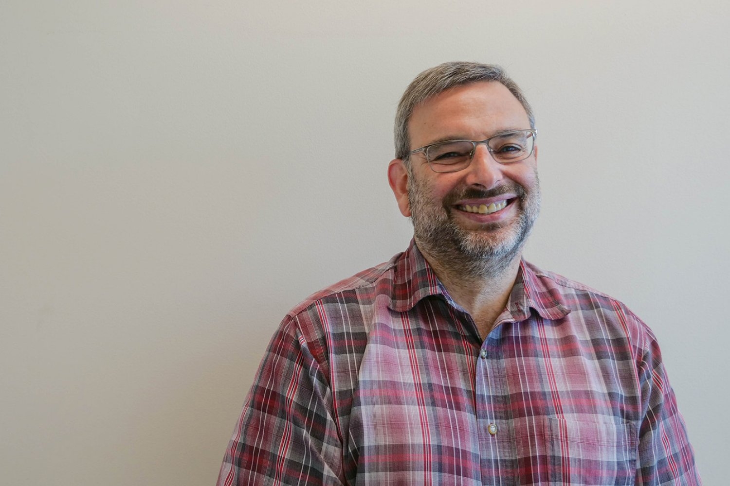
Daniel Lew uses fungal model systems to ask how cells orient their activities in space, including oriented growth, cell wall remodeling, and organelle segregation. Different cells take on an astonishing variety of shapes, which are often critical to be able to perform specialized cell functions like absorbing nutrients or contracting muscles. Lew studies how different cell shapes arise and how cells control the spatial distribution of their internal constituents, taking advantage of the tractability of fungal model systems, and addressing these questions using approaches from cell biology, genetics, and computational biology to understand molecular mechanisms.
Lew received a bachelor’s degree in genetics from Cambridge University followed by a PhD in molecular biology from Rockefeller University. After postdoctoral training at the Scripps Research Institute, he joined the Duke University faculty in 1994. Lew joins MIT as a professor of biology with tenure.
Eluned Smith uses rare beauty decays measured with the LHCb detector at CERN to search for new fundamental particles at mass scales above the collision energy of the Large Hadron Collider (LHC). Her group leverages data to elucidate the physics of beauty quarks, whose behavior cannot be explained by the Standard Model of particle physics. In doing so, her work aims to resolve whether the anomalies are misunderstood quantum chromodynamics or the first sign of beyond-the-Standard-Model-physics at the LHC.
Smith joins MIT as an assistant professor in the Department of Physics and the Laboratory for Nuclear Science. She earned her undergraduate and doctoral degrees at Imperial College London, which she completed in 2017. She did her first postdoc at RWTH Aachen before winning an Ambizione Fellowship from the Swiss National Science Foundation at the University of Zürich.
Gaia Stucky de Quay explores topographic signals and landscape evolution, in order to both de-convolve and quantify primary driving forces such as tectonics, climate, and local geological processes. She integrates fieldwork, lab work, modeling, and remote sensing to improve our quantitative understanding of such processes at compelling geological sites such as Martian valleys and lakes, the surfaces of icy moons, and volcanic islands in the Atlantic Ocean.
Stucky de Quay joins the Department of Earth, Atmospheric and Planetary Sciences as an assistant professor. Most recently, she was a Daly Postdoctoral Fellow at Harvard University. Previously, she was a postdoc at the University of Texas at Austin and a visiting student at the University of Chicago. Stucky de Quay earned her MS from the University College of London and a PhD from Imperial College London.
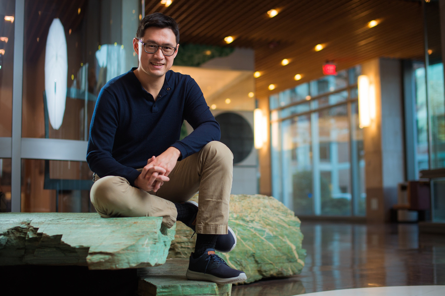
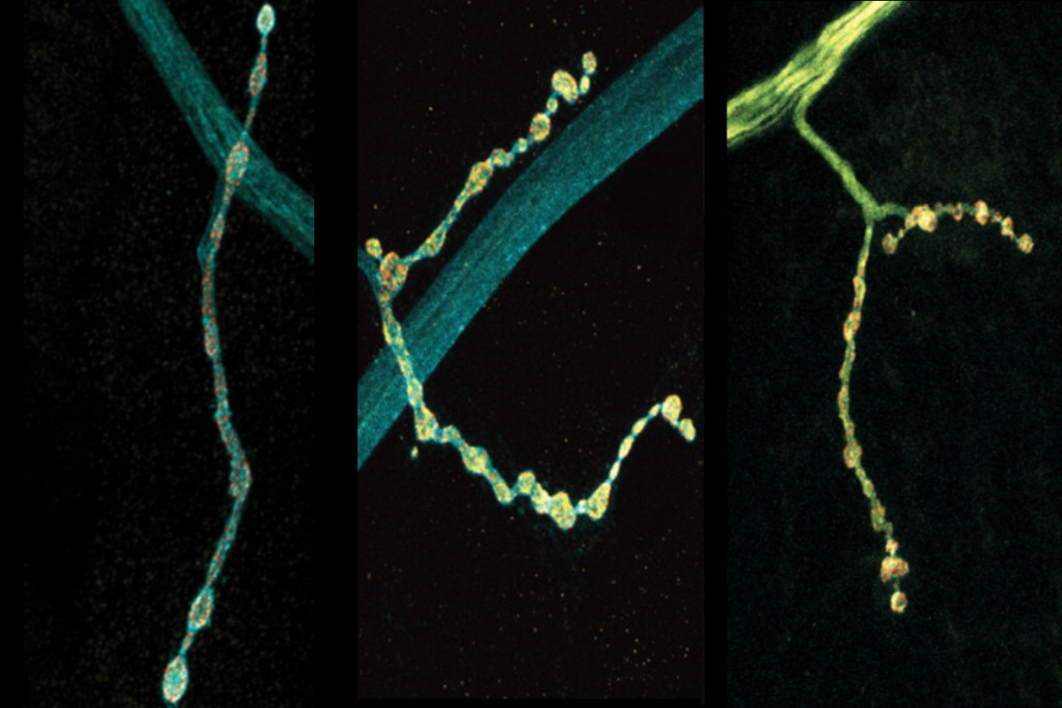
Brandon “Brady” Weissbourd uses the jellyfish, Clytia hemisphaerica, to study nervous system evolution, development, regeneration, and function. With a foundation is in systems neuroscience, his lab uses genetic and optical techniques to examine how behavior arises from the activity of networks of neurons; they investigate how the Clytia nervous system is so robust; and they use Clytia’s evolutionary position to make inferences about the ultimate origins of nervous systems.
Weissbourd received a BA in human evolutionary biology from Harvard University in 2009 and a PhD from Stanford University in 2016. He then completed postdoctoral research at Caltech and The Howard Hughes Medical Institute. He joins MIT as an assistant professor in the Department of Biology and an investigator in The Picower Institute for Learning and Memory.
This fall, the School of Science welcomes nine new faculty members.
Facundo Batista studies the fundamental biology of the immune system to develop the next generation of vaccines and therapeutics. B lymphocytes are the fulcrum of immunological memory, the source of antibodies, and the focus of vaccine development. His lab has investigated how, where, and when B cell responses take shape. In recent years, the Batista group has expanded into preclinical vaccinology, targeting viruses including HIV, malaria, influenza, and SARS-CoV-2.
Batista is an MIT professor of biology with tenure as well as the associate director and scientific director of the Ragon Institute of MGH, MIT, and Harvard. He received his PhD from the International School of Advanced Studies in Trieste, Italy, and his undergraduate degree from the University of Buenos Aires, Argentina. Prior to MIT, Batista was a tenured member of the Francis Crick Institute, a professor at Imperial College London, and a professor of microbiology and immunology at Harvard Medical School.
Anna-Christina Eilers is an observational astrophysicist. Her research focuses on the formation of the first galaxies, quasars, and supermassive black holes in the early universe, during an era known as the Cosmic Dawn. In particular, Eilers is interested in the growth of the first supermassive black holes which reside in the center of luminous, distant galaxies known as quasars, to understand how black holes evolve from small stellar remnants to billion-solar-mass black holes within very short amounts of cosmic time.
Previously, Eilers received a bachelor’s degree in physics from the University of Goettingen, a master’s degree in astrophysics from the University of Heidelberg, and a PhD in astrophysics from the Max Planck Institute for Astronomy in Heidelberg. In 2019, she was awarded a NASA Hubble Fellowship and the Pappalardo Fellowship to continue her research at MIT. Eilers remains at MIT as an assistant professor in the Department of Physics and the MIT Kavli Institute for Astrophysics and Space Research.
Masha Elkin combines catalyst development, natural products synthesis, and machine learning to tackle important chemical challenges. Her group develops new transition metal catalysts that enable efficient bond disconnections and access to value-added compounds, leveraging these transformations for the synthesis of bioactive natural products that address outstanding needs in human health, and uses computational tools to explore all possible molecules and accelerate reaction discovery.
Elkin joins MIT as the D. Reid (1941) and Barbara J. Weedon Career Development Assistant Professor of Chemistry. She earned her bachelor’s degree in chemistry from Washington University in St. Louis in 2014, and her PhD from Yale University in 2019, then began as a postdoc at the University of California at Berkeley.
Mikhail Ivanov’s research has developed at the interface of theoretical physics and data analysis, bridging state-of-the-art theoretical ideas with observational data. The overarching aim of his research is to use Effective Field Theory in combination with astrophysical data in order to resolve fundamental challenges of modern physics, such as the nature of dark matter, dark energy, inflation, and gravity.
Ivanov joins MIT as an assistant professor in the Department of Physics and the Center for Theoretical Physics in the Laboratory for Nuclear Science. He obtained his PhD from the École Polytechnique Fédérale de Lausanne in 2019. During his PhD studies, he spent a year at the Institute for Advanced Study in Princeton, New Jersey, as a fellow of the Swiss National Science Foundation. Subsequently, he was a postdoc at New York University and a NASA Einstein Fellow at the Institute for Advanced Study.
Efforts to target pathogenic proteins with drugs or chemical probes can often be analogized to a lock and key, where the protein target is the “lock” and the molecule is the “key.” However, what happens when the target is flexible or lacks a defined structure? In all living things, molecular chaperone proteins have evolved to support proper folding of these moving targets. Yet, protein misfolding and aggregation is a hallmark of many myopathies and neurodegenerative diseases. Oleta Johnson uses chemical and biophysical tools to understand and tune the activity of molecular chaperone proteins in protein misfolding diseases. Thus, her research group will reveal the molecular underpinnings of molecular chaperone dysfunction in a broad array of disorders including Huntington’s disease and Parkinson’s disease. These tools and finding will be further developed to develop novel treatments for patients of these diseases.
Johnson joins the Department of Chemistry as an assistant professor. She earned her bachelor’s degree in biochemistry from Florida Agricultural and Mechanical University in 2013, and her PhD from the University of Michigan in 2018. Prior to MIT, Johnson completed postdoctoral research at the University of California at San Francisco.
Nicole Xike Nie is an isotope geo/cosmochemist using the chemical and isotopic compositions of extraterrestrial materials to understand the formation of our solar system. Her research is driven by fundamental questions about the origin and evolution of the early solar system. Leveraging geochemical methods, she wants to understand questions such as why all planetary bodies are depleted of volatile elements when their building block materials aren’t, and why the Earth’s chemical signatures are distinct from other planetary bodies.
Nie joins MIT as an assistant professor in the Department of Earth, Atmospheric and Planetary Sciences. Nie received a BS in geology from China University of Geosciences in 2010, an MS in geochemistry from Chinese Academy of Sciences in 2013, and a PhD in geo/cosmochemistry from the University of Chicago in 2019. After graduating she was a Carnegie Postdoc Fellow at Carnegie Institution for Science and a postdoc researcher at Caltech.
Tristan Ozuch works in the field of geometric analysis and focuses on Einstein manifolds and Ricci flows. His work has shed light on the moduli space of Einstein metrics in four dimensions, addressing questions that have lingered since the 1980s. These questions originated from the systematic study of Einstein’s equations and their degenerations since the 1970s, in both physics and mathematics.
After receiving a bachelor’s degree, master’s degree, and PhD from École Normale Supérieure, Tristan Ozuch joined MIT as a C.L.E. Moore Instructor of Mathematics. He continues in the Department of Mathematics as an assistant professor.
Climate scientist Talia Tamarin-Brodsky’s research is driven by questions on the interface between weather and climate. In her work, Tamarin-Brodsky combines theory, computational methods, and observational data to study Earth’s climate and weather and how they respond to climate change. Her interests include atmospheric dynamics, temperature variability, weather and climate extremes, and subseasonal-to-seasonal predictability. For example, she studies how nonlinear wave breaking events in the upper atmosphere influence surface weather and extremes, and the mechanisms shaping the spatial distribution of Earth’s near-surface temperature.
Tamarin-Brodsky received a bachelor’s degree in mathematics and geophysics as well as a master’s in physics from Tel Aviv University, Israel, before earning her PhD from the Weizmann Institute. She completed a postdoctoral project at the University of Reading, U.K., and a postdoctoral fellowship at Tel Aviv University. She joins the Department of Earth, Atmospheric and Planetary Studies as an assistant professor.
John Urschel PhD ’21 is a mathematician focused on matrix analysis and computations, with an emphasis on theoretical results and provable guarantees for practical problems. His research interests include numerical linear algebra, spectral graph theory, and topics in theoretical machine learning.
Urschel earned bachelor’s and master’s degrees in mathematics from Pennsylvania State University, then completed a PhD in mathematics at MIT in 2021. He was a member of the Institute for Advanced Study and a junior fellow at Harvard University before returning to MIT as an assistant professor of mathematics this fall.