Pew recognizes four MIT researchers for innovation in biomedical science

Biophysicist Ibrahim Cissé and cell biologist Gene-Wei Li honored as Pew Scholars; postdocs Ana Fiszbein and María Inda are named Pew Latin American Fellows.

Julia C. Keller | School of Science
June 20, 2017

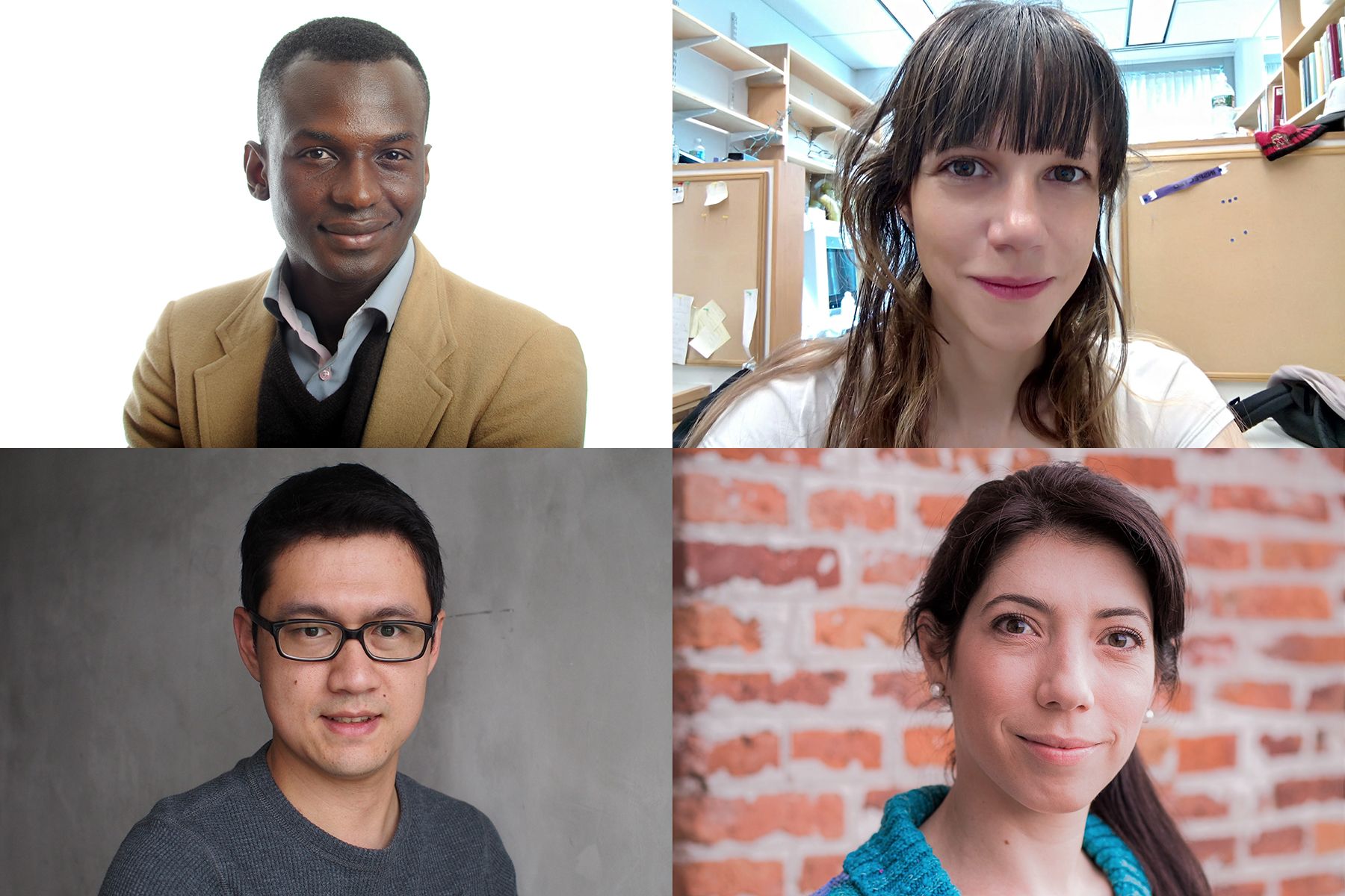
The Pew Charitable Trusts has named Ibrahim Cissé, assistant professor of physics, and Gene-Wei Li, assistant professor of biology, as 2017 Pew Scholars in the Biomedical Sciences. In addition, two postdocs, Ana Fiszbein and María E. Inda, were named to the 2017 class of Pew Latin American Fellows in the Biomedical Sciences in computational biology and synthetic biology, respectively.

The Pew Scholars program encourages early-career scientists to pursue innovative research to advance the understanding of human biology and disease. This year, 22 Pew Scholars will receive $240,000 over four years and gain inclusion into a select community of scientists that includes three Nobel Prize winners, five MacArthur Fellows, and five recipients of the Albert Lasker Medical Research Award. The applicants, who conduct research in all areas of biomedical sciences, must be nominated by one of 180 invited institutions. To date, the program has invested in more than 900 scholars.

“Pew’s biomedical programs not only provide young scientists with the flexibility to pursue creative ideas; they also spark interdisciplinary thinking and collaborations that can open new paths in the search for answers,” says Craig C. Mello, who won the 2006 Nobel Prize for physiology or medicine, was a 1995 Pew Scholar, and chairs the Pew Scholars National Advisory Committee.

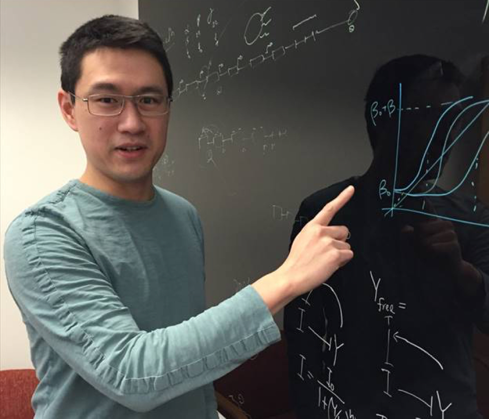
Cissé, the MIT Class of 1922 Career Development Assistant Professor, says “support from Pew at an early stage is great encouragement in pushing my lab further at the frontiers of different fields.”

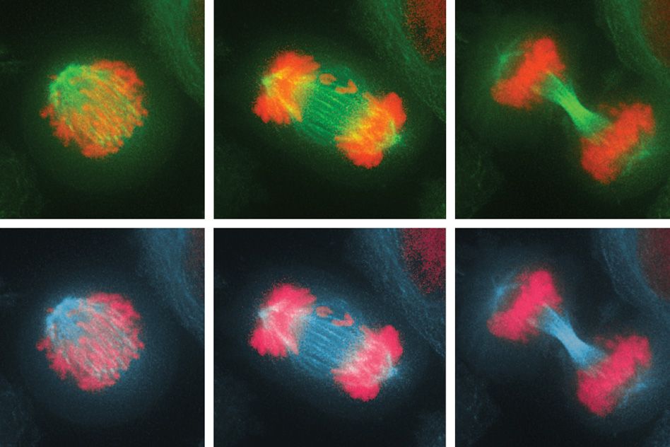
Cissé’s research group is investigating the fundamental processes involved in gene activation. Using a combination of techniques in cell and molecular biology, biochemistry, genomics, and super-resolution microscopy, he will continue his investigations of the behaviors of the enzyme involved in the transcription of DNA to RNA molecules. The enzyme, RNA polymerase II, has been well-studied in vitro, but Cissé’s work looks at these transient biological interactions within living cells. His findings will deepen the understanding of these processes, disruptions in which are linked to human disease, including most cancers.

Li’s research looks at evolution of cells’ production of proteins to answer a fundamental biological question of how cells specify how much of each type of protein to produce. In Li’s Quantitative Biology Lab, researchers have developed a technique for measuring the precise production rates of every protein in a cell. Combining this approach with other techniques in cell, molecular, and computational biology, Li is comparing a broad range of organisms across evolutionary distances to determine whether all of their proteins are maintained at some preferred level. By artificially perturbing the quantities of selected proteins, Li can explore the mechanisms cells use to reestablish the proper protein balance to better understand when misregulation occurs that leads to disease.

“The success of my research hinges on close integration between expertise in biological and physical sciences, as well as constant stimulation from both disciplines,” says Li, the Helen Sizer Career Development Assistant Professor. “The Pew scholarship will also provide a unique opportunity to interact with the brightest young minds in the biomedical sciences outside my field that will elevate my research to unanticipated levels.”

Each year, current scholars come together to discuss their research and learn from peers in fields outside of their own. “I am looking forward to interacting with other Pew scholars, many of whom are also working on paradigm-shifting ideas,” says Cissé.

Rebecca W. Rimel, president and CEO of The Pew Charitable Trusts calls the scholars an “impressive group” that has demonstrated “the curiosity and courage that drive great scientific advances, and we are excited to help them fulfill their potential.”

The Pew Latin American Fellows program, meanwhile, is intended to support postdocs from Latin America. Winners are awarded two years of funding to conduct research at laboratories and academic institutions in the United States.

The program also provides additional funding to awardees who return to Latin America to launch their own research labs after the completion of their fellowships. About 70 percent of program participants have taken advantage of this incentive and are conducting work on regional and global health challenges in nine Latin American countries, according to The Pew Charitable Trusts.

“Almost 150 young scientists have returned to their home countries and established independent research labs, providing critical groundwork for biomedical research across Latin America,” says Torsten N. Wiesel, the 1981 Nobel laureate in physiology or medicine and chair of the Latin American Fellows National Advisory Committee.

Ten Pew Latin American Fellows were named this year. The fellowship provides a $30,000 salary stipend to support two years of research, as well as an additional $35,000 for laboratory equipment should the fellow return to Latin America to start his or her own lab. Since the program’s inception in 1990, the program has supported almost 150 young Latin American scientists.

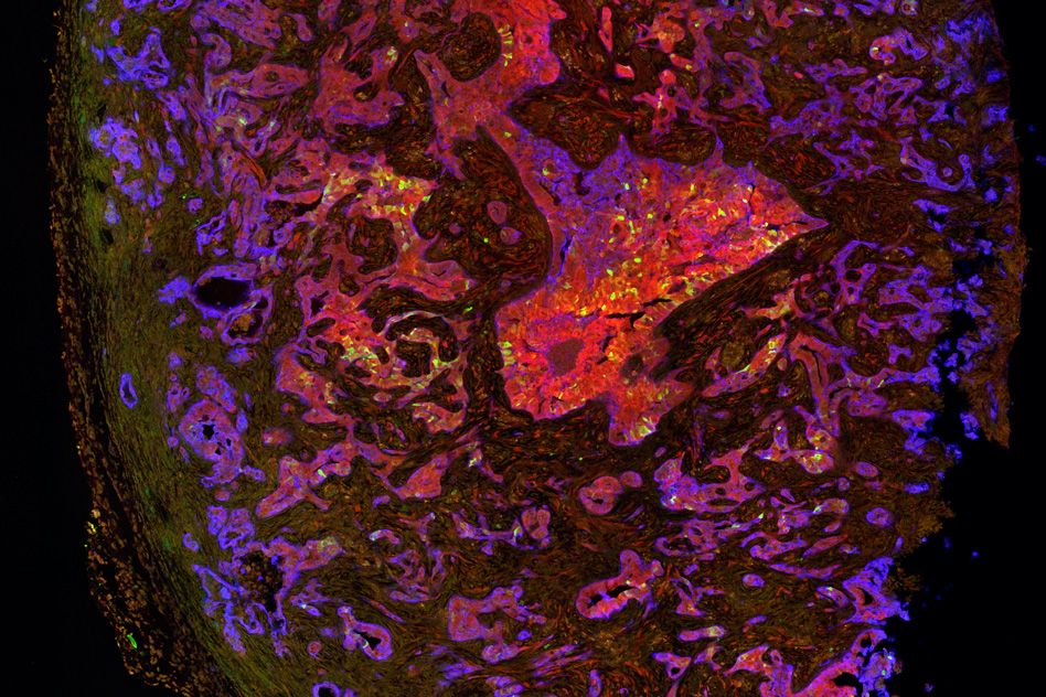
Ana Fiszbein is a postdoc working in the Burge Lab, where she researches the role that changes in gene splicing could play in the biology of normal and tumor cells, with the goal of revealing novel targets for cancer therapeutics. “I am very honored to receive this award, it is a privilege and also a responsibility,” she says. Fiszbein is working with Professor Christopher B. Burge, of the departments of Biology and Biological Engineering and the Broad Institute of MIT and Harvard.

“Ana is an exceptionally talented molecular biologist and independent thinker who came to my lab very well trained from her PhD in Alberto Kornblihtt’s lab,” Burge says. “She has developed very interesting hypotheses about the mechanistic connections between transcription and RNA splicing.”

The activity of genes can be regulated on many levels, including how often DNA is read to produce an RNA, where within the gene that reading begins, and which of the gene’s segments are represented in the RNA molecules that ultimately direct the formation of protein. Tumor cells harbor genetic changes that can alter all three of these points of control. However, little is known about the control of these regulatory processes or how they might be interconnected. Fiszbein is working on a sequence study of RNA in different species, with a sequence analysis of human cancer genomes to identify RNAs that may be present more often in cancer cells. She will then assess whether those RNA segments are co-regulated with the sites where the reading of a gene begins.

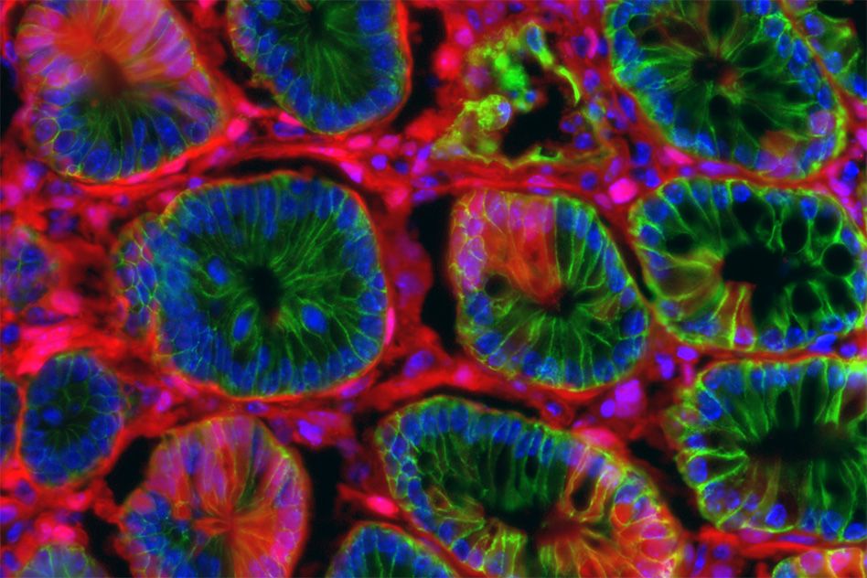
Inda is a postdoc working in the lab of Timothy Lu, an assistant professor leading the Synthetic Biology Group in the departments of Electrical Engineering and Computer Science and Biological Engineering. In the Lu Lab, she will work on the development of novel noninvasive strategies, for the early diagnosis and alleviation of inflammation in intestinal disorders, such as inflammatory bowel disease (IBD).

“The fellowship provides me a unique opportunity to learn the practical and theoretical underpinnings of cutting-edge research in the synthetic biology field for diagnosis and treatment of serious ailments,” she says.

A variety of bacteria inhabit healthy human intestines, and members of the Lu laboratory have been working to commandeer some of these microbes for use as sentinels that could patrol the gut and secrete therapeutic molecules in areas that appear inflamed. Inda plans to equip bacteria with biosensors that recognize the molecular markers of IBD — and then trigger the release of anti-inflammatory compounds. She will then assess the engineered microbes’ ability to distinguish between diseased and healthy tissue and to treat inflammation in an animal model of IBD.

Wiesel has high praise for the quality of this year’s Latin American Scholars. “The 2017 class is again made up of researchers of outstanding promise who will no doubt continue to enhance the growing biomedical research community in the region,” he says.

How cells combat chromosome imbalance

Biologists discover the immune system can eliminate cells with too many or too few chromosomes.

Anne Trafton | MIT News Office
June 19, 2017

Most living cells have a defined number of chromosomes: Human cells, for example, have 23 pairs. As cells divide, they can make errors that lead to a gain or loss of chromosomes, which is usually very harmful.

For the first time, MIT biologists have now identified a mechanism that the immune system uses to eliminate these genetically imbalanced cells from the body. Almost immediately after gaining or losing chromosomes, cells send out signals that recruit immune cells called natural killer cells, which destroy the abnormal cells.

The findings raise the possibility of harnessing this system to kill cancer cells, which nearly always have too many or too few chromosomes.

“If we can re-activate this immune recognition system, that would be a really good way of getting rid of cancer cells,” says Angelika Amon, the Kathleen and Curtis Marble Professor in Cancer Research in MIT’s Department of Biology, a member of the Koch Institute for Integrative Cancer Research, and the senior author of the study.

Stefano Santaguida, a research scientist at the Koch Institute, is the lead author of the paper, which appears in the June 19 issue of Developmental Cell.

“A downward spiral”

Before a cell divides, its chromosomes replicate and then line up in the middle of the cell. As the cell divides into two daughter cells, half of the chromosomes are pulled into each cell. If these chromosomes fail to separate properly, the process leads to an imbalanced number of chromosomes in the daughter cells — a state known as aneuploidy.

When aneuploidy occurs in embryonic cells, it is almost always fatal to the organism. For human embryos, extra copies of any chromosome are lethal, with the exceptions of chromosome 21, which produces Down syndrome; chromosomes 13 and 18, which lead to developmental disorders known as Patau and Edwards syndromes; and the X and Y sex chromosomes, extra copies of which may cause various disorders but are not usually lethal.

In recent years, Amon’s lab has been exploring an apparent paradox of aneuploidy: When normal adult cells become aneuploid, it impairs their ability to survive and proliferate; however, cancer cells, which are nearly all aneuploid, can grow uncontrollably.

“Aneuploidy is highly detrimental in most cells. However, aneuploidy is highly associated with cancer, which is characterized by upregulated growth. So, a very important question is: If aneuploidy hampers cell proliferation, why are the vast majority of tumors aneuploid?” Santaguida says.

To try to answer that question, the researchers wanted to find out more about how aneuploidy affects cells. Over the past few years, Santaguida and Amon have been studying what happens to cells immediately after they experience a mis-segregation of chromosomes, leading to imbalanced daughter cells.

In the new study, they investigated the effects of this imbalance on the cell division cycle by interfering with the process of proper chromosome attachment to the spindle, the structure that holds chromosomes in place at the cell’s equator before division. This interference leads some chromosomes to lag behind and get shuffled into the two daughter cells.

The researchers found that after the cells underwent their first division, in which some of the chromosomes were unevenly distributed, they soon initiated another cell division, which produced even more chromosome imbalance, as well as significant DNA damage. Eventually, the cells stopped dividing altogether.

“These cells are in a downward spiral where they start out with a little bit of genomic mess, and it just gets worse and worse,” Amon says.

“This paper very convincingly and clearly shows that when chromosomes are lost or gained, initially cells can’t tell if their chromosomes have mis-segregated,” says David Pellman, a professor of pediatric oncology at Dana-Farber Cancer Institute who was not involved in the study. “Instead, the imbalance of chromosomes leads to cellular defects and an imbalance of proteins and genes that can significantly disrupt DNA replication and cause further damage to the chromosomes.”

Targeting aneuploidy

As genetic errors accumulate, aneuploid cells eventually become too unstable to keep dividing. In this senescent state, they start producing inflammation-inducing molecules such as cytokines. When the researchers exposed these cells to immune cells called natural killer cells, the natural killer cells destroyed most of the aneuploid cells.

“For the first time, we are witnessing a mechanism that might provide a clearance of cells with imbalanced chromosome numbers,” Santaguida says.

In future studies, the researchers hope to determine more precisely how aneuploid cells attract natural killer cells, and to find out whether other immune cells are involved in clearing aneuploid cells. They would also like to figure out how tumor cells are able to evade this immune clearance, and whether it may be possible to restart the process in patients with cancer, since about 90 percent of solid tumors and 75 percent of blood cancers are aneuploid.

“At some point, cancer cells, which are highly aneuploid, are able to evade this immune surveillance,” Amon says. “We have really no understanding of how that works. If we can figure this out, that probably has tremendous therapeutic implications, given the fact that virtually all cancers are aneuploid.”

The research was funded, in part, by the National Institutes of Health, the Kathy and Curt Marble Cancer Research Fund, the American Italian Cancer Foundation, a Fellowship in Cancer Research from Marie Curie Actions, the Italian Association for Cancer Research, and a Koch Institute Quinquennial Cancer Research Fellowship.

Biologists identify key step in lung cancer evolution

Blocking the transition to a more aggressive state could offer a new treatment strategy.

Anne Trafton | MIT News Office
May 10, 2017

Lung adenocarcinoma, an aggressive form of cancer that accounts for about 40 percent of U.S. lung cancer cases, is believed to arise from benign tumors known as adenomas.

MIT biologists have now identified a major switch that occurs as adenomas transition to adenocarcinomas in a mouse model of lung cancer. They’ve also discovered that blocking this switch prevents the tumors from becoming more aggressive. Drugs that interfere with this switch may thus be useful in treating early-stage lung cancers, the researchers say.

“Understanding the molecular pathways that get activated as a tumor transitions from a benign state to a malignant one has important implications for treatment. These findings also suggests methods to prevent or interfere with the onset of advanced disease,” says Tyler Jacks, director of MIT’s Koch Institute for Integrative Cancer Research and the study’s senior author.

The switch occurs when a small percentage of cells in the tumor begin acting like stem cells, allowing them to give rise to unlimited populations of new cancer cells.

“It seems that the stem cells are the engine of tumor growth. They’re endowed with very robust proliferative potential, and they give rise to other cancer cells and also to more stem-like cells,” says Tuomas Tammela, a postdoc at the Koch Institute and lead author of the paper, which appears in the May 10 online edition of Nature.

Tumor stem cells

In this study, the researchers focused on the role of a cell signaling pathway known as Wnt. This pathway is usually turned on only during embryonic development, but it is also active in small populations of adult stem cells that can regenerate specific tissues such as the lining of the intestine.

One of the Wnt pathway’s major roles is maintaining cells in a stem-cell-like state, so the MIT team suspected that Wnt might be involved in the rapid proliferation that occurs when early-stage tumors become adenocarcinomas.

The researchers explored this question in mice that are genetically programmed to develop lung adenomas that usually progress to adenocarcinoma. In these mice, they found that Wnt signaling is not active in adenomas, but during the transition, about 5 to 10 percent of the tumor cells turn on the Wnt pathway. These cells then act as an endless pool of new cancer cells.

In addition, about 30 to 40 percent of the tumor cells begin to produce chemical signals that create a “niche,” a local environment that is necessary to maintain cells in a stem-cell-like state.

“If you take a stem cell out of that microenvironment, it rapidly loses its properties of stem-ness,” Tammela says. “You have one cell type that forms the niche, and then you have another cell type that’s receiving the niche cues and behaves like a stem cell.”

While Wnt has been found to drive tumor formation in some other cancers, including colon cancer, this study points to a new kind of role for it in lung cancer and possibly other cancers such as pancreatic cancer.

“What’s new about this finding is that the pathway is not a driver, but it modifies the characteristics of the cancer cells. It qualitatively changes the way cancer cells behave,” Tammela says.

“It’s a very nice paper that points to the influence of the microenvironment in tumor growth and shows that the microenvironment includes factors secreted by a subset of tumor cells,” says Frederic de Sauvage, vice president for molecular oncology research at Genentech, who was not involved in the study.

Targeting Wnt

When the researchers gave the mice a drug that interferes with Wnt proteins, they found that the tumors stopped growing, and the mice lived 50 percent longer. Furthermore, when these treated tumor cells were implanted into another animal, they failed to generate new tumors.

The researchers also analyzed human lung adenocarcinoma samples and found that 70 percent of the tumors showed Wnt activation and 80 percent had niche cells that stimulate Wnt activity. These findings suggest it could be worthwhile to test Wnt inhibitors in early-stage lung cancer patients, the researchers say.

They are also working on ways to deliver Wnt inhibitors in a more targeted fashion, to avoid some of the side effects caused by the drugs. Another possible way to avoid side effects may be to develop more specific inhibitors that target only the Wnt proteins that are active in lung adenocarcinomas. The Wnt inhibitor that the researchers used in this study, which is now in clinical trials to treat other types of cancer, targets all 19 of the Wnt proteins.

The research was funded by the Janssen Pharmaceuticals-Koch Institute Transcend Program, the Lung Cancer Research Foundation, the Howard Hughes Medical Institute, and the Cancer Center Support grant from the National Cancer Institute.

New model could speed up colon cancer research

Introducing genetic mutations with CRISPR offers a fast and accurate way to simulate the disease.

Anne Trafton | MIT News Office
May 1, 2017

Using the gene-editing system known as CRISPR, MIT researchers have shown in mice that they can generate colon tumors that very closely resemble human tumors. This advance should help scientists learn more about how the disease progresses and allow them to test new therapies.

Once formed, many of these experimental tumors spread to the liver, just like human colon cancers often do. These metastases are the most common cause of death from colon cancer.

“That’s been a missing piece in the study of colon cancer. There is really no reliable method for recapitulating the metastatic progression from a primary tumor in the colon to the liver,” says Omer Yilmaz, an MIT assistant professor of biology, a member of MIT’s Koch Institute for Integrative Cancer Research, and the lead senior author of the study, which appears in the May 1 issue of Nature Biotechnology.

The study builds on recent work by Tyler Jacks, the director of the Koch Institute, who has also used CRISPR to generate lung and liver tumors in mice.

“CRISPR-based technologies have begun to revolutionize many aspects of cancer research, including building mouse models of the disease with greater speed and greater precision. This study is a good example of both,” says Jacks, who is also an author of the Nature Biotechnology paper.

The paper’s lead authors are Jatin Roper, a research affiliate at the Koch Institute and a gastroenterologist at Tufts Medical Center, and Tuomas Tammela, a research scientist at the Koch Institute.

Mimicking human tumors

For many years, cancer biologists have taken two distinct approaches to modeling cancer. One is to grow immortalized human cancer cells known as cancer cell lines in a lab dish. “We’ve learned a lot by studying these two-dimensional cell lines, but they have limitations,” Yilmaz says. “They don’t really reproduce the complex in vivo environment of a tumor.”

Another widely used technique is genetically engineering mice with mutations that predispose them to develop cancer. However, it can take years to breed such mice, especially if they have more than one cancer-linked mutation.

Recently, researchers have begun using CRISPR to generate cancer models. CRISPR, originally discovered by biologists studying the bacterial immune system, consists of a DNA-cutting enzyme called Cas9 and short RNA guide strands that target specific sequences of the genome, telling Cas9 where to make its cuts. Using this process, scientists can make targeted mutations in the genomes of living animals, either deleting genes or inserting new ones.

To induce cancer mutations, the investigators package the genes for Cas9 and the RNA guide strand into viruses called lentiviruses, which are then injected into the target organs of adult mice.

Yilmaz, who studies colon cancer and how it is influenced by genes, diet, and aging, decided to adapt this approach to generate colon tumors in mice. He and members of his lab were already working on a technique for growing miniature tissues known as organoids — three-dimensional growths that, in this case, accurately replicate the structure of the colon.

In the new paper, the researchers used CRISPR to introduce cancer-causing mutations into the organoids and then delivered them via colonoscopy to the colon, where they attached to the lining and formed tumors.

“We were able to transplant these 3-D mini-intestinal tumors into the colon of recipient mice and recapitulate many aspects of human disease,” Yilmaz says.

More accurate modeling

Once the tumors are established in the mice, the researchers can introduce additional mutations at any time, allowing them to study the influence of each mutation on tumor initiation, progression, and metastasis.

Almost 30 years ago, scientists discovered that colon tumors in humans usually acquire cancerous mutations in a particular order, but they haven’t been able to accurately model this in mice until now.

“In human patients, mutations never occur all at once,” Tammela says. “Mutations are acquired over time as the tumor progresses and becomes more aggressive, more invasive, and more metastatic. Now we can model this in mice.”

To demonstrate that ability, the MIT team delivered organoids with a mutated form of the APC gene, which is the cancer-initiating mutation in 80 percent of colon cancer patients. Once the tumors were established, they introduced a mutated form of KRAS, which is commonly found in colon and many other cancers.

The scientists also delivered components of the CRISPR system directly into the colon wall to quickly model colon cancer by editing the APC gene. They then added CRISPR components to also edit the gene for P53, which is commonly mutated in colon and other cancers.

“These new approaches reduce the time frame to develop genetically engineered mice from two years to just a few months, and involve very basic gene engineering with CRISPR,” Roper says. “We used P53 and KRAS to demonstrate the principle that the CRISPR editing approach and the organoid transplantation approach can be used to very quickly model any possible cancer-associated gene.”

In this study, the researchers also showed that they could grow tumor cells from patients into organoids that could be transplanted into mice. This could give doctors a way to perform “personalized medicine” in which they test various treatment options against a patient’s own tumor cells.

Fernando Camargo, a professor of stem cell and regenerative biology at Harvard University, says the study represents an important advance in colon cancer research.

“It allows investigators to have a very flexible model to look at multiple aspects of colorectal cancer, from basic biology of the genes involved in progression and metastasis, to testing potential drugs,” says Camargo, who was not involved in the research.

Yilmaz’ lab is now using these techniques to study how other factors such as metabolism, diet, and aging affect colon cancer development. The researchers are also using this approach to test potential new colon cancer drugs.

The research was funded by the Howard Hughes Medical Institute, the National Institutes of Health, the Department of Defense, the V Foundation V Scholar Award, the Sidney Kimmel Scholar Award, the Pew-Stewart Trust Scholar Award, the Koch Institute Frontier Research Program through the Kathy and Curt Marble Cancer Research Fund, the American Federation of Aging Research, and the Hope Funds for Cancer Research.

From MSRP student to MIT professor
Justin Chen
March 13, 2017

The biology department welcomes Eliezer Calo back to MIT

By Justin Chen


As the newest faculty member of the MIT biology department, Eliezer Calo is working in Building 68, the same building where he was first inspired to become a scientist. Professor Calo’s relationship with MIT began eleven years ago when he was a chemistry major at the University of Puerto Rico with hazy career aspirations. Encouraged by his instructors to attend a Minority Access to Research Careers (MARC) conference, Calo came across a booth advertising the MIT Summer Research Program (MSRP). Even though Calo initially associated MIT with engineering and math, he applied for and received a summer internship position in Professor Stephen Bell’s lab studying DNA replication. “Experiencing the scope of MIT’s biological research and seeing how collaborative and enthusiastic people were about biology was eye opening,” Calo says. “That was the summer I decided to do a PhD.”

MSRP launched Calo’s scientific career and cemented his love for MIT. After graduating from the University of Puerto Rico, he returned to MIT’s biology department for graduate school and earned a PhD under the mentorship of Professor Jackie Lees. He then moved to Stanford University for postdoctoral training with Professor Joanna Wysocka. He began his faculty position at MIT in January and became an extramural member of the Koch Institute in March.

“We are thrilled to welcome Eliezer back to MIT as a faculty member,” says Biology Department head Alan Grossman. “He and the two other new faculty members, Professors Stefani Spranger and Sebastian Lourido, exemplify the energy and cutting edge research in the department. We eagerly anticipate many years of exciting discoveries from their labs.”

Now leading his own lab, Calo seeks to understand how cells assemble ribosomes and the roles they play in development and in disease. Ribosomes, intricate molecular machines, create building blocks of the body by translating the genome into proteins.  In order to sustain growth, human cells assemble millions of ribosomes. When defects in ribosome assembly occur during embryonic development, cells are unable to grow and divide, leading to developmental disorders.

One such disorder is Treacher Collins syndrome, which arises from a genetic alteration that impairs the expression of a gene named Treacle,whose protein product assists in ribosome assembly.  Surprisingly, although Treacle is expressed in most cells during early embryo development, the mutation affects only the nascent face: individuals have smaller facial bones making up their cheeks and jaws.

“Treacher Collins and other syndromes caused by abnormal ribosome assembly and function challenge our understanding of the ribosome,” Calo explains. “We think of ribosomes as constitutively expressed molecular machines required only for protein synthesis. These diseases, however, suggest that ribosomes might unexpectedly have very specific developmental roles as well.”

Describing how a single genetic mutation warps cell biology and triggers disease is a difficult task. In the case of Treacher Collins Syndrome, the precise mechanism remains unknown but scientists have identified two potential factors.  First, cells destined to become facial bones grow quickly during development and may be especially sensitive to reduced protein production. Second, new research suggests that Treacher Collins may also be caused by defective ribosomes activating cancer suppressor pathways, leading to slower cell division and cell death.

To design a simplified model of Treacher Collins syndrome, Calo has used CRISPR gene editing technology to introduce disease-relevant mutations into human embryonic stem cells in culture. The cells are then grown and differentiated into the specific facial tissues affected by Treacher Collins syndrome. These in vitro cell communities allow Calo to closely observe abnormalities as they arise during development and better understand how decreased protein levels, tumor suppressor pathways, or other factors yet to be discovered contribute to cell death.

To determine whether the results in cultured cells apply to whole organisms, Calo plans to validate his findings in zebrafish. Mutant zebrafish, like humans, have craniofacial defects and allow researchers to screen chemicals that may lessen facial anomalies. By working with human embryonic stem cells in culture and then testing the findings in zebrafish, Calo has created a powerful two-pronged approach to understand Treacher Collins and address fundamental questions of ribosome biology and disease.

As Calo establishes his new laboratory, he is also reprising a familiar role of instructor and mentor. While performing graduate research, he served as a teaching assistant for MIT’s introductory biology course (7.01) and as a program assistant for MSRP. Calo, who still runs into former students in New York, Boston, and Stanford, enjoys learning about their accomplishments and future goals. Now a professor, Calo will inspire the next generation of biologists by advising graduate students and MSRP researchers. “My MSRP experience shaped the course of my scientific career, so I look forward to having MSRP students working in my lab,” Calo says. “I want them to experience what it is like to do research at MIT.”

Posted: 12.5.17
Meet Gene-Wei Li
May 4, 2016

Gene-Wei Li is the newest member of the MIT Department of Biology. He opened his lab on the second floor in building 68 about one year ago. But who is Gene? Born in California and raised in Taiwan, Gene fell in love with math and physics and a boyhood dream to figure out quantum teleportation. It was not until he arrived at Harvard that he discovered the field of biophysics. “As a physicist I like thinking about numbers and when I came to Harvard I suddenly realized there was so much biophysics going on in a diversity of labs,” Gene says of his years in graduate school.

In his thesis project, Gene was looking at how transcription factors find their target through single molecule imaging in bacterial cells. He became focused on protein dynamics. Do transcription factors diffuse through cytosol and randomly land on DNA or do they scan through in a directional manner? He discovered they do a bit of both. “As a cell you would optimize the amount of transcription factors searching at any given time and the number of sites. You would not want to crowd the DNA,” Gene explains smiling.

Despite his work in a biological system, Gene admits he still saw himself primarily as a physicist at the beginning of his postdoc. “When I started my postdoc at the Weissman lab at UCSF, I did not even know what ubiquitin was,” he laughs. That was soon to change. At UCSF, Gene utilized a novel method called ribosome profiling which enables the study of protein synthesis rates by looking at ribosome density. “In my postdoc, I was lucky to get a paper published early on and so I had an opportunity to explore what I enjoyed. Quantification is always hard so I decided to see whether there is a good metric to measure, and found a striking result that density corresponds to stoichiometry really well. All the subunits are made in proportion to their stoichiometry. While this makes intuitive sense it was not necessarily obvious before,” Gene describes his postdoctoral experience. What about single subunit proteins? “No protein acts alone,” Gene replies, “we need to look at a whole system — enzymes could be diffusing but receive substrates and the amount of enzyme matters. Make enough but not too much because that would be wasteful.”

Are there physical and quantitative principles behind the precise control of transcription and translation? How do cells fine-tune their RNA and protein production to result in correct stoichiometric complexes? And importantly, if a cell is engineered in the lab to express exogenous proteins are there detrimental effects? Gene’s growing team at MIT (currently, two graduate students, a technician, an undergraduate, and a joint postdoc) are focused on cracking precisely these key questions. “As a mentor, my philosophy is to be supportive but leave freedom for students and postdocs to explore on their own too. In graduate school, I was stuck on a project for two years but was also allowed to follow side stories that both eventually went to fruition.”

Gene’s lab uses bacteria (E.coli and B. subtilis) in their experimental work. “Their operons are surprisingly conserved despite a billion years’ separation. The power is in comparison though – even though the gene order and protein stoichiometry are conserved, these bugs use different tricks of post-transcriptional controls to get the same amount of proteins,” Gene says of his model organisms. Being a young faculty in the MIT Department of Biology is a very humbling experience because it has so much history, he adds. “Boris Magasanik from this department was one of the pioneers of bacterial physiology — we know the system much better now, we can quantitate it better too but he laid the foundation. My lab space is formerly Alexander Rich’s who discovered polysomes — now we are stretching polysomes individually and looking at the actual distribution of ribosomes along the mRNA.”

In his free time, Gene enjoys traveling with his wife though it has become more difficult with the recent birth of their son (congratulations!) and his three-year old brother. He loves meeting people of different backgrounds and thinking about science from different perspectives. “It takes a while to adjust from postdoc to faculty – becoming a manager, accountant, grant writer, colleague, mentor – leaving less time for research,” he says. “The nice thing about the MIT Biology Department is that I can knock on any door and ask for advice on things big and small.”