The need to know

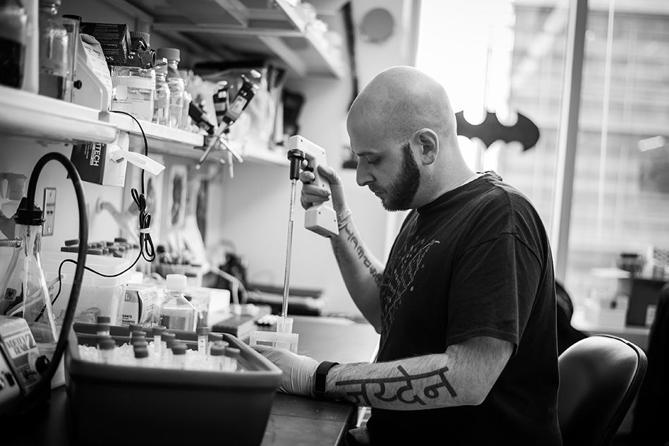
Driven by curiosity, former auto mechanic Ryan Kohn now pursues a PhD in biology.

Bridget E. Begg | Office of Graduate Education
December 18, 2017

The name of Ryan Kohn’s son, Jayden, is tattooed in Hindi on his left outer forearm. Other tattoos on his inner arms declare “Respect” and “Loyalty.” A Latin phrase balances the tableau on his right outer forearm: “Many fear their reputation. Few their conscience.”

Kohn may stand out in the corporate milieu of Kendall Square, but he feels home at MIT. No one has ever judged me,” he says. “For as rigorous scientifically and academically as MIT is, it can be such a laid-back place. I’ve always felt included, if I wanted to be.”

Kohn, now a PhD candidate in the Jacks Lab at MIT’s Koch Institute for Integrative Cancer Research, has overcome a challenging adolescence, colored by economic difficulties and punctuated by personal loss. These hardships developed in him a resilient curiosity that made an unexpected cultural match between MIT and Kohn, a father and former mechanic from Boyertown, Pennsylvania.

Compelled to seek answers

After being placed in an alternative high school outside of Philadelphia for insubordination, Kohn graduated with a 1.8 GPA. His son was born three years later, while Kohn worked for six and a half years as a mechanic and manager at a Dodge dealership. After losing his job during the Great Recession, he decided to go back to school, attending his local community college on a premed track before transferring to Kutztown University after two years.

Kohn attributes some of his troubled youth to early tragedy. His older sister, Nicole, died from sepsis when she was a senior in college, just 10 days after 9/11; on the morning of her funeral, Kohn’s grandfather passed away from colon cancer. Kohn felt compelled to understand why and how these illnesses happened to his loved ones, and found himself spending his time googling the immune system, the inflammatory response, and cancer.

This habit remained with him. Kohn recalls scouring the internet again and again to understand illness when it arose near him, from his own son’s immunoglobulin A deficiency to the early-onset multiple sclerosis of a friend. Though he admits he did not yet have the core scientific knowledge to fully grasp what he read at the time, Kohn says he needed, deeply, to try.

At Kutztown University, Kohn met his undergraduate mentor Angelika Antoni, a professor who taught both oncology and immunology. According to Kohn, Antoni constantly encouraged him to pursue his curiosity despite the college’s lack of laboratory resources. In fact, Antoni paid for laboratory reagents with her own credit card, while Kohn wrote his own grants and subscribed to well-known biology journals out of his own pocket because journal access was not available through Kutztown.

These challenges shaped Kohn as an experimental biologist, requiring him to precisely understand the mechanisms of experimental techniques in order to reconstruct them in the most creative and inexpensive ways possible. Perhaps most importantly, this small-college experience cultivated Kohn’s persistent curiosity.

Diving into cancer research

In his current position at the Jacks Lab, Kohn studies cancer immunotherapy, the use of a cancer patient’s own immune system to fight cancer cells. To do this, Kohn uses a mouse model of lung cancer that mimics the natural development of human cancer: Mutations identical to those found in many human cancers are triggered in the mouse, causing a tumor to arise that originates from the mouse’s own cells. These mice, like human cancer patients, have an immune system that can recognize the cancer as aberrant. Kohn’s work focuses modifying mouse immune cells to identify and attack a tumor.

Kohn is excited by the translational potential of his work, but also eagerly defends basic research at MIT when he encounters skepticism about its practicality in his conservative hometown.

Kohn often draws on metaphors in these types of conversations. He may leverage car talk, for example, to explain why there will never be a single cure for cancer: “So your ‘check engine’ light always presents the same way … but there’s literally a multitude of different things that can [cause] it. It could be a loose gas cap for the evaporative emissions system that set it off, it could be a misfire because of a bad spark plug, it could be a catalytic converter.”

Likewise, cancer can be caused by many possible biological errors that lead to an overgrowth of cells, Kohn explains. “So just like there will never be a cure for ‘check engine light,’ there will never be a [single] cure for cancer.”

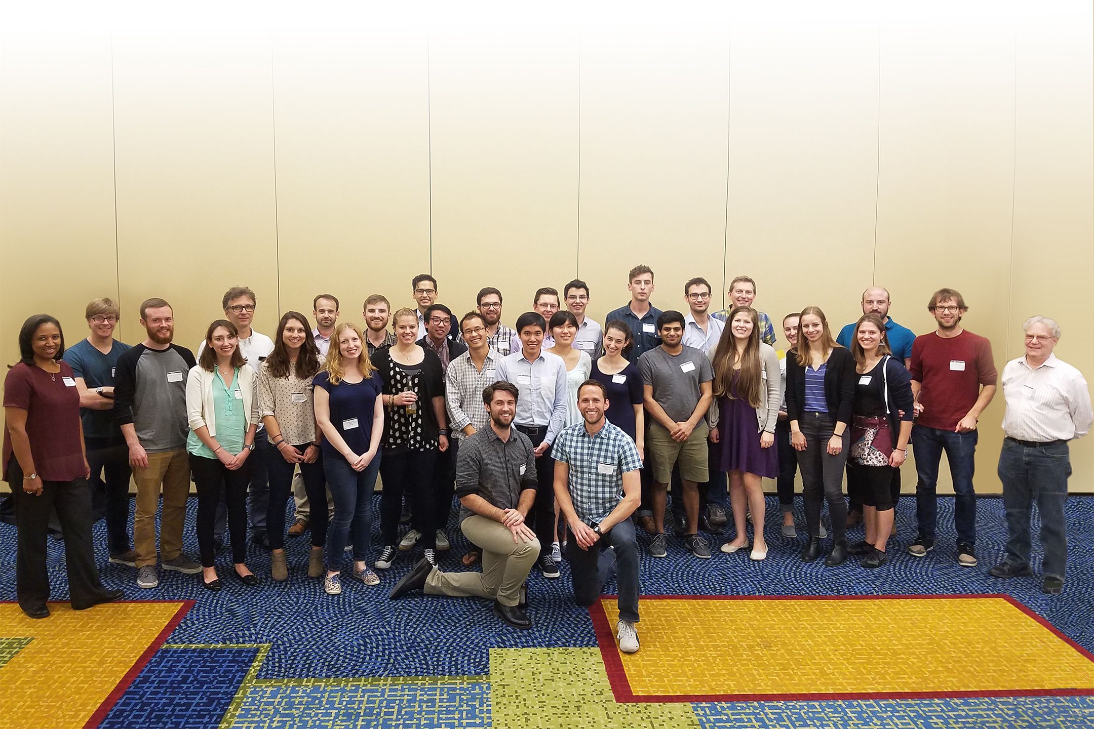
Perhaps unsurprisingly, Kohn embraces the scientific freedom of the research in his lab. His advisor, Tyler Jacks, director of the Koch Institute, an HHMI investigator, and a David H. Koch Professor of Biology at MIT, is frequently in high demand, but Kohn says he has felt fully supported in his work — including in the bold ideas and unconventional projects he undertakes in his free time.

Jacks remains accessible despite his busy schedule, according to Kohn, and his emphasis on mentorship has inspired the postdocs in the lab to mentor the graduate students. The Jacks Lab also enjoys a thriving social environment. Kohn regularly attends casual weekend parties held by his labmates, and every other year Jacks organizes a cross-campus themed scavenger hunt for which the whole lab dresses in elaborate costumes.

“Real conversations about ideas”

Outside of lab, Kohn calls himself a homebody and prefers to relax after a full day, often with a beer and a movie. He spends much of this down time with his partner Ruthlyn, whether they are exploring the Boston area or talking with friends and colleagues at local pubs.

Kohn speaks about these conversations with genuine excitement: “You meet so many different people, every religion, every gender identity, every country, every language, and you just meet these people and you get to have these cool conversations … these real conversations about ideas. Because that’s really what you want, right?”

He enthusiastically notes that, in contrast to his largely homogenous hometown, more than 200 countries are represented at MIT. Kohn says the diversity and ideals of MIT reflect his own worldview.

Despite his deep sense of belonging on campus, leaving home did lay an exceptional burden on Kohn: Twelve-year-old Jayden remains in Pennsylvania with his mother, over 300 miles away.

Kohn speaks about his son with immense pride, describing Jayden as not only an extremely talented baseball player, but as a positive, energetic, and deeply mature young person. Kohn recounts with admiration, and a trace of relief, that despite the difficulty of the distance, Jayden said his father’s coming to MIT was the right thing to do.

As for his own parents, Kohn finally feels that all the headaches he has given them over the years have been worthwhile. His intense desire for knowledge has driven him through many obstacles, connected him with like minds from all over the world, and still shows no signs of waning.

Kohn has a reputation in his lab for asking questions, big and small. Asked if he’s ever afraid to admit what he doesn’t know, he says no: “I want to know … and that’s really what it comes down to.”

Epigenetic rheostat helps uncover how gene regulation is inherited and maintained
December 14, 2017

While our genome contains a vast repertoire of genes that are responsible for virtually all of the cellular and developmental processes life requires, it is the complex dance of regulating their expression that is vital for genetic programs to be executed successfully. Genes must be turned on and off at appropriate times or, in some cases, never turned on or off at all.

Methylation—the addition of chemical tags to DNA—typically reduces the expression of methylated genes. In many cases, DNA methylation can be thought of as roadblocks on a gene. The more methylated a gene is, the less likely it is that it will be active. Such genetic demarcations are critical to ensure that genes involved in particular stages of development are active at the right time, for example. Methylation is essential for proper cellular function, and its dysregulation is associated with diseases, such as cancer in humans. Despite its importance, little is known about how critical methylation patterns are inherited or maintained. Whitehead Institute Member Mary Gehring and her lab have identified a mechanism important for maintaining methylation, that when disrupted, results in the demethylation of large sections of the Arabidopsis plant’s genome. Their work is described this week in the journal Nature Communications.

Using an unusual gene in the plant Arabidopsis, Gehring is teasing apart the mechanisms that underpin methylation. By breaking this unique gene’s “circuit,” Gehring and Ben Williams, a postdoctoral researcher in her lab, have gained important insights into how methylation is maintained, including a surprising finding that previously erased methylation can be restored under certain circumstances.

In order to better understand methylation’s heritability, Gehring and Williams looked closely at an anomaly, the ROS1 gene in Arabidopsis plants, which encodes a protein that removes methylation from its own gene as well as others. Previously, Gehring and Williams had determined that ROS1 methylation actually functions in the complete opposite way from the existing paradigm—unlike most genes, when a short section of this gene is methylated, the gene is actually activated instead of inactivated. Conversely, if it is methylated, the gene is turned on. As a result, ROS1 can act as a rheostat for the Arabidopsis genome: As methylation increases, ROS1 turns on and begins removing methyl groups, and as methylation decreases, ROS1 shuts off and reduces its demethylating activity.

In the current research, Williams altered methylation at ROS1 so that its activity was uncoupled from methylation levels in the genome, in order to see what effects such a change would have on methylation throughout the entire genome. When he analyzed the plants’ methylation, it was haywire. Methylation was lost throughout the genome and progressively decreased in subsequent generations, except in a particular part of the genome called the heterochromatin—genomic areas that are strongly repressed. Interestingly, Williams found that, despite the alteration of the ROS1regulatory circuit, these heterochromatic sections of the genome actually regain their methylation and approach full methylation by the fourth generation— the same time point by which the rest of the genome has lost much of its methylation .

The researchers determined that the ROS1 circuit they uncovered is important for methylation homeostasis because it causes heritable loss of methylation when disrupted.  And yet methylation returns at some locations, albeit not immediately, suggesting that Arabidopsis enlists multiple mechanisms to maintain methylation homeostasis. Gehring and Williams are intrigued by that delay in remethylation and are working to identify its cause as well as other mechanisms that may also be at work regulating this critical process.

This work was supported by the National Institute of General Medical Sciences of the National Institutes of Health (R01GM112851).

Written by Nicole Giese Rura
* * *
Mary Gehring’s primary affiliation is with Whitehead Institute for Biomedical Research, where her laboratory is located and all her research is conducted. She is also an associate professor of biology at Massachusetts Institute of Technology.
* * *
Full Citation:
“Stable transgenerational epigenetic inheritance requires a DNA methylation-sensing circuit”
Nature Communications, December 14, 2017.
Ben P. Williams (1) and Mary Gehring (1,2).
1. Whitehead Institute for Biomedical Research, Cambridge, MA 02142
2. Department of Biology, Massachusetts Institute of Technology, Cambridge, MA 02139
Celebrating a decade of interdisciplinary microbiology

The Microbiology Graduate PhD Program spans 50 labs across 10 departments and divisions, offering a broad approach to microbial science and engineering.

Raleigh McElvery | Department of Biology
December 12, 2017

Ten years ago, MIT launched the Microbiology Graduate PhD Program. Today, it boasts 28 alumni and 33 current students, and offers a broad, interdisciplinary approach to microbial science and engineering. Between five and eight trainees enroll each year and can choose among more than 50 labs spanning 10 departments and divisions — from biology and biological engineering to chemical engineering and physics.

Many diverse disciplines are rooted in microbiology. Basic scientists use microorganisms as model systems to understand fundamental biological processes. Engineers leverage microorganisms to create new manufacturing processes and energy sources. Even ecologists, biomedical researchers, and earth scientists dedicate their careers to investigating the role of microbes in our ecosystems, on our bodies, and on our planet. In sum, the study of microbiology permeates so many research areas that no single department at MIT could house them all.

The idea for an interdisciplinary microbiology program first came to Alan Grossman, head of the Department of Biology, while he was recovering from a heart transplant back in 2006.

“There were people scattered all over MIT who were doing microbial science and engineering, but there was no mechanism to connect them or give students outside those departments easy access to the labs,” Grossman says. “I began by talking to a few faculty members in order to gauge general interest, before pitching it to a handful of department heads and forming a committee. Everyone was very excited about it, and it really grew from the ground up.”

The Committee on Graduate Programs approved his proposal in May 2007, and the first cohort of eight students began in the fall of 2008. Martin Polz, co-director since 2015 and professor of civil and environmental engineering, sat on Grossman’s initial committee.

“MIT’s program is unique from most other microbiology programs because it’s so interdisciplinary,” Polz says. “Many microbiology programs across the country are associated with medical schools and focused primarily on pathogenesis. The students who apply here really appreciate the breadth of our program, and it has become a fixture at MIT over the years.”

Kristala Prather, co-director since 2013 and professor of chemical engineering, said the program also provides an opportunity to bring life scientists and engineers together to tackle research questions.

“I find there is a difference in the way engineers and scientists approach research problems,” Prather says. “Each approach has rigor, but having both perspectives breeds a richer set of discussions than just hearing from one discipline alone.”

During the past 10 years, Prather has watched a thriving and diverse community unite, spurred by a common interest in the microbial world.

Nathaniel Chu, who matriculated in 2014, said the program allows him to sample different disciplines while still maintaining a close affiliation with his advisor’s home department, Biological Engineering. As part of Eric Alm’s lab, Chu studies the interaction between the gut microbiome and immune system, and how imbalances in that delicate relationship can trigger conditions such as Type 2 diabetes, obesity, and inflammatory bowel disease.

“The program provides flexibility to explore your research interests, and my advisor has given me a lot of space to conceive and manage my own projects,” Chu says. “I’ve been able to interact with a diverse set of individuals within the microbiology circle, including clinical partners, immunologists, geneticists, bioinformaticians, and computational biologists.”

Jacquin Niles, incoming co-director, was a junior faculty member in Department of Biological Engineering when Grossman first proposed the idea. He says the students — past and present — are the heart of the program.

“A lot has changed over the 10 years the program has been in existence, but the caliber of students has remained consistent,” Niles says. “If I had to emphasize any particular aspect of the program, the students would be numbers one, two, and three. Each generation has been exceptional, and they are all very much on top of their research game.”

Michael Laub, co-director from 2012 to 2015 and professor of biology, adds that the early students deserve much credit for the program’s success. “They took a chance on a brand-new initiative, and as a result we ended up attracting ambitious, risk-taking, and creative folks who really paved the way for current students,” he says.

Alumni pursue a variety of careers, ranging from academia to industry. Some join existing institutions or companies. Others start their own.

Mark Smith PhD ’14 was a member of the second graduating class. Like Chu, he was one of Alm’s advisees, studying networks of gene exchange within the human microbiome, and building statistical models to determine the role of environment in various gut-related diseases. Smith went on to co-found a nonprofit organization known as OpenBiome, harnessing the microbiome to cure recurrent Clostridium difficile infections. In 2016, he co-founded another company, Finch Therapeutics Group, focused on scaling and commercializing clinical treatments for diseases rooted in the microbiome. In 2017, he was named to the Forbes 30 Under 30 list for science.

“OpenBiome and Finch Therapeutics were really a translation of the initial work that was done through the microbiology program, and a step toward developing those tools to improve human health,” Smith says. “The program taught me the foundational work I’ve come to rely on in almost every aspect of my job today.”

Like Smith, Jacob Rubens PhD ’16 aims to apply his training at MIT to help develop new products. After working in Timothy Lu’s lab — straddling the realms of biological engineering and electrical engineering — Rubens joined Flagship Pioneering, a company that starts, funds, and runs breakthrough biotechnology startups in Cambridge, Massachusetts. Rubens was also named to the Forbes 30 Under 30 list for science in 2017.

During the six years that Rubens was at MIT, he watched the microbiology cohort grow from roughly 20 to a force permeating more labs across campus than he could count.

“It’s heartwarming to see people bringing a microbiological perspective into all these different spaces, and influencing cutting-edge research across the Institute,” he says. “As a microbiology student, you become an integrator and synthesizer of many different viewpoints, and a node to foster cross-talk between disciplines.”

As Niles prepares to assume the role of co-director in July 2018 and usher in the program’s second decade, he intends to maintain its integrity and structure.

“The program has matured into what it is today thanks to a lot of previous, careful thought,” he says. “The students have indicated that there is a lot of value in the structure that we’ve refined over the years, and so my goal is to continue that positive momentum.”

Researchers establish long-sought source of ocean methane

An abundant enzyme in marine microbes may be responsible for production of the greenhouse gas.

Anne Trafton | MIT News Office
December 7, 2017

Industrial and agricultural activities produce large amounts of methane, a greenhouse gas that contributes to global warming. Many bacteria also produce methane as a byproduct of their metabolism. Some of this naturally released methane comes from the ocean, a phenomenon that has long puzzled scientists because there are no known methane-producing organisms living near the ocean’s surface.

A team of researchers from MIT and the University of Illinois at Urbana-Champaign has made a discovery that could help to answer this “ocean methane paradox.” First, they identified the structure of an enzyme that can produce a compound that is known to be converted to methane. Then, they used that information to show that this enzyme exists in some of the most abundant marine microbes. They believe that this compound is likely the source of methane gas being released into the atmosphere above the ocean.

Ocean-produced methane represents around 4 percent of the total that’s discharged into the atmosphere, and a better understanding of where this methane is coming from could help scientists better account for its role in climate change, the researchers say.

“Understanding the global carbon cycle is really important, especially when talking about climate change,” says Catherine Drennan, an MIT professor of chemistry and biology and Howard Hughes Medical Institute Investigator. “Where is methane really coming from? How is it being used? Understanding nature’s flux is important information to have in all of those discussions.”

Drennan and Wilfred van der Donk, a professor of chemistry at the University of Illinois at Urbana-Champaign, are the senior authors of the paper, which appears in the Dec. 7 online edition of Science. Lead authors are David Born, a graduate student at MIT and Harvard University, and Emily Ulrich, a graduate student at the University of Illinois at Urbana-Champaign.

Solving the mystery

Many bacteria produce methane as a byproduct of their metabolism, but most of these bacteria live in oxygen-poor environments such as the deep ocean or the digestive tract of animals — not near the ocean’s surface.

Several years ago, van der Donk and University of Illinois colleague William Metcalf found a possible clue to the mystery of ocean methane: They discovered a microbial enzyme that produces a compound called methylphosphonate, which can become methane when a phosphate molecule is cleaved from it. This enzyme was found in a microbe called Nitrosopumilus maritimus, which lives near the ocean surface, but the enzyme was not readily identified in other ocean microbes as one would have expected it to be.

Van der Donk’s team knew the genetic sequence of the enzyme, known as methylphosphonate synthase (MPnS), which allowed them to search for other versions of it in the genomes of other microbes. However, every time they found a potential match, the enzyme turned out to be a related enzyme called hydroxyethylphosphonate dioxygenase (HEPD), which generates a product that is very similar to methylphosphonate but cannot be cleaved to produce methane.

Van der Donk asked Drennan, an expert in determining chemical structures of proteins, if she could try to reveal the structure of MPnS, in hopes that it would help them find more variants of the enzyme in other bacteria.

To find the structure, the MIT team used X-ray crystallography, which they performed in a special chamber with no oxygen. They knew that the enzyme requires oxygen to catalyze the production of methylphosphonate, so by eliminating oxygen they were able to get snapshots of the enzyme as it bound to the necessary reaction partners but before it performed the reaction.

The researchers compared the crystallography data from MPnS with the related HEPD enzyme and found one small but critical difference. In the active site of both enzymes (the part of the protein that catalyzes chemical reactions), there is an amino acid called glutamine. In MPnS, this glutamine molecule binds to iron, a necessary cofactor for the production of methylphosphonate. The glutamine is fixed in an iron-binding orientation by the bulky amino acid isoleucine, which is directly below the glutamine in MPnS. However, in HEPD, the isoleucine is replaced by glycine, and the glutamine is free to rearrange so that it is no longer bound to iron.

“We were looking for differences that would lead to different products, and that was the only difference that we saw,” Born says. Furthermore, the researchers found that changing the glycine in HEPD to isoleucine was sufficient to convert the enzyme to an MPnS.

An abundant enzyme

By searching databases of genetic sequences from thousands of microbes, the researchers found hundreds of enzymes with the same structural configuration seen in their original MPnS enzyme. Furthermore, all of these were found in microbes that live in the ocean, and one was found in a strain of an extremely abundant ocean microbe known as Pelagibacter ubique.

“This exciting result builds on previous, related studies showing that the metabolism of the methylphosphonate can lead to the formation of methane in the oxygenated ocean. Since methane is a potent greenhouse gas with poorly understood sources and sinks in the surface ocean, the results of this study will serve to facilitate a more comprehensive understanding of the methylphosphonate cycle in nature,” says David Karl, a professor of oceanography at the University of Hawaii, who was not involved in the research.

It is still unknown what function the MPnS enzyme and its product serve in ocean bacteria. Methylphosphonates are believed to be incorporated into fatty molecules called phosphonolipids, which are similar to the phospholipids that make up cell membranes.

“The function of these phosphonolipids is not well-established, although they’ve been known to be around for decades. That’s a really interesting question to ask,” Born says. “Now we know they’re being produced in large quantities, especially in the ocean, but we don’t actually know what they do or how they benefit the organism at all.”

Another key question is how the production of methane by these organisms is influenced by environmental conditions in the ocean, including temperature and pollution such as fertilizer runoff.

“We know that methylphosphonate cleavage occurs when microbes are starved for phosphorus, but we need to figure out what nutrients are connected to this, and how is that connected to the pH of the ocean, and how is it connected to temperature of the ocean,” Drennan says. “We need all of that information to be able to think about what we’re doing, so we can make intelligent decisions about protecting the oceans.”

The research was funded by the National Institutes of Health and the Howard Hughes Medical Institute.

Rethinking transcription factors and gene expression

Study shows that, like proteins, genomes must fold appropriately to function properly and that some transcription factors provide the structural support.

Nicole Giese Rura | Whitehead Institute
December 7, 2017

Transcription — the reading of a segment of DNA into an RNA template for protein synthesis — is fundamental for nearly all cellular processes, including growth, responding to stimuli, and reproduction. Now, Whitehead Institute researchers have upended our understanding of how transcription is controlled and the role of transcription factors in the process.

The paradigm shift, described in an article online on Dec. 7 in the journal Cell, hinges on a small protein that plays a key role in genome structure and gives us new insights into how changes in the control of transcription and gene expression can lead to disease.

Transcription has several important players that must all be in the right place at the right time: the transcription machinery, transcription factors, promoters, and enhancers.  According to the existing model, transcription factors are proteins that bind to enhancer regions of the genome and recruit the transcription machinery to the promoter DNA regions, which then initiate the genes’ transcription.

“We’ve always assumed that the role of transcription factors was to recruit the transcription machinery to genes to turn them on or turn them off,” says Richard Young, a Whitehead Insistute member and professor of biology at MIT. “But we never imagined that the transcription factors we’ve studied for three decades actually contribute to the genome’s structure. And as a consequence, they regulate genes. So we now look at genomes like proteins: They have to fold up appropriately in order to control genes.”

Scientists have known that the genome’s structure — how it bends and folds — is essential for efficiently compressing two meters of DNA into each human cell, which is the equivalent of packing a strand ten football fields long into a space the size of a marble. Yet until recently, researchers have not had the tools necessary to appreciate this architecture’s importance in fine control of gene expression or study the genome’s structure at sites ready for transcription.

In 2014, Young and his lab determined that portions of the genome reside in loop-based structures, creating insulated neighborhoods that bring enhancers, promoters, and genes into close proximity. Each loop is tied at the top by a pair of molecules, called CTCF, that are bound together. This structure is essential for proper gene control: If the loop structure is broken, gene expression is altered, and cells can become diseased or die.

In the current research, Young along with co-first authors Abraham Weintraub and Charles Li took a closer look at a protein that is well known but not well understood: Yin Yang 1 (YY1). Hundreds of scientific papers have linked YY1 dysfunction to diseases such as viral infections, cancer, and arthritis, and yet the studies produced seemingly contradictory observations of YY1’s function.

According to Young and colleagues, YY1 is a unique transcription factor that occupies both enhancers and promoters, is essential for cell survival, and is found in almost every cell type in humans and mice. Like CTCF, YY1 can also pair with itself and bind to DNA to form loops that enhance DNA transcription.

“YY1 is expressed broadly, and it is necessary for establishing enhancer-promoter loops in multiple cell types,” says Weintraub. “That’s its job, not recruiting the transcription apparatus. When the structure created by YY1 is removed, the genome is no longer folded properly, gene control is lost and transcription of the affected genes is significantly diminished, which can cause dysfunction.”

This model of YY1’s function could account for its association with a number of disparate diseases. Earlier this year, scientists reported YY1 syndrome — a genetic syndrome causing cognitive disabilities in people with mutations in their YY1 gene.

According to Young, YY1 is probably not the only transcription factor with this loop-forming role, and his lab will be searching for additional factors with similar functions.

“YY1 is most likely just the first one, and there are probably a bunch of collaborators that have similar roles,” says Young. “Instead of the classic function that we thought these transcription factors had — interacting with the transcription apparatus and giving instructions on how much or how little of a gene’s transcript to produce — they are bringing together regulatory elements with the gene. The whole job of these transcription factors is just making structure. We are realizing that the things that form physical structures are much more important than we had appreciated.”

The researchers’ work was supported by the National Institutes of Health, the Ludwig Graduate Fellowship funds, the National Science Foundation, the American Cancer Society, a Margaret and Herman Sokol Postdoctoral Award, the Damon Runyon Cancer Research Foundation, and the Cancer Research Institute. The Whitehead Institute has filed a patent application based on this study.

Revealing an imperfect actor in plant biotechnology

Whitehead Institute researchers detect the chemical mistakes of a common herbicide-resistance enzyme, then successfully re-engineer it for enhanced precision.

Nicole Davis | Whitehead Institute
November 29, 2017

A research team led by MIT’s Whitehead Institute for Biomedical Research has harnessed metabolomic technologies to unravel the molecular activities of a key protein that enables plants to withstand a common herbicide.

Their findings reveal how the protein — a kind of catalyst or enzyme first isolated in bacteria and introduced into plants such as corn and soybeans in the 1990s — can sometimes act imprecisely, and how it can be successfully re-engineered to be more precise. The new study, which appears online in the journal Nature Plants, raises the standards for bioengineering in the 21st century.

“Our work underscores a critical aspect of bioengineering that we are now becoming technically able to address,” says senior author Jing-Ke Weng, a member of the Whitehead Institute and an assistant professor of biology at MIT. “We know that enzymes can behave indiscriminately. Now, we have the scientific capabilities to detect their molecular side effects, and we can leverage those insights to design smarter enzymes with enhanced specificity.”

Plants provide an extraordinary model for scientists to study how metabolism changes over time. Because they cannot escape from predators or search for new food sources when supplies run low, plants must often grapple with an array of environmental insults using what is readily available — their own internal biochemistry.

“Although they appear to be stationary, plants have rapidly evolving metabolic systems,” Weng explains. “Now, we can gain an unprecedented view of these changes because of cutting-edge techniques like metabolomics, allowing us to analyze metabolites and other biochemicals on a broad scale.”

Key players in this evolutionary process, and a major focus of research in Weng’s laboratory, are enzymes. Traditionally, these naturally occurring catalysts have been viewed as mini-machines, taking the proper starting material (or substrate) and flawlessly converting it to the correct product. But Weng and other scientists now recognize that they make mistakes, often by latching on to an unintended substrate.

“This concept, known as enzyme promiscuity, has a variety of implications, both in enzyme evolution and more broadly, in human disease,” Weng says.

It also has implications for bioengineering, as Bastien Christ, a postdoctoral fellow in Weng’s laboratory, and his colleagues recently discovered.

Christ, then a graduate student in Stefan Hörtensteiner’s lab at the University of Zurich in Switzerland, was studying a particular strain of the flowering plant Arabidopsis thaliana as part of a separate project when he made a puzzling observation. He found that two biochemical compounds were present at unusually high levels in the plant’s leaves.

Strangely, these compounds (called acetyl-aminoadipate and acetyl-tryptophan) weren’t present in any of the normal, so-called wild type plants. As he and his colleagues searched for an explanation, they narrowed in on the source: an enzyme, called BAR, that was engineered into the plants as a kind of chemical beacon, enabling scientists to more readily study them.

But BAR is more than just a tool for scientists. It is also one of the most commonly deployed traits in genetically modified crops such as soybeans, corn, and cotton, enabling them to withstand a widely-used herbicide (known as phosphinothricin or glufosinate).

For decades, scientists have known that BAR, originally isolated from bacteria, can render the herbicide inactive by tacking on a short string of chemicals, made of two carbons and one oxygen (also called an acetyl group). As the researchers describe in their Nature Plants paper, BAR has a promiscuous side, and can work on other substrates, too, such as the amino acids tryptophan and aminoadipate (a lysine derivative).

That explains why they can detect the unintended products (acetyl-tryptophan and acetyl-aminoadipate) in crops genetically engineered to carry BAR, such as soybeans and canola.

Their research included detailed studies of the BAR protein, including crystal structures of the protein bound to its substrates. This provided them with a blueprint for how to strategically modify BAR to make it less promiscuous, and favor only the herbicide as a substrate and not the amino acids. Christ and his colleagues created several versions that lack the non-specific activity of the original BAR protein.

“These are natural catalysts, so when we borrow them from an organism and put them into another, they may not necessarily be perfect for our purposes,” Christ says. “Gathering this kind of fundamental knowledge about how enzymes work and how their structure influences function can teach us how to select the best tools for bioengineering.”

There are other important lessons, too. When the BAR trait was first evaluated by the U.S. Food and Drug Administration (FDA) in 1995 for use in canola, and in subsequent years for other crops, metabolomics was largely non-existent as a technology for biomedical research. Therefore, it could not be applied toward the characterization of genetically engineered plants and foods, as part of their regulatory review. Nevertheless, acetyl-aminoadipate and acetyl-tryptophan, which are normally present in humans, have been reviewed by the FDA and are safe for human and animal consumption.

Weng and his colleagues believe their study makes a strong case for considering metabolomics analyses as part of the review process for future genetically engineered crops.

“This is a cautionary tale,” Weng says.

The work was supported by the Swiss National Science Foundation, the EU-funded Plant Fellows program, the Pew Scholar Program in the Biomedical Sciences, and the Searle Scholars Program.

Muscle plays surprising role in tissue regeneration

Whitehead Institute researchers have pinpointed distinct muscle subsets that orchestrate and pattern regrowth.

Nicole Davis | Whitehead Institute
November 22, 2017

Researchers at the Whitehead Institute have illuminated an important role for different subtypes of muscle cells in orchestrating the process of tissue regeneration.

In a paper appearing online today in Nature, they reveal that a subtype of muscle fibers in flatworms is required for triggering the activity of genes that initiate the regeneration program. Notably, in the absence of these muscles, regeneration fails to proceed. Another type of muscle, they report, is required for giving regenerated tissue the proper pattern — for example, forming one head instead of two.

“One of the central mysteries in organ and tissue regeneration is: How do animals initiate all of the cellular and molecular steps that lead to regeneration?” says senior author Peter Reddien, a member of Whitehead Institute, professor of biology at MIT, and investigator with the Howard Hughes Medical Institute. “We’ve helped answer this question by revealing a surprising molecular program that operates within a subgroup of muscle cells that helps establish the molecular information required for proper tissue regeneration after injury.”

For more than a decade, Reddien and the researchers in his laboratory have studied the biological mechanisms that underlie regeneration in a tiny flatworm called planarians. These worms possess some impressive regenerative capabilities: When sliced in two, each piece of the worm can regrow the body parts needed to form two complete organisms. In previous studies, Reddien’s team identified a set of always-on genes, known as position control genes (PCGs), that provide cells with region-specific instructions, like a set of GPS coordinates, that tell cells where they are in the body, and thus what body part to regenerate. Interestingly, PGCs are active in planarian muscle cells, suggesting muscle may play a major role in the regeneration process.

“This discovery raised a lot of questions about how muscle participates in this process,” Reddien says.

In planarians, there are a handful of muscle cell types. For example, if you imagine the worms as simple cylindrical tubes, there are longitudinal muscle fibers, which run head-to-tail along the tubes’ long axis. There are also circular fibers, which are perpendicular to the longitudinal fibers and hug the tubes’ outer circumference.

To assess the roles of these different muscle cell types in regeneration, first author Lucila Scimone and her colleagues needed a method to selectively remove them. When myoD, a gene found specifically in the longitudinal fibers, is inhibited, those fibers fail to form. Similarly, the nkx1-1 gene marks the circular fibers, and when its function is reduced, they do not develop. Using these genes as molecular scalpels, Scimone and her co-authors could test the effects of ablating these distinct muscle groups on regeneration.

Surprisingly, when the longitudinal fibers were removed, the results were dramatic. The worms live quite normally, but when they are injured (the head removed, for example) they cannot regenerate the missing parts.

“This is an amazing result; it tells us that these longitudinal fibers are essential for orchestrating the regeneration program from the very beginning,” says Scimone, a scientist in Reddien’s lab.

As the researchers dug deeper into the finding, they learned that the functions of two critical genes are disrupted when longitudinal fibers are missing. These genes, called notum and follistatin, are known for their fundamental roles in regeneration, controlling head-versus-tail decisions and sustained cell proliferation, respectively, following tissue injury.

In addition to this essential role for longitudinal fibers, the research team also uncovered a key role for circular fibers. When these muscles are missing, planarians are able to regenerate missing body parts, but what regrows is abnormally patterned. For example, two heads may be regenerated within a single outgrowth, instead of one.

These results underscore an important and previously unappreciated role for muscle, widely known for its contractile properties, in instructing the tissue regeneration program. The Whitehead researchers will continue to probe the role of different muscle cell types in planarian regeneration and also explore whether other animals with regenerative capabilities possess a similar muscle-localized program for conferring positional information.

“It’s hard to understand what limits humans’ abilities to regenerate and repair wounds without first knowing what mechanisms are enabling some animals, like planarians, to do it so amazingly well,” Reddien says.

This work was supported by the National Institutes of Health, Howard Hughes Medical Institute, and the Eleanor Schwartz Charitable Foundation.

Of highways, engines, and chromosomes

Whitehead researchers unravel fundamental molecular machinery that propels chromosome movement

November 16, 2017

Each day, billions of cells in the human body undergo a vital ritual, wherein one cell divides to form two. This process, known as cell division, is as beautiful as it is essential, undergirding the body’s growth in times of both health and disease. Despite the fact that cell division (or “mitosis”) has been a basic topic in high school biology classes for the past 70 years, the mechanisms by which cells conduct this critical event remain poorly understood. In particular, there are lingering uncertainties about how chromosomes — large units of DNA that include our genes — get properly allocated so that both daughter cells receive intact, complete copies of their genetic blueprint.

“People have been watching chromosomes move, align, and segregate for more than a century — it’s such a fundamental aspect of biology,” says Iain Cheeseman, a member of the Whitehead Institute for Biomedical Research and an associate professor of biology at Massachusetts Institute of Technology. “It’s also much more elegant and complicated than we ever anticipated.”

Cheeseman and members of his Whitehead laboratory have discovered many of the molecular movers and shakers that ensure chromosomes get to the right place at the right time. These components assemble together — like the parts of an engine — to establish robust connections with chromosomes and ultimately power their movement within cells. “The most important unanswered question in the field is how do these components work together? That is, how do you build a machine that is more than the sum of its parts?” he says.

Now, in two recent papers in the journals eLife and Current Biology, Cheeseman and his colleagues help shed light on this central question.

Back to the drawing board

As recently as 15 years ago, scientists assumed that in dividing cells, chromosomes move the way many other cellular objects move — transported by tiny molecular motors. These mini-motors are specifically designed to travel along roads made from rod-like structures called microtubules. Like a car cruising on a highway, they can carry cargo over long distances. Although such microtubule-based vehicles seemed a logical suspect, when scientists inactivate them in human cells, chromosomes can still move and segregate just fine. So something else must contribute the necessary molecular muscle.

“We basically had to throw out the major hypothesis that was out there and go back to the drawing board,” says Cheeseman.

Over the last several years, he and other scientists in the field have helped develop a clearer view of how this process works and who the key players are that enable a very different type of movement. Consider a car sitting motionless on a highway. Rather than revving its own engine to generate motion, the highway itself moves, shrinking or growing while the car hangs on. “It is a radically different way of imagining this movement process,” says Cheeseman. “An important part of my lab’s mission has been to figure out how do you build a motor like that? What are the factors required and how do they act?”

Of course, the highway — or more precisely, the microtubule — must grow and shrink as needed. But even more important, there must also be an apparatus that can enable chromosomes to hold on to such a dynamic structure. As Cheeseman and his colleagues have uncovered, this coupling requires a suite of highly sophisticated molecular players.

Building an unusual machine

Cheeseman and his laboratory have focused on three key groups or complexes of proteins that play essential roles in chromosome segregation in human cells. These components assemble together to form a kind of molecular tether point on chromosomes (called the kinetochore) where microtubules attach.

Diagram of the kinetochore/microtube interface
Courtesy: David Kern/Whitehead Institute

Among this trio of parts, the most critical is the Ndc80 complex. “It is the major connection between the kinetochore and the microtubule,” says Cheeseman. As a postdoc, he discovered the biochemical properties that enable this Ndc80 complex to grab on to microtubules, research that sparked his lab’s quest to study the various pieces of the kinetochore machinery and how they work.

While Ndc80 forms a critical linkage, it lacks some key capabilities, like processivity — the ability to keep ahold of something while it moves. In a series of papers, one published in 2009, another in 2012, and a new one in Current Biology, Cheeseman’s team revealed that Ska1 can perform this crucial function. That is, it has the biochemical capacity to enable chromosomes to hang onto microtubules while they grow and as they shrink, an activity that it can impart to the Ndc80 complex. “These are pretty powerful properties,” says Cheeseman.

Diving even deeper into Ska1’s bag of tricks, Julie Monda and Ian Whitney, lead authors of the Current Biology paper, went on to decipher the precise molecular features that enable the complex’s dynamic capabilities, uncovering multiple surfaces that associate with microtubules and enable Ska1 to undergo something akin to molecular somersaults. These somersaults are what allow it to maintain its association with microtubules.

The third complex, Astrin-SKAP, also plays a unique role. As Cheeseman’s team described in their recent eLife paper, led by first author David Kern, it serves as a master stabilizer — like a final drop of superglue to secure everything in place. “It’s the last thing that comes in and helps lock down these interactions, so you can stabilize and maintain them,” says Cheeseman.

Uncovering its role was no easy feat. Astrin-SKAP proved to be rather temperamental biochemically, complicating Kern’s efforts to purify and manipulate it in the laboratory. Also, as he and his colleagues discovered, a tiny piece of the structure had previously gone undetected; it works alongside the rest of the complex and is required for its normal function. Perhaps the most important revelation was that Astrin-SKAP doesn’t just work alone — it also coordinates with Ndc80. “This is an important finding for how we think about these components as a whole,” saysCheeseman.

Although questions remain about how all of these parts work together and how other pieces may come into play, Cheeseman believes these studies provide an exciting start. “The first human kinetochore component wasn’t identified until 1987, when many of the other key processes in the cell had already been intensively studied,” he says. “There are so many exciting questions that are accessible now that we have these tools and knowledge.”

Now, he and his colleagues will continue to meld approaches in cell biology and biochemistry to decode the inner workings of the kinetochore. That includes understanding how the various components operate not only in individual cells, but also in multicellular organisms.

“We are currently thinking a lot about the physiological context— that is, what matters to cells and to an organism,” says Cheeseman. “The work that our lab and others have conducted over the past two decades has given us a molecular handle on this problem. I’m excited to be able to apply these finding to understanding the ways that cell division is altered in development and in disease states.”

Written by Nicole Davis
* * *
Iain Cheeseman’s primary affiliation is with Whitehead Institute for Biomedical Research, where his laboratory is located and all his research is conducted. He is also an associate professor of biology at Massachusetts Institute of Technology.
* * *
Full citations:
“Astrin-SKAP complex reconstitution reveals its kinetochore interaction with microtubule-bound Ndc80”
eLife 2017;6:e26866 August 25, 2017. DOI: 10.7554/eLife.26866
David M Kern (1,2), Julie K Monda (1,2), Kuan-Chung Su (1), Elizabeth M Wilson-Kubalek (3), and Iain M Cheeseman (1,2).
1. Whitehead Institute for Biomedical Research, 455 Main Street, Cambridge, MA 02142, USA
2. Department of Biology, Massachusetts Institute of Technology, Cambridge, MA 02142, USA
3. Department of Cell Biology, The Scripps Research Institute, La Jolla, CA 92037, USA
“Microtubule tip tracking by the spindle and kinetochore protein Ska1 requires diverse tubulin-interacting surfaces”
Current Biology, online November 16, 2017. DOI: 10.1016/j.cub.2017.10.018
Julie K. Monda (1,2,6), Ian P. Whitney (1,6), Ekaterina V. Tarasovetc (3,4), Elizabeth Wilson-Kubalek (5), Ronald A. Milligan (5), Ekaterina L. Grishchuk (3), and Iain M. Cheeseman (1,2).
1. Whitehead Institute for Biomedical Research, 455 Main Street, Cambridge, MA 02142, USA
2. Department of Biology, Massachusetts Institute of Technology, Cambridge, MA 02142, USA
3. Department of Physiology, Perelman School of Medicine, University of Pennsylvania, Philadelphia, PA 19104, USA
4. Center for Theoretical Problems of Physicochemical Pharmacology, Russian Academy of Sciences, Moscow, Russia
5. Department of Cell Biology, The Scripps Research Institute, La Jolla, CA 92037, USA
6. These authors contributed equally
A scientific approach to writing fiction

Megan Miranda '02 graduated from MIT intent on pursuing a career in biotechnology. Instead, she became a New York Times best-selling author.

Jay London | MIT Alumni Association
November 15, 2017

Megan Colpitts Miranda ’02, who graduated from MIT with a degree in biology, intended to pursue a career in biotechnology. Instead, she became a successful fiction author whose book, “All the Missing Girls,” is a New York Times best-seller. Both careers share a trial-and-error approach to achieving success, she believes.

“There are a lot of similarities in the process,” Miranda says. “Each book draft is an experiment where I can assess what’s working and what’s not. You start with a blank slate; then each step gets you closer to a solution.”

Miranda worked in biotech in Boston for two years after graduation before moving with her husband, Luis Miranda ’01, to North Carolina, where she spent two years as a high school science teacher.

“Teaching put me back in touch with the elements that made me initially fall in love with science,” she says. “That love of science kind of funneled into writing my first books, which all contained weird scientific elements in their plots.”

Miranda began writing full time after the birth of her two children. After a few years of proposals, rewrites, and revisions, her first book, the young-adult thriller “Fracture,” was released in 2012. Six other books quickly followed, including “Hysteria,” “Vengeance,” “Soulprint,” and “The Safest Lies.” But the one to make the biggest impact has been “All the Missing Girls,” a story about the disappearance of two young women that was named editors’ choice by The New York Times Book Review and one of The Wall Street Journal’s “5 Killer Books for 2016.”

Miranda’s most recent work, “The Perfect Stranger,” was published by Simon and Schuster this year. And her next young-adult book, “Fragments of the Lost,” is due out in early 2018.

Miranda credits the thematic elements of her young-adult books, in part, to her coursework at MIT, where she mixed bioengineering with a steady dose of anthropology and literature.

“My first books combined biology and anthropology,” she says. “They are different sides of the same interests. Biology is the science element, confirmed by process of experiment, while anthropology is the human element.”

MIT’s “fail-forward” mentality also helped lay the groundwork for her literary career. “At MIT, I learned not to fear failure,” Miranda says. “MIT is the type of place where you need self-discipline and a willingness to take risks and try a different approach. Writing is no different.”

Miranda lives near Charlotte with her husband, a senior manager at Accenture, and their 11-year-old daughter and nine-year-old son. She enjoys connecting with readers through school and library visits, and she offers Skype Q&A sessions to book clubs and classes.

This article originally appeared in the September/October 2017 issue of MIT Technology Review.

Mary Clare Beytagh: Finding poetry in medicine

MIT senior and aspiring physician aims to tell stories that humanize the patients behind medical statistics.

Fatima Husain | MIT News correspondent
November 12, 2017

When MIT senior Mary Clare Beytagh isn’t performing research at the Koch Institute for Integrative Cancer Research or writing poetry, she can be found in ballet class at the Harvard Dance Center, continuing her 15 years of intensive dance training.

For Beytagh, ballet provides a reprieve from the hustle and bustle of academics and research. Her twice-a-week classes are “a nice way to de-stress and think about things,” including flashbacks to exciting moments on stage as a preprofessional ballerina, and fond memories with friends.

On days without dance class, Beytagh goes running. The two activities are “sort of antithetical to each other,” she notes. However, she makes it work. Beytagh is majoring in biology and literature at MIT — two fields that, like running and ballet, rarely intersect. But Beytagh aims to change that.

Running start on research

The summer before Beytagh’s senior year in high school, her teachers encouraged her to apply to a research program at the University of Texas Southwestern Medical Center.

The eight-week program took Beytagh out of the the classroom and into to the lab of Kathryn O’Donnell-Mendell, a cancer researcher studying B-cell lymphoma. The program was Beytagh’s first experience with scientific and medical research, and she was hooked.

She continued the research into her senior year of high school and submitted a paper to the prestigious Siemens Competition in Math, Science, and Technology.

While working in the lab, she met an MD-PhD student who opened Beytagh’s eyes to the possibility of pursuing medicine and cancer research simultaneously. When Beytagh applied to college, she looked for schools that emphasized undergraduate research. MIT topped her list.

“MIT rises above everyone else in that aspect,” she says. During an on-campus visit, she took part in a tour that allowed her to learn about the different types of research performed at the Institute. By the end of the tour, Beytagh knew MIT was the right fit. “These are my people,” she recalls thinking.

Upon the advice of her research advisor at UT Southwestern, after Beytagh arrived at MIT she sought out Tyler Jacks, professor of biology and director of the Koch Institute.

Beytagh has worked in the Jacks Lab since her second semester at the Institute. She and the other researchers are developing mouse models for cancer that recapitulate more aspects of the human disease. One goal, for example, is to have the tumors grow in the same locations in the animals as they do in humans.

Last year, Beytagh was invited to speak at the American Association for Cancer Research meeting. There, she presented her research alongside postdocs and early-career cancer biologists.

“That was a cool experience,” she says, “But then, it was back to the lab immediately!”

Documenting experiences

Outside of the lab, Beytagh enjoys expressing herself through her writing as a literature major.

During her sophomore Independent Activities Period (IAP), she traveled to Madrid to study Spanish literature. Her class was taught by MIT professors Stephen Tapscott and Margery Resnick. It examined post-Spanish Civil War novels and poetry — and captivated Beytagh.

After IAP ended, Beytagh continued studying poetry in Tapscott’s course 21L.487 (Modern Poetry). During the class, distinguished American poet Martha Collins visited and performed a poetry reading.

The visit had such an impact on Beytagh that she embarked upon an exercise inspired by one of Collins’ poetry series. The experiment lasted 21 days, during which Beytagh wrote poetic snapshots of each day within a set of predetermined rules.

“I’m a person who likes rules, but within those rules finds creativity,” Beytagh says.

On the 21st day, poetry morphed from hobby to emotional necessity. She found out her good friend had been diagnosed with Hodgkin’s lymphoma. At that moment, her poetry “became catharsis.”

She decided to declare literature as her second major.

“I had been flirting with the idea, but I had never committed,” she says, “Then, at the end of [sophomore] year, I committed.”

“This is it,” she says, recounting her reasoning, “These professors are amazing. I’m having a great time. It’s enriching me as a person.”

Bringing backstories to the forefront

Beytagh often integrates her research and other undergraduate experiences into her writing.

During her junior year IAP, she did an externship in the Yale School of Medicine’s emergency medicine department, with Charles Wira, III. She worked on developing a new risk score system for patients experiencing sepsis, but it was what she witnessed while shadowing in the emergency room that transformed her outlook.

“The most timely and impactful thing I saw there was the nature of the opioid epidemic,” she says, “You can read all you want in The New York Times and look at graphs — but that’s just statistics.”

That winter, she witnessed two to three patients coming into the emergency room for opioid overdoses each day she was there.

“What you don’t get in a graph,” she points out, “are the backstories of all these people.”

After that experience, she began to write about patients she saw and interacted with, in her poetry. In the long term, Beytagh hopes to become a science writer as well as a physician-scientist, telling stories that humanize patients and focus on the social and economic determinants of health.

Though she plans to study cancer biology in an MD-PhD program, she hopes to end up at an institution that allows her to take on other projects such as epidemiological research on opioid addiction.

Facilitating leadership

After a recommendation from her roomates freshman year, Beytagh joined the Leadership Training Institute, an organization which provides leadership training and mentorship to underprivileged Boston area high school students. The institute runs a 12-week program for 50 students each spring.

As the director of the program, Beytagh aims to reach students who are shy but passionate about community service and leadership, and works to provide them with transformative experiences.

“It’s always very gratifying when the students [graduate from the program],” she says. “They say, ‘You guys have made me realize that I not only want to keep service as a part of high school, but as a part of my career and onward.’”

“That gives you chills,” Beytagh says. “If you can spark that in someone and make them realize having an others-focused heart is the way to live life, it can only be good for our world.”