‘What Were you Thinking?’

How brain circuits integrate many sources of context to flexibly guide behavior

Picower Institute
September 29, 2021

Mating is instinctual for a mouse but sometimes, for instance when his potential partner smells sick, a male mouse will keep away. When Mark Hyman Jr. Career Development Associate Professor Gloria Choi and colleagues published a study in Nature in April revealing how this primal form of social distancing occurs, they provided an exquisite (and timely) example of how brain circuits factor context into behaviors, making them adaptive and appropriate even when they are innate, or “hardwired.” 

When the odor of illness enters the mouse’s nose, that stimulates neurons in its vomeronasal organ to send an electrical signal through a nerve to the brain’s olfactory bulb. Cells there, Choi’s team discovered, relay the signal on to neurons in a region called the cortical amygdala that govern the mating instinct. Finally, completing the health-preserving circuit that will inhibit the mating instinct, those neurons pass on the message to brethren in the neighboring medial amygdalar nucleus. In so doing, this sequence feeds a sensory context, the female’s ill odor, into a circuit to override the default context of an internal state, the instinct to mate. The researchers even showed that by artificially stimulating cortical amygdala neurons they could prevent a mouse from mating with a healthy partner and by artificially silencing those same cells they could make a mouse mate with an ill-smelling one.

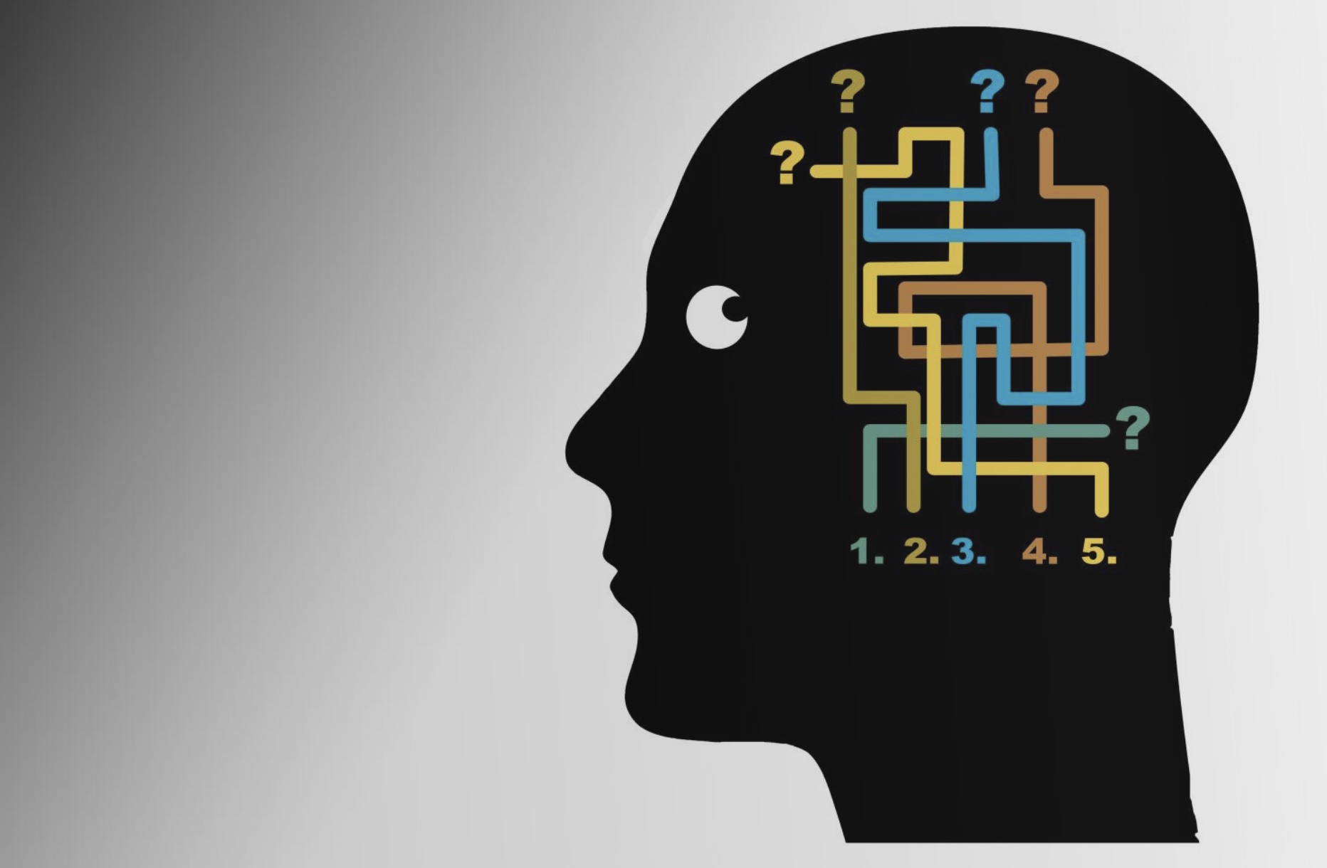
As you can learn below, the brain has much greater flexibility in how it operates than the electrical circuits that power your house or even the chips that drive your cell phone. But fundamentally it is the routing of electrical signals from neuron to neuron that forms the basis not only for how we behave, but also how we match behavior appropriately to the circumstances we encounter, Choi said.

“The closest component to behaviors and internal states, and changes in those, are still believed to be neurons and circuits,” she said.

Understanding how brain circuits produce behavior is an exciting area of neuroscience research, including in many Picower Institute labs. Their studies are helping to elucidate how the brain’s anatomy is arranged to process information, and how the many dimensions of flexibility that the central nervous system overlays upon that infrastructure can integrate context to guide appropriate behavior. Context, after all, comes from many sources in many forms—from the senses, like scents and sounds and sights; from internal states, like mating drive or hunger or sleepiness; and even from time and place and from what we’ve learned and remember.

So what were you thinking when you did “this” instead of “that”? You were thinking about the context and relying on your brain’s ability to account for it.

Chemical control

The popular “circuit” metaphor makes it easy to think of neurons as merely switches and wires that pass electrical transmissions from one point to another. And indeed they do that, although instead of being screwed and soldered to metal contacts, they use molecules called neurotransmitters to send signals across tiny junctions called synapses. But if that were all that was going on, the brain would be pretty static and it is anything but. Many members of the Institute’s faculty study how learning occurs and memories are formed when the brain changes its synapses to create or edit circuit connections, but none of that is strictly necessary for existing circuits to flexibly control behaviors that we’ve already learned or that are innate. The brain has other ways to flexibly change how it operates. Choi’s team, for instance, found that the behavioral change of inhibiting mating could not occur without the cortical amygdala neurons also sending a chemical, thyrotrophin releasing hormone (TRH), to the medial amygdalar nucleus neurons. 

In the lab of Lister Brothers Associate Professor Steven Flavell, researchers study how internal states and behaviors emerge and change using a worm so simple that its complete, invariant “wiring diagram” has been completely mapped out for decades. Yet even in C. elegans, with its exact total of 302 neurons, scientists are still discovering how the animal adapts its actions to survive and thrive in a world of ever-changing contexts.

“Since 1986, that wiring diagram has been staring at researchers,” Flavell quipped. “Many of the small circuits embedded in the wiring diagram have been closely studied, while others haven’t. But a key question that we are trying to answer is how does the whole system work. How are these circuits coupled together to give rise to so-called ‘brain states’?”

In several studies Flavell has shown how a small number of neurons encode contexts and then signal that those circumstances are afoot by releasing chemicals called “neuromodulators” to many other neurons, giving rise to a brain state. Just as TRH may be doing in the circuit Choi uncovered, neuromodulators such as serotonin and dopamine, which are also ubiquitous in humans, add an extra dimension of tuning that can change, or “modulate,” how hardwired circuits process information and output behaviors, Flavell said. Neuromodulators can make neurons more or less electrically excitable given the same degree of input, Flavell explained. They can also make transmission at individual synapses more or less effective.

“The physical connections are like a roadmap, but the way that traffic is actually flowing on the road, the way that neurons are coupled to each other, is dynamic and changes with the animal’s context,” Flavell said. Neuromodulators are one way to make that happen.

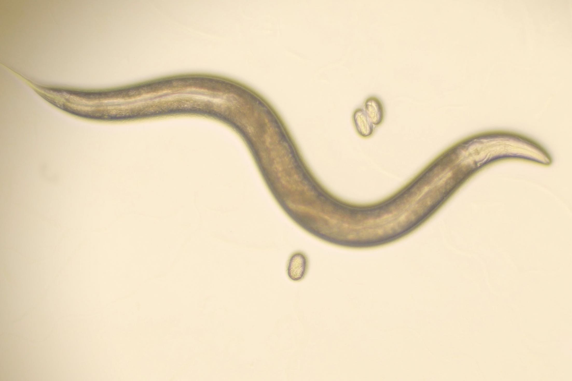
For instance, in a 2019 paper in Cell, Flavell’s lab showed how a hungry worm knows to slow down and savor a patch of yummy bacteria when it finds one. A single neuron called NSM extends a little tendril called a neurite into the worm’s pharynx. Equipped with bacterial sensors (that turn out to also be present in the human intestine), the neurite detects when the worm has started to ingest and mash up its food. NSM releases serotonin, which finds its way to many of the neurons in worm’s brain that control locomotion. Upon sensing the serotonin, they hit the brakes.

In a more recent study in bioRxiv, the lab takes their investigation of neuromodulators even further. The study characterizes exactly how serotonin release from NSM modulates that activity of specific neurons in the C. elegans brain. In addition, Flavell’s group found that a neuron called AIA integrates information from sensory neurons about the smell of food. NSM can help determine what it does with that information, depending on whether it detects that the worm is eating or not. If it is, the smell of food (detected by AIA) reinforces that it should stick around to continue dining, a state maintained with serotonin. If the worm isn’t eating, the food smells signal that the animal should go exploring to find the source of that enticing odor. AIA, in that case, can instead trigger neurons that produce a different neuromodulator, called PDF, that cause the worm to start roaming (toward the food odor). Even in the simple circuitry of C. elegans, context changes how neurons interact, giving the animal flexibility to process sensory information.

That neurons capable of emitting neuromodulators can exert far-flung influence over behavior is illustrated by research in Newton Professor Mriganka Sur’s lab, too. There Sur’s team has a focus on a deeply situated, tiny brain region called the locus coeruleus (LC) that happens to supply most of the brain’s norepinephrine. Classically, neuroscientists have regarded norepinephrine from the LC as increasing the brain’s internal state of general arousal, but recent research in the Sur lab suggests it has profound, context-dependent effects on learning and behavior.

For instance, members of the lab have trained mice to expect a reward if they push a lever after hearing a high-pitched tone; the mice also receive an unexpected and irritating puff of air if they mistakenly press the lever after a low-pitched tone. By varying the loudness of the tones, the researchers can also vary the certainty the mice have about what tone they heard. Sur’s lab has found that the louder a high-pitched tone, the more norepinephrine a mouse will send to the motor cortex, which plans movement, before pushing the lever – as if greater certainty prompts it more strongly to push the lever. 

Once the lever has been pushed and the mouse gets its feedback of reward or air puff, LC neurons producing norepinephrine then act to fine-tune learning by calling attention to any surprising feedback, Sur’s team has seen. For instance, if the tone was high pitched and faint, but the mouse took the risk to push the lever, the neurons will send a burst of norepinephrine to the prefrontal cortex to note that pleasant surprise. The biggest post-push surge of the neuromodulator, however, occurs when the mouse guesses wrong: that norepinephrine release to the prefrontal cortex appears to signal that the adverse result must be noted. Sure enough, Sur said, the team has seen that the mouse’s performance typically improves after making an error. The LC’s neuromodulatory actions may contribute to that behavioral improvement, though more research is needed to prove it.

Sur’s is not the only research in The Picower Institute showing that the LC communicates with the prefrontal cortex to improve task performance, though. Last November in the Proceedings of the National Academy of Sciences, Picower Professor Susumu Tonegawa’s lab showed that LC norepinephrine neurons connect via distinct circuits to two different parts of the prefrontal cortex to endow mice with both the ability to curb impulses (i.e. to not “jump the gun” when waiting to perform tasks) and to ignore distractions, such as false cues. 

Rhythms among regions

Much as the Sur and Tonegawa labs have been investigating the LC, Fairchild Professor Matt Wilson’s lab studies how a different region appears to be a key hub for integrating contexts such as location, motion and memories of reward into behaviors such as navigation: the lateral septum (LS). As rats learn to find and return to the location of a reward in a maze, the lab’s extensive measurements of electrical activity among neurons in the LS shows that those cells are taking in and processing crucial contextual input from many other regions. The LS then appears to package that context to help direct the rat’s navigational plans and actions.

Over the past two years, Wilson and former graduate student Hannah Wirtshafter have published papers in Current Biology and in eLife showing that populations of LS neurons distinctively encode place information coming from the hippocampus, reward information coming from the ventral tegmental area and speed and acceleration information coming from the brainstem. The encoding is apparent in changes in the timing and rate at which the neurons “fire,” or electrically activate, in these different contexts. Some LS neurons, for example, become especially active specifically when the rat nears the reward location. In a new article published in Neuroscience and Biobehavioral Reviews in July, Wilson and Wirtshafter combined their observations with those of other labs to propose that the lateral septum packages all this contextual information into an “integrated movement value signal.”

“The lateral septum has a ton of different inputs,” Wirtshafter said. “What could the animal be doing with place-related firing that’s reward modulated and then velocity and acceleration? The answer, we think, based on where the LS outputs to, is that it is sending a signal about the context and whatever reward is part of that context. It includes what movement needs to be done and whether that movement is worth it in that context.”

While there are ample signs in the research that neuromodulators such as dopamine help the LS communicate about contexts like the feeling of reward, the studies also highlight the key role of another mechanism of flexibility: brain rhythms. Also known as brain waves or oscillations, these rhythms arise from the coordinated fluctuation of electrical activity among neurons that are working in concert. They allow neurons in brain regions to broadcast information and neurons in other regions to tune into those broadcasts, so that they can work together to perform a function, Wilson said.  

“These brain dynamics ensure that whoever is sending the information and whoever is receiving the information are doing it at the same time,” Wilson said.

In fact, Picower Professor Earl Miller, who has published numerous studies on how brain rhythms guide the flow of information across the many regions of the brain’s cortex, uses much the same kind of traffic analogy in talking about the function of rhythms that Flavell uses when talking about neuromodulators. Much as those chemicals can, oscillations also flexibly direct the flow of information on the network of “roads” that physical circuit connections create. The traffic metaphor perhaps combines well with the broadcasting one: Just like drivers who tune into a radio traffic report can decide to take an alternate route when they hear about an accident ahead, neurons in a brain region may act differently when they tune into new contextual information coming in from another brain region.

Wilson and Wirtshafter’s research, for example, demonstrates that lateral septum neurons tune into the hippocampus’s broadcast of location information via a specific “theta” frequency of brain waves. In particular, movement through a place is represented by the phase (peak or trough) of the theta waves with which neurons spike. 

“In the hippocampus, the phase at which a cell fires during theta can communicate information about the current, prospective, or retrospective spatial location,” Wilson and Wirtshafter wrote in their article. “For instance, …firing of individual hippocampus place cells begins on a particular phase of theta rhythm and progressively shifts forward as the animal moves through the place field.”

So maybe you are not a mouse deciding whether to mate or a rat rooting through a maze for a treat, but you are a person who has stayed out late at a friend’s house. Your internal state is that you are tired. You could head out on long drive home to the reward of your clean, warm bed, or you could sleep on your friend’s notably mustier couch and explain it your spouse the next morning. Then you remember from the drive to your friend’s place earlier, that there was an all-night rest stop along the highway where you could get coffee. Whether you decide to take the wheel or your friend’s offer of the couch will come from how a combination of neuromodulators and rhythms route information along circuits through key brain regions to integrate all this context—your internal state of tiredness, the memory of where that rest stop was, and the reward of your bed (or the punishment of an angry spouse who might ask “What were you thinking?”). Your brain gives you all the flexibility you need.

J-WAFS announces 2021 Solutions program grants for commercialization of water and food technologies

This year’s projects address mobile evaporative vegetable preservation, portable water filtration, and dairy waste reduction.

Susanna Maize | Abdul Latif Jameel Water and Food Systems Lab
August 29, 2021

Today, the Abdul Latif Jameel Water and Food Systems Lab (J-WAFS) at the Massachusetts Institute of Technology announced the 2021 J-WAFS Solutions grant recipients. The J-WAFS Solutions program aims to propel MIT water and food-related research toward commercialization. Grant recipients receive one year of financial support, as well as mentorship, networking, and guidance from industry experts, to begin their journey into the commercial world — whether that be in the form of bringing innovative products to market or launching cutting-edge startup companies.

This year, three projects will receive funding across water, food, and agriculture spaces. The winning projects will advance nascent technologies for off-grid refrigeration, portable water filtration, and dairy waste recycling. Each provides an efficient, accessible solution to the respective challenge being addressing.

Since the start of the Solutions program in 2015, the grants have provided instrumental support in creating a number of key MIT startups that focus on major water and food challenges. A 2015-2016 Solutions grant helped the team behind Via Separations develop their business plan to massively decarbonize industrial separations processes. Other successful Solutions alumni include researchers who created a low-cost water filter made from tree branches and the team that launched the startup Xibus Systems, which is developing a handheld food safety sensor.

“New technological advances are being made at MIT every day, and J-WAFS Solutions grants provide critical resources and support for these technologies to make it to market so that they can transform our local and global water and food systems,” says J-WAFS executive director, Renee Robins. “This year’s grant recipients offer innovative tools that will provide more accessible food storage for smallholder farmers in places like Africa, safer drinking water, and a new approach to recycling food waste,” Robins notes. She adds, “J-WAFS is excited to work with these teams, and we look forward to seeing their impact on the water and food sectors.”

The J-WAFS Solutions program is implemented in collaboration with Community Jameel, the global philanthropic organization founded by MIT alumnus Mohammed Jameel, and is supported by the MIT Venture Mentoring Service and the iCorps New England Regional Innovation Node at MIT.

Read more about the 2021 J-WAFS Solutions grantee projects below.

Mobile evaporative cooling rooms for vegetable preservation
Food waste is a persistent problem across food systems supply chains, as 30-50% of food produced is lost before it reaches the table. The problem is compounded in areas without access to the refrigeration necessary to store food after it is harvested. Hot and dry climates in particular struggle to preserve food before it reaches consumers. A team led by Daniel Frey, faculty director for research at MIT D-Lab and professor of mechanical engineering, has pioneered a new approach to enable farmers to better preserve their produce and improve access to nutritious food in the community. The team includes Leon Glicksman, professor of building technology and mechanical engineering, and Eric Verploegen, a research engineer in MIT D-Lab.

Instead of relying on traditional refrigeration with high energy and cost requirements, the team is utilizing forced-air evaporative cooling chambers. Their design, based on retrofitting shipping containers, will provide a lower-cost, better performing solution enabling farmers to chill their produce without access to power. The research team was previously funded by J-WAFS through two different grants in 2019 to develop the off-grid technology in collaboration with researchers at the University of Nairobi and the Collectives for Integrated Livelihood Initiatives (CInI), Jamshedpur. Now, the cooling rooms are ready for pilot testing, which the MIT team will conduct with rural farmers in Kenya and India. The MIT team will deploy and test the storage chambers through collaborations with two Kenyan social enterprises and an NGO in Gujarat, India.

Off-grid portable ion concentration polarization desalination unit
Shrinking aquifers, polluted rivers, and increased drought is making fresh drinking water increasingly scarce, driving the need for improved desalination technologies. The water purifiers market, which was $45.0B in 2019, is expected to grow to $90.1B in 2025. However, current products on the market are limited in scope, in that they are designed to treat water that is already relatively low in salinity, and do not account for lead contamination or other technical challenges. A better solution is required to ensure access to clean and safe drinking water in the face of water shortages.

A team led by Jongyoon Han, professor of biological engineering and electrical engineering at MIT, has developed a portable desalination unit that utilizes an ion concentration polarization process. The compact and lightweight unit has the ability to remove dissolved and suspended solids from brackish water at a rate of one liter per hour, both in installed and remote field settings. The unit was featured in an award-winning video in the 2021 J-WAFS World Water Day Video Competition: MIT Research for a Water Secure Future. The team plans to develop the next-generation prototype of the desalination unit alongside a mass-production strategy and business model.

Converting dairy industry waste into food and feed ingredients
One of the trendiest foods in the last decade, Greek yogurt, has a hidden dark side: acid whey. This low-pH, liquid by-product of yogurt production has been a growing problem for producers as untreated disposal of the whey can pose environmental risks due to its high-organic content and acidic odor. With an estimated three million tons of acid whey generated in the U.S. each year, MIT researchers saw an opportunity to turn waste into a valuable resource for our food systems. Led by the Willard Henry Dow Professor in Chemical Engineering, Gregory Stephanopoulos, and Anthony J. Sinskey, professor of microbiology, the researchers are utilizing metabolic engineering to turn acid whey into carotenoids, the yellow and orange organic pigments found naturally in carrots, autumn leaves, and salmon. The team is hoping that these carotenoids can be utilized as food supplements or feed additives to make the most of what otherwise would have been wasted.

So-called “junk” DNA plays a key role in speciation
Eva Frederick | Whitehead Institute
August 23, 2021

More than 10 percent of our genome is made up of repetitive, seemingly nonsensical stretches of genetic material called satellite DNA that do not code for any proteins. In the past, some scientists have referred to this DNA as “genomic junk.”

Over a series of papers spanning several years, however, Whitehead Institute Member Yukiko Yamashita and colleagues have made the case that satellite DNA is not junk, but instead has an essential role in the cell: it works with cellular proteins to keep all of a cell’s individual chromosomes together in a single nucleus.

Now, in the latest installment of their work, published online July 24 in the journal Molecular Biology and Evolution, Yamashita and former postdoctoral fellow Madhav Jagannathan, currently an assistant professor at ETH Zurich, Switzerland, take these studies a step further, proposing that the system of chromosomal organization made possible by satellite DNA is one reason that organisms from different species cannot produce viable offspring.

“Seven or eight years ago when we decided we wanted to study satellite DNA, we had zero plans to study evolution,” said Yamashita, who is also a professor of biology at the Massachusetts Institute of Technology and an investigator with the Howard Hughes Medical Institute. “This is one very fun part of doing science: when you don’t have a preconceived idea, and you just follow the lead until you bump into something completely unexpected.”

The origin of species: DNA edition 

Researchers have known for years that satellite DNA is highly variable between species. “If you look at the chimpanzee genome and the human genome, the protein coding regions are, like, 98 percent, 99 percent identical,” she says. “But the junk DNA part is very, very different.”

“These are about the most rapidly evolving sequences in the genome, but the prior perspective has been, ‘Well, these are junk sequences, who cares if your junk is different from mine?’” said Jagannathan.

But as they were investigating the importance of satellite DNA for fertility and survival in pure species, Yamashita and Jagannathan had their first hint that these repetitive sequences might play a role in speciation.

When the researchers deleted a protein called Prod that binds to a specific satellite DNA sequence in the fruit fly Drosophila melanogaster, the flies’ chromosomes scattered outside of the nucleus into tiny globs of cellular material called micronuclei, and the flies died. “But we realized at this point that this [piece of] satellite DNA that was bound by the Prod protein was completely missing in the nearest relatives of Drosophila melanogaster,” Jagannathan said. “It completely doesn’t exist. So that’s an interesting little problem.”

If that piece of satellite DNA was essential for survival in one species but missing from another, it could imply that the two species of flies had evolved different satellite DNA sequences for the same role over time.  And since satellite DNA played a role in keeping all the chromosomes together, Yamashita and Jagannathan wondered whether these evolved differences could be one reason different species are reproductively incompatible.

“After we realized the function [of satellite DNA in the cell], the fact that satellite DNA is quite different between species really hit like lightning,” Yamashita said. “All of a sudden, it became a completely different investigation.”

A tale of two fruit fly species

To study how satellite DNA differences might underlie reproductive incompatibility, the researchers decided to focus on two branches of the fruit fly family tree: the classic lab model Drosophila melanogaster, and its closest relative, Drosophila simulans. These two species diverged from each other two to three million years ago.

Researchers can breed a Drosophila melanogaster female to a Drosophila simulans male, “but [the cross] generates very unhappy offspring,” Yamashita said. “Either they’re sterile or they die.”

Yamashita and Jagannathan bred the flies together, then studied the tissues of the offspring to see what was leading these “unhappy” hybrids to drop like flies. Right away they noticed something interesting: “When we looked at those hybrid tissues, it was very clear that their phenotype was exactly the same as if you had disrupted the satellite DNA [-mediated chromosomal organization] of a pure species,” Yamashita said. “The chromosomes were scattered, and not encapsulated in a single nucleus.”

Furthermore, the researchers could create a healthy hybrid fly by mutating certain genes in the parent flies called “hybrid incompatibility genes,” which have been shown to localize to satellite DNA in the cells of pure species.  Via these experiments, the researchers were able to demonstrate how these genes affect chromosomal packaging in hybrids, and pinpoint the cellular phenotypes associated with them for the first time. “I think for me, that is probably the most critical part of this paper,” Jagannathan said.

Taken together, these findings suggest that because satellite DNA mutates relatively frequently, the proteins that bind the satellite DNA and keep chromosomes together must evolve to keep up, leading each species to develop their own “strategy” for working with the satellite DNA. When two organisms with different strategies interbreed, a clash occurs, leading the chromosomes to scatter outside of the nucleus.

In future studies, Yamashita and Jagannathan hope to put their model to the ultimate test: if they can design a protein that can bind the satellite DNA of two different species and hold the chromosomes together, they could theoretically ‘rescue’ a doomed hybrid, allowing it to survive and produce viable offspring.

This feat of bioengineering is likely years off. “Right now it’s just a pure conceptual thing,” Yamashita said. “In doing this tinkering, there’s probably a lot of specifics that will have to be solved.”

For now, the researchers plan to continue investigating the roles of satellite DNA in the cell, armed with their new knowledge of the part it plays in speciation. “To me, the surprising part of this paper is that our hypothesis was correct,” Jagannathan said. “I mean, in retrospect, there are so many ways things could have been inconsistent with what we hypothesized, so it’s kind of amazing that we’ve sort of been able to chart a clear path from start to finish.”

Summer students thrive in Picower labs

Undergraduates from colleges across the country gain scientific training, mentorship and experience as participants in the MIT Summer Research Program

Picower Institute
August 11, 2021

A college student could imagine many ways to spend a summer, but for 11 undergraduates at universities from the Caribbean to California, an uncommon passion for science and an eagerness for immersion in current, world-class research made joining Picower Institute labs a compelling choice.

At a bustling poster session in early August where they presented their work, it was clear that the students hailing from underrepresented or disadvantaged backgrounds and non-research intensive home institutions, made the most of their participation in the MIT Summer Research Program (MSRP) in Biology and Brain and Cognitive Neuroscience. They said the experience, skills, contacts, and inspiration they gained can advance their academic ambitions.

Performing experiments to study possible treatments for the developmental vision disorder amblyopia in the lab of Picower Professor Mark Bear gave Alysa Alejandro-Soto, a student at the University of Puerto Rico Mayaguez an inspiring exposure to fundamental lab neuroscience that can also have direct, future relevance to patients, she said.

“I really wanted to do neuroscience research but I hadn’t been able to do it in my undergraduate studies,” she said of her work alongside postdoctoral mentor Hector de Jesús-Cortés. “I want to do an MD/PhD and it’s really exciting for me to see how this could be applied clinically.”

Hanoka Belai said that her summer in the lab of Latham Family Associate Professor Myriam Heiman has recharged her interest in pursuing a neuroscience degree. A biotechnology major at Roxbury Community College, Belai said her research with postdoctoral mentor Brent Fitzwalter to advance a novel strategy for treating the terminal neurodegenerative condition Huntington’s disease was exciting because she wants to learn science to help people.

Heiman said she was delighted to host two MSRP students this summer. Along with Belai, she and graduate student Preston Ge also welcomed Rim Bozo who is on her way to Dartmouth College after graduating from Pioneer Charter School Of Science near Boston. Bozo said the chance to go from high school labs to working on studies of Parkinson’s disease at MIT provided exceptional preparation for college.

“They are both outstanding young scientists,” Heiman said. “The MSRP students are always very motivated and eager to learn so we always look forward to working with them.”

In all, eight Picower labs hosted at least one MSRP student.

Paola Alicea-Román from the University of Puerto Rico, Humacao, first worked with the Bear lab last summer, but could only do so virtually because of the Covid-19 pandemic. Even though her research applying a deep learning algorithm to assess the vision of mice with amblyopia was computational, she said, she reveled in the chance to be in the lab in person this year. Being there not only allowed her to help shape the experiments providing the data, it also gave opportunities to network with professors, fellow women in science and graduate students.

Relationships and mentorship are an especially important component of MSRP for many students who participate. Sonia Okekenwa, a student at Fisk University in Nashville, said a particularly valuable aspect of her work in the lab of Picower Professor Li-Huei Tsai was the frequent dialogue she had with postdoctoral mentor Vishnu Dileep and other lab members, who challenged her to think deeply about what she was finding out in her research mapping where DNA breaks open to enable neuronal processes (see p. 2).

Miriam Goras of Arizona State, who worked in the lab of William R. and Linda R. Young Professor Elly Nedivi with postdoc Baovi Vo, said she similarly valued the challenge of having to figure out genuine problems with no pre-determined answers. For instance, during her work this summer studying the molecular biology of treatment for bipolar disorder, she had to dig into the scientific literature to troubleshoot biochemical methods in the optimization of her cell culture experiment.

Several other Picower MSRP students, who included Jordina Pierre of the University of the Virgin Islands, Miguel Coste of Notre Dame, Joshua Powers of George Washington University, Patricia Pujols of Bayamon Central University, and Hanna Caris of Pomona College said they also valued the exposure, training, and guidance they gained through MSRP, which is coordinated by Director of Diversity and Science Outreach Mandana Sassanfar.

Powers, in fact, was back for his fourth summer after first engaging with MIT programs as a high schooler. His experience in the Flavell lab with postdoc Cassi Estrem has helped him clarify that he wants to pursue research as a career.

“The Flavell lab continues to show support for me, to teach me and go out of their way to make sure I’m keeping up with them for my benefit,” he said.

For Powers and his colleagues, these have been summers well spent.

Unusual Labmates: Fruit flies
Greta Friar | Whitehead Institute
August 4, 2021

All the buzz in the lab

On a sunny summer morning in Cambridge, Massachusetts, Mariyah Saiduddin walked into a room and was met by the sight of thousands of fruit flies. For most people, this would be an emergency: time to call an exterminator, take out the trash, and scrub the room from top to bottom. However, this room full of flies is part of Whitehead Institute Director Ruth Lehmann’s lab, where fruit flies are seen not as pests but as valuable research tools—and are safely contained in vials. Saiduddin is a graduate student researcher in Lehmann’s lab who uses a fraction of the flies in the room in her research.

The flies found in Lehmann’s lab, and in the adjacent lab run by Whitehead Institute Member Yukiko Yamashita, are not exactly like their less-beloved wild counterparts. Fruit flies have been used in research for more than a century, and in that time, they have been engineered to become powerful, malleable models capable of answering questions in many areas of research. The most common species used in research is Drosophila melanogaster, often referred to simply as “Drosophila.” The researchers who use flies call themselves Drosophilists, and their community around the world works together to maintain a rich variety of flies and create new tools with which to manipulate those flies. In the past century, work in fruit flies has led to six Nobel Prizes in Physiology or Medicine, and has shed light on topics from the basics of genetics, to the principles of embryonic development, to circadian rhythms, to the immune system, to a plethora of diseases.

A very fly model organism

Fruit flies became a go-to research tool during the explosion of genetics research around the turn of the 20th century. What makes them such a good model organism? First of all, they are easy and relatively cheap to raise in large numbers. They have short lifespans and quick reproduction times, so researchers can rapidly breed and study multiple generations. Fruit flies are ready to reproduce—growing from embryo to larva to adult—in under two weeks and then can lay hundreds of eggs in a matter of days.

Fruit flies and humans have enough similarities in their genetics and development that research in fruit flies often reflects human biology. In particular, when it comes to genetics, fruit flies have more commonalities with humans than they have differences. Nearly three-quarters of the genes that cause diseases in humans have an equivalent gene—one derived from the same ancestral gene—in fruit flies.
Not only are fruit flies naturally suited for research, but over the years they have been engineered to become even better research subjects. One of the earliest improvements to fruit flies as a research model came from researchers discovering that they could create flies with genetic mutations that change things like a fly’s eye color, wing shape, body shape, or the bristles on its thorax. Researchers began selectively breeding different lines of fruit flies to have these distinctive physical traits or “markers,” which make them easy to tell apart.
Researchers can tie a visible genetic marker, such as curly wings, to a genetic mutation that they are studying that may not be visible, so that they can easily sort their flies. For example, Whitehead Institute researchers using flies to study mutations that affect the germ cells, the set of cells that make or become eggs and sperm, cannot tell just by looking at a fly whether it has a mutation that affects germ cells, but if they tie inheritance of the mutation to the curly wing marker, then the flies with the desired mutation become easy to identify.
Drosophila biologists have adapted tools from other model organisms to control and study essentially any gene in flies. The Gal4-UAS system, which was developed based on a gene and gene regulator found in yeast, is now commonly used in flies. The system lets researchers activate a gene only in certain tissues or sets of cells. For example, researchers may want to know what part of the brain a certain gene is active in, so they will use the Gal4-UAS system to express green fluorescent protein (GFP) only in cells that would also normally express the gene of interest, allowing researchers to map gene activity based on fluorescence. Or, Gal4-UAS can be used to turn off a gene involved in embryonic development, to see what changes when that gene is not active. This is a common approach that researchers use to figure out a gene’s function; it works in the same way that one could deduce the purpose of brakes on a car by taking the brakes off and observing the car being unable to stop when its driver steps on the brake pedal. Gal4 lines can be developed and tested quickly, in a couple of months, and because the Drosophilist community tends to share their resources, as soon as one lab has developed a Gal4 line for a gene, any fruit fly lab can use that tool to ask their own research questions. Thousands of Gal4 lines are maintained in centralized collections, making them very easy to access.

The embryonic days of fly research

The transformation of fruit flies from wild pests into top notch research tools began in the early 1900s. Charles W. Woodworth, at Harvard University at the turn of the 20th century, is credited with being the first researcher to breed Drosophila in large numbers and with suggesting that the species could be used to study genetics, then a new field of research.[2] Thomas Hunt Morgan, at Columbia University, was one of several researchers to follow Woodworth in using fruit flies for his research, and it was Morgan who really established fruit flies as a model organism, through both his own success and that of the students who came out of his lab and the soon-famous Columbia fly room.

For his research, Morgan bred fruit flies until one developed a mutation, white eyes (most fruit flies’ eyes are red), and then continued breeding the mutant and its descendants to track patterns in inheritance of the white-eyes trait. With these experiments, Morgan showed that genes, which had recently been established as the smallest units of inheritance, are organized on chromosomes, cellular structures each one of which contains a certain, consistent set of genes. One of Morgan’s students, Alfred Sturtevant, expanded on this work, showing that the genes on each chromosome can be mapped in a specific linear order. The proof of the chromosome theory of inheritance won Morgan the 1933 Nobel Prize in Physiology or Medicine and the mutation that Morgan identified, white, is still used as a marker in fruit flies today. Morgan and the scientists who came through his lab continued to do groundbreaking research, demonstrating the potency of fruit flies as a model, and soon flies became a popular research tool.

Research in fruit flies has led to five further Nobel Prizes since Morgan’s, including the 1995 prize awarded to Edward B. Lewis, Christiane Nüsslein-Volhard and Eric Wieschaus for their discoveries regarding “the genetic control of early embryonic development.” These three researchers identified and discovered the function of key genes involved in determining and carrying out the blueprint for a fly’s body during development. Nüsslein-Volhard and Wieschaus systematically mutated many flies in order to discover the genes involved in body patterning. After they introduced lots of mutations, they observed what happened to the flies, and then they determined which genes had been mutated to cause the effects to body patterning that they observed. Using this strategy, they identified and characterized many key genes involved in guiding the development of an embryo into a segmented body.

Lewis, meanwhile, identified and determined the function of what would come to be known as homeotic genes, the genes that determine which specific body parts grow in each body segment: these genes essentially determine the blueprint for the fly’s body, and—as Lewis showed—when mutated lead to some very unusual body plans. Collectively, these researchers’ discoveries illuminated both the genetics and the evolution of the body plan in flies—work that was quickly extrapolated to other species, including humans, whose development occurs in a similar fashion.

Lewis, Nüsslein-Volhard and Wieschaus’ work set the stage for future researchers such as Lehmann and Yamashita who study development in flies. In fact, Nüsslein-Volhard had a direct influence on Lehmann: Lehmann trained with her as a graduate student. Nüsslein-Volhard’s later work provided important insights into the morphogen gradients that help guide the developing embryo in assembling itself correctly, and included an in-depth gene screen of zebrafish using the same extensive process of mutation and observation that she and Wieschaus had used in Drosophila.

Researchers at Whitehead Institute are using fruit flies to answer a wide variety of questions. In previous years, former Whitehead Institute Member Terry Orr-Weaver used Drosophila to study the process of cell division during development. She looked at questions such as what determines cell size, what regulates the transition from egg to embryo, and how DNA is accurately replicated and sorted into dividing cells. Whitehead Institute Member David Bartel, also a professor of biology at the Massachusetts Institute of Technology (MIT) and an HHMI investigator, studies RNA and has done some of that research in flies. His work has improved our understanding of how tiny regulatory RNAs called microRNAs target and initiate the destruction of the RNAs that code for proteins in many species, including flies. He used these insights to create TargetScanFly, a database that provides researchers around the world with fly microRNAs’ predicted targets. Bartel’s lab also recently discovered how some microRNAs are rapidly degraded in Drosophila cells and how other types of small regulatory RNAs are protected from this degradation. In other studies of gene regulation in flies, performed in collaboration with Orr-Weaver, the Bartel lab identified the RNAs that are first produced by the developing embryo and determined why RNAs of some genes are much better than those of others at producing proteins in fly oocytes and early embryos.

Lehmann and Yamashita use fruit flies to study germ cells, the cells set aside to make or become eggs and sperm—as did Orr-Weaver. The germ line is set aside from the rest of the body’s cells early on, and has the rare property of being, essentially, immortal: all of the other cell lines in the body will eventually die with it, but germ cells survive to become offspring, which contain new germ cells, and so on through the generations.

Lehmann, who is also a professor of biology at MIT, studies how germ cells are set aside, how they migrate during development, and how the germ line is maintained into adulthood. During development of the gonads, cells must work in perfect sync. They follow cues, many of which are still unknown, to ensure that every cell—including germ cells—ends up in the right place at the right time to form functioning, fertile ovaries or testes. Lehmann’s research has shed light on how these processes occur. Lehmann’s lab also studies how RNA is regulated and organized within germline-specific granules inside of the cells. Another interest of the lab is the inheritance of mitochondria, structures inside of cells that provide energy, which are passed down through the generations exclusively through the female germline. Mitochondria carry their own genomic DNA, and this could accumulate deleterious mutations over time; Lehmann’s lab has helped to determine how the germline manages to selectively inherit mitochondria that are mutation-free or healthy.

Yamashita, who is also a professor of biology at MIT and an investigator with the Howard Hughes Medical Institute, studies many aspects of germ cell biology in the context of the adult Drosophila testis. Germline stem cells divide asymmetrically, so one dividing stem cell produces one differentiated cell that will go on to become sperm and one new stem cell that will replenish the germline. Yamashita’s lab has studied what distinguishes the cellular components that are inherited by the new stem cell versus the differentiating cell, as well as how the stem cell is able to identify and retain those components. If this asymmetrical division goes awry, then the germline could be lost. Yamashita’s lab also studies a type of repetitive DNA, which contains many repeats of the same sequences of nucleotides (DNA building blocks) that do not code for any genes. This genetic material was once considered “junk DNA.” They found that this “junk” actually helps to ensure that each germ cell contains the proper number of chromosomes, which is necessary in order to produce viable offspring. The lab continues to look at how repetitive DNAs are maintained and at their roles in germ cell development.

Fortunately for researchers in Lehmann and Yamshita’s labs, the fly research world is one that promotes resource sharing. As they seek to better understand development and related topics, Whitehead Institute researchers have many rich resources to draw from. FlyBase is an online database of Drosophila genetic and molecular data, which contains the complete annotated Drosophila melanogaster genome. The site also has educational resources, community networking links, images and videos, and more. Fly researchers can order the flies they need from stock centers that maintain thousands of lines with different genetic variations, suitable for different research questions. Two of the main stock centers are in Bloomington, Indiana and Kyoto, Japan.

“The fly community, from the very beginning on, has been an example of sharing before publication and exchanging tools and ideas, which is how the best science happens,” Lehmann says.

Fly researchers regularly gather at meetings such as the Annual Drosophila Research Conference, regional Drosophila meetings, and the biennial Crete meeting for principal investigators—which has been held every other year for more than four decades—to exchange ideas and to foster trust and collaboration in the community.

In the typical communal spirit of Drosophilists, Lehmann and Yamashita have physically joined their labs and share a fly room—a room full of microscopes and tools for examining the fruit flies. In order to pursue their inquiries, researchers in both labs allocate a portion of their time to an important activity: keeping their flies alive and breeding.

Drosophilist: A day in the life

Each researcher in the lab keeps their own stock of flies. The flies live in vials that are partially filled with wet, packed down food—typically a mixture of yeast, cornmeal, agar and a few other ingredients. The tops of the vials are sealed with cotton swabs.

Adult flies rest on the sides of the vial, and lay their eggs in the food. Larva hatch and live in the food mixture as they grow through several molts. Eventually, they crawl out of the food and form pupae on the sides of the vials. In a few days, adult flies emerge from the pupal casings and soon begin to mate, continuing the cycle.

A regular part of work in a fly lab is “flipping flies,” or transferring flies into a new vial with fresh food. This has to be done regularly to keep the vials from overcrowding as the fly population expands. Researchers in fly labs soon become adept at flipping vials.

Researchers also flip flies or clear vials of adults in preparation for breeding specific crosses. If a researcher wants to make sure that a specific line of females flies mates with a specific line of males, then they need to use virgin females. That’s because female flies can store sperm and use it to fertilize their eggs later, so the offspring of a non-virgin female could be from the male she just mated with or from a male she mated with as long as two weeks ago.

The most common way for researchers to ensure that the females they use for breeding are virgins is to clear a vial of all of its adult flies, and then wait for new adults to hatch out of their pupal casings. Adult flies cannot mate for the first few hours of their lives, and if kept in chilly conditions—18 degrees Celsius—then they cannot mate for 18 hours. Therefore, a researcher can clear a vial of adult flies, leave it for up to 18 hours, and then collect all of the adult females from the vial, confident that they are virgins.

Taking a closer look

When the researchers want to get a close-up view of their specimens, they take them out of the vials and examine them under the microscope. In order to do this, they dose the flies with carbon dioxide (CO2), which keeps them asleep on a pad under the microscope.

Researchers use gentle tools to maneuver the unconscious flies. The Lehmann lab uses one type of common fly mover: paintbrushes. The Yamashita lab uses another: feathers.

Each lab member has their own brush or feather that feels best in their hands—the Yamashita lab also selects their feathers based on color.

“You learn everyone’s favorite color quickly,” Fingerhut says—hers is purple. Most of the feathers are of the craft store variety, but Yamashita uses real bird feathers (sanitized for lab use).

Under the microscope, it’s very easy to tell the flies apart by their genetic markers. It’s also possible to view the stages of fruit fly development from start to finish.

“In the stages that we’re studying, the embryo will undergo massive morphological changes that you can watch happen over a few hours using video imaging. There’s just so much of what’s going on during those few hours that we don’t know anything about, but you can see it with your own eyes easily with the microscope,” Saiduddin says.

In the following video, Saiduddin captured germ cell precursors forming in a Drosophila embryo. They appear in the posterior pole, shown on the right. Images were captured every 30 seconds.

“When you look at the testis or the ovary, you can see single cell resolution, so you can see what’s happening in all the different cell types, kind of all at once, and you can get a picture of what genes are important at what stage of germ cell development,” Fingerhut says. “You can see the whole process going on in one snapshot when you look at a single tissue, like you can see the stem cells, and their niche, and then you can see every stage up until a mature sperm that’s ready to go on to fertilization.”

Fruit flies may seem like a nuisance when they suddenly start multiplying in the kitchen trash, but the contributions they have made to science are multitudinous. With the tools to manipulate their genomes, their short generation time, large numbers, and easily observable development, fruit flies make for an excellent model organism, and the success of Drosophila research over the decades bears that out. They have been instrumental in shaping our understanding of genetics, development, health and disease, and more. The many flies inside of Whitehead Institute will help answer important questions about how life begets new life, and how new life develops during its early stages.

Rewiring cell division to make eggs and sperm
Whitehead Institute
July 30, 2021

To create eggs and sperm, cells must rewire the process of cell division. Mitosis, the common type of cell division that our bodies use to grow everything from organs to fingernails and to replace aging cells, produces two daughter cells with the same number of chromosomes and approximately the same DNA sequence as the original cell. Meiosis, the specialized cell division that makes egg and sperm in two rounds of cell division, creates four granddaughter cells with new variations in their DNA sequence and half as many chromosomes in each cell. Meiosis uses most of the same cellular machinery as mitosis to achieve this very different outcome; only a few key molecular players prompt the rewiring from one type of division to another. One such key player is the protein Meikin, which is found exclusively in cells undergoing meiosis.

New research from Whitehead Institute Member Iain Cheeseman, graduate student Nolan Maier and collaborators Professor Michael Lampson and senior research scientist Jun Ma at the University of Pennsylvania demonstrates how Meikin is elegantly controlled, and sheds light on how the protein acts to serve multiple roles over different stages of meiosis. The findings, which appear in Developmental Cell on July 30, reveal that Meikin is precisely cut in half midway through meiosis. Instead of this destroying the protein, one half of the molecule, known as C-Meikin, goes on to play a critical role as a previously hidden protein actor in meiosis.

“Cells have this fundamental process, mitosis, during which they have to divide chromosomes evenly or it will cause serious problems like cancer, so the system has to be very robust,” Maier says. “What’s incredible is that you can add one or two unique meiotic proteins like Meikin and dramatically change the whole system very quickly.”

Helping chromosomes stick together

During both mitosis and meiosis, sister chromatids — copies of the same chromosome — pair up to form the familiar “X” shape that we recognize as a chromosome. In mitosis, each chromatid—each half of the X — is connected to a sort of cellular fishing line and these lines reel the chromatids to opposite ends of the cell, where the two new cells are formed around them. However, in the first round of division in meiosis, the sister chromatids stick together, and one whole “X” is reeled into each new cell. Meikin helps to achieve this different outcome by ensuring that, while the chromosomes are being unstuck from each other in preparation for being pulled apart, each pair of sister chromatids stays glued together in the right place. Meikin also helps ensure that certain cellular machinery on the sister chromatids is fused so that they will connect to the same line and be reeled together to the same side of the cell.

More specifically, when chromosomes are first paired up, they are glued together by adhesive molecules in three regions: the centromere, or center of the X, where Meikin localizes; the region around the center; and the arms of the X. In the first round of meiosis, Meikin helps to keep the glue in the region around the center intact, so the sister chromatids will stick together. Simultaneously, Meikin helps to prime the center region to be unglued, while a separate process unglues the arms. This ungluing allows the chromosomes to separate and be prepared for later stages of meiosis.

Cheeseman and Maier initially predicted that Meikin’s role ended after meiosis I, the first round of meiotic cell division. In meiosis II, the second round of cell division, the cells being created should end up with only one sister chromatid each, and so the chromatids must not be kept glued together. Maier found that near the end of meiosis I, Meikin is cleaved in two by an enzyme called Separase, the same molecule that cleaves the adhesive molecules gluing together the chromosomes. At first, this cleavage seemed like the end of Meikin and the end of this story.
A hidden role for a hidden proteinHowever, unexpectedly, the researchers found that cells lacking Meikin during the second half of meiosis do not divide properly, prompting them to take another look at what happens to Meikin after it gets cleaved. They found that Separase cleaves Meikin at a specific point — carving it with the precision of a surgeon’s scalpel — to create C-Meikin, a previously unknown protein that turns out to be necessary for meiosis II. C-Meikin has many of the same properties as the intact Meikin molecule, but it is just different enough to take on a different role: helping to make sure that the chromosomes align properly before their final division.

“There’s a lot of protein diversity in cells that you would never see if you don’t go looking for it, if you only look at the DNA or RNA. In this case, Separase is creating a completely different protein variant of Meikin than can function differently in meiosis II,” says Cheeseman, who is also a professor of biology at Massachusetts Institute of Technology. “I’m very excited to see what we might discover about other hidden protein forms in cell division.”

Recombining ideas

Answering the question of Meikin’s role and regulation throughout meiosis required a close collaboration and partnership between Maier and Lampson lab researcher Ma – the Lampson lab being experts on studying meiosis using mouse models. Working with mouse oocytes (immature egg cells), Ma was able to reveal the behaviors and critical contributions of Meikin cleavage in meiotic cells in mice. Both labs credit the close exchange with helping them to get a deeper understanding of how cells rewire for meiosis.

“It was a pleasure working together to understand how some of the specialized meiotic functions that are necessary for making healthy eggs and sperm are controlled,” Lampson says.

Finally, once cells have completed these specialized meiotic divisions, the researchers found that it was critical for oocytes to fully eliminate Meikin. The researchers determined that, after meiosis two, C-Meikin is degraded by another molecule (the anaphase-promoting complex or APC/C)—this time for good. With Meikin gone and the rewiring of cell division reversed, eggs and sperm are ready for mitosis; should they fuse and form an embryo, that is the next cell division they will undergo. The researchers note that the way Meikin is regulated by being broken down — first into C-Meikin and then completely — may help cells to organize their timing during meiosis. Breaking apart a protein is an irreversible step that creates a clear demarcation between before and after in a multi-step process.The researchers hope that by uncovering the intricacies of meiosis, they may shed light on what happens when the creation of eggs and sperm goes wrong, and so perhaps contribute to our understanding of infertility. Cheeseman also hopes that by studying how mitotic processes are rewired for meiosis, his lab can gain new insights into the original wiring of mitosis.

Mapping the cellular circuits behind spitting

Roundworms change the flow of material in and out of their mouths in response to bright light, revealing a new way for neurons to control muscle cells.

Raleigh McElvery
July 23, 2021

For over a decade, researchers have known that the roundworm Caenorhabditis elegans can detect and avoid short-wavelength light, despite lacking eyes and the light-absorbing molecules required for sight. As a graduate student in the Horvitz lab, Nikhil Bhatla proposed an explanation for this ability. He observed that light exposure not only made the worms wriggle away, but it also prompted them to stop eating. This clue led him to a series of studies that suggested that his squirming subjects weren’t seeing the light at all — they were detecting the noxious chemicals it produced, such as hydrogen peroxide. Soon after, the Horvitz lab realized that worms not only taste the nasty chemicals light generates, they also spit them out.

Now, in a study recently published in eLife, a team led by former graduate student Steve Sando reports the mechanism that underlies spitting in C. elegans. Individual muscle cells are generally regarded as the smallest units that neurons can independently control, but the researchers’ findings question this assumption. In the case of spitting, they determined that neurons can direct specialized subregions of a single muscle cell to generate multiple motions — expanding our understanding of how neurons control muscle cells to shape behavior.

“Steve made the remarkable discovery that the contraction of a small region of a particular muscle cell can be uncoupled from the contraction of the rest of the same cell,” says H. Robert Horvitz, the David H. Koch Professor of Biology at MIT, a member of the McGovern Institute for Brain Research and the Koch Institute for Integrative Cancer Research, Howard Hughes Medical Institute Investigator, and senior author of the study. “Furthermore, Steve found that such subcellular muscle compartments can be controlled by neurons to dramatically alter behavior.”

A roundworm spits after it is exposed to the nasty-tasting hydrogen peroxide produced by bright light. Video by Steve Sando.

Roundworms are like vacuum cleaners that wiggle around hoovering up bacteria. The worm’s mouth, also known as the pharynx, is a muscular tube that traps the food, chews it, and then transfers it to the intestines through a series of “pumping” contractions.

Researchers have known for over a decade that worms flee from UV, violet, or blue light. But Bhatla discovered that this light also interrupts the constant pumping of the pharynx, because the taste produced by the light is so nasty that the worms pause feeding. As he looked closer, Bhatla noticed the worms’ response was actually quite nuanced. After an initial pause, the pharynx briefly starts pumping again in short bursts before fully stopping — almost like the worm was chewing for a bit even after tasting the unsavory light. Sometimes, a bubble would escape from the mouth, like a burp.

After he joined the project, Sando discovered that the worms were neither burping nor continuing to munch. Instead, the “burst pumps” were driving material in the opposite direction, out of the mouth into the local environment, rather than further back into the pharynx and intestine. In other words, the bad-tasting light caused worms to spit. Sando then spent years chasing his subjects around the microscope with a bright light and recording their actions in slow motion, in order to pinpoint the neural circuitry and muscle motions required for this behavior.

“The discovery that the worms were spitting was quite surprising to us, because the mouth seemed to be moving just like it does when it’s chewing,” Sando says. “It turns out that you really needed to zoom in and slow things down to see what’s going on, because the animals are so small and the behavior is happening so quickly.”

To analyze what’s happening in the pharynx to produce this spitting motion, the researchers used a tiny laser beam to surgically remove individual nerve and muscle cells from the mouth and discern how that affected the worm’s behavior. They also monitored the activity of the cells in the mouth by tagging them with specially-engineered fluorescent “reporter” proteins.

They saw that while the worm is eating, three muscle cells towards the front of the pharynx called pm3s contract and relax together in synchronous pulses. But as soon as the worm tastes light, the subregions of these individual cells closest to the front of the mouth become locked in a state of contraction, opening the front of the mouth and allowing material to be propelled out. This reverses the direction of the flow of the ingested material and converts feeding into spitting.

The team determined that this “uncoupling” phenomenon is controlled by a single neuron at the back of the worm’s mouth. Called M1, this nerve cell spurs a localized influx of calcium at the front end of the pm3 muscle likely responsible for triggering the subcellular contractions.

M1 relays important information like a switchboard. It receives incoming signals from many different neurons, and transmits that information to the muscles involved in spitting. Sando and his team suspect that the strength of the incoming signal can tune the worm’s behavior in response to tasting light. For instance, their findings suggest that a revolting taste elicits a vigorous rinsing of the mouth, while a mildly unpleasant sensation causes the worm spit more gently, just enough to eject the contents.

In the future, Sando thinks the worm could be used as a model to study how neurons trigger subregions of muscle cells to constrict and shape behavior — a phenomenon they suspect occurs in other animals, possibly including humans.

“We’ve essentially found a new way for a neuron to move a muscle,” Sando says. “Neurons orchestrate the motions of muscles, and this could be a new tool that allows them to exert a sophisticated kind of control. That’s pretty exciting.”

Former Horvitz lab graduate student Steve Sando studies the neurons that allow roundworms to taste the chemicals produced by light — and then spit them out.

Citation:
“An hourglass circuit motif transforms a motor program via subcellularly localized muscle calcium signaling and contraction”
eLife, online July 2, 2021, DOI: 10.7554/eLife.59341
Steven R Sando, Nikhil Bhatla, Eugene L Q Lee, and H. Robert Horvitz