A tale of two projects

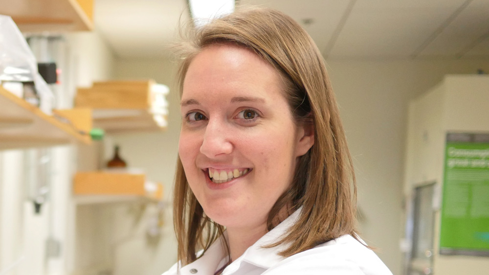
Graduate student Julie Monda has spent five years investigating two divergent aspects of cell division, revealing some unexpected results and new research questions.

Raleigh McElvery
July 23, 2018

To sixth year graduate student Julie Monda, dividing cells are among the most beautiful things she’s ever seen. Watching the tiny, delicate spheres split into identical versions of themselves also provides her with a visual readout for her experiments — will the process continue if she removes a certain piece of a certain protein? Will the genetic material still distribute equally between the two cells? Which molecules are crucial for cell division, and how are they regulated?

Our cells are constantly dividing in order to grow and repair themselves, although some (like skin cells) do so more often than others, say, in the brain. This process, known as mitosis, is the primary focus of Iain Cheeseman’s lab, situated in the Whitehead Institute for Biomedical Research. Most of the research in the Cheeseman lab involves the kinetochore, a group of proteins located on the chromosome where the arms join. During mitosis, long, fibrous structures, known as microtubules, attach to the kinetochore to pull apart the duplicated chromosomes as the parent cell splits in half, ensuring each daughter cell receives an exact copy of the parent’s genetic blueprint.

Before she arrived at MIT Biology in the fall of 2012, Monda worked as a research technician at St. Jude Children’s Research Hospital in Memphis, Tennessee in the lab of Brenda Schulman PhD ’96 . As she recalls, she always “preferred performing hands-on research techniques at the lab bench over being in a classroom.” So she surprised even herself when she chose MIT’s graduate program in biology precisely because it requires all first-year students to take a full course load their fall semester before beginning lab rotations.

“That structure seemed useful given that I studied biochemistry as an undergraduate at the University of Tulsa, and the degree requirements were weighted more towards chemistry than biology,” she says. “Plus, when you’re only taking classes, you spend more time interacting with your classmates. It creates a close-knit community that extends throughout your entire graduate career and beyond.”

Monda ultimately selected the Cheeseman lab because it married her interests in biochemistry and cell biology.

“The research in this lab focuses on various elements of kinetochore function and cell division, but everyone is generally working on their own distinct questions,” she explains. “I knew I would have an area that was mine to explore. It’s both exciting and challenging because no one else is thinking about your projects to the extent that you are.”

Monda’s story is a tale of two projects: one focused on the interface between the kinetochore and the array of microtubules known as the mitotic “spindle,” and another project that ended up taking both her and the lab in a slightly new direction.

The first, concerning kinetochore-microtubule interactions, represented a collaboration with former lab technician Ian Whitney. For this endeavor, Monda investigated a protein complex called Ska1, found at the outer kinetochore.

The Ska1 complex is located where the kinetochore and microtubule meet. Ska1’s role, Monda explains, is to allow the kinetochores to remain attached to the spindle during chromosome segregation, even as the microtubules that compose the spindle begin to disassemble (as they must do).

“We wanted to know how the kinetochore hangs onto this polymer that is essentially falling apart,” Monda explains. “Long story short, we ended up defining specific surfaces within the Ska1 complex that are important for holding on to the microtubule as it shrinks, and — as we were surprised to note — also as it grows”

Although Ska1 only requires a single point of contact to bind a microtubule, Monda and Whitney pinpointed multiple surfaces on Ska1 that are required to allow it to remain associated with the microtubules as they disassemble and reassemble themselves.

While her Ska1 project was very much in line with the types of questions that the Cheeseman lab traditionally pursues, Monda also worked on another endeavor that “began as a side project and slowly evolved into a more full-time effort.” This project involves a motor protein called dynein, which helps to align the chromosomes and position the spindle during mitosis.

Dynein piqued Monda’s interest because of its role in mitosis, as well as its importance throughout the entire cell cycle. Motor proteins are molecules powered by the release of chemical energy that move along surfaces, sometimes transporting cargo, sometimes performing other essential tasks. Dynein is a motor protein that walks in one direction along microtubules, even when the microtubules latch onto the kinetochore to yank apart the chromosomes during mitosis.

But dynein doesn’t act alone. There are a number of additional proteins that also play a key role in coordinating its activity and localization. Monda is studying two of these accessory regulatory proteins, Nde1 and NdeL1, which bind to dynein and help promote some of its functions. She wanted to understand how Nde1 and NdeL1 interact with dynein to activate it. Although Nde1 and NdeL1 are nearly identical in function, Monda discovered that Nde1 (but not NdeL1) binds to another complex: the 26S proteasome.

The proteasome degrades proteins within the cell, influencing virtually all aspects of cellular function, including DNA synthesis and repair, transcription, translation, and cell signaling. Given its ubiquity, it has remained a point of interest among the scientific community for years. And yet, before Monda’s research, the interaction between Nde1 and the proteasome had apparently gone unnoticed. Researchers have long studied Nde1 in relation to dynein, but it’s possible that the interaction between Nde1 and the proteasome represents a new function for Nde1 unrelated to dynein regulation. In fact, Monda’s finding may have implications for understanding the development of the human brain.

“It’s clear that patients with mutations in Nde1 have much more severe neurodevelopmental defects than scientists would have predicted,” Monda says, “so it’s possible that this new interaction between Nde1 and the proteasome could help to explain why Nde1 is so important in the brain.”

Her most recent results have been published in Molecular Biology of the Cell.

“I’ve found some exciting results over the past few years,” Monda says, “and even though a lot of my research has gone in a direction that’s not strictly mitosis-related, Iain has been great about allowing me to follow the science wherever it leads. We want to know what these proteins are actually doing, both in terms of this new interaction and also more broadly within the cell.”

Monda intends to submit and defend her thesis this summer, and assume a postdoctoral position at the University California, San Diego in the fall. Although she’s been watching cells divide for years now, the process still retains its grandeur.

“Often times biologists investigate questions at scales where we can’t really see what we’re studying as we study it,” she says. “But having this visual readout makes it more tangible; I feel like I can better appreciate what exactly it is that I’m trying to understand, as well as the beauty and complexity of the processes that sustain life.”

Sharpening the edges of cancer chemotherapy
Nicole Davis | Whitehead Institute
July 11, 2018

Cambridge, MA — Tackling unsolved problems is a cornerstone of scientific research, propelled by the power and promise of new technologies. Indeed, one of the shiniest tools in the biomedical toolkit these days is the genome editing system known as CRISPR/Cas9. Whitehead Institute Member David Sabatini and his colleagues pioneered the use of this tool as a foundation for large-scale genetic screens in human cells, turning up a treasure trove of new insights into cellular metabolism, in both normal cells and cancer cells.

When Naama Kanarek, a postdoc in Sabatini’s laboratory, pondered how to apply these state-of-the-art CRISPR/Cas9 screens to her own research, her thoughts turned to a classic cancer chemotherapy drug, methotrexate, which has been in clinical use for nearly seven decades. Often used to treat a form of pediatric leukemia, known as acute lymphoblastic leukemia (ALL), the drug, when deployed as part of a multifaceted treatment plan, can be highly effective. But its power comes at a cost. Because methotrexate can damage not only cancer cells but also healthy tissues, it must be administered with great care. For children who receive high doses of the drug, a mainstay of ALL treatment, that can mean several days spent in the hospital with rigorous clinical monitoring.

In other forms of cancer, methotrexate’s efficacy is more uncertain. For example, in pediatric osteosarcoma, only 65 percent of patients respond. Unfortunately, there is currently no way for doctors to pinpoint who will and who will not.

“From a scientific standpoint, methotrexate is quite special because it was the first metabolic drug to be developed, but much of its biology remains to be discovered — particularly what drives these different responses in patients,” Kanarek says. “So, this is really one of these old, classic questions that has been lingering in the field for some time. We thought we could learn something new.”

And they did. In the July 11 online issue of the journal Nature, Kanarek, Sabatini, and their colleagues report the findings of a CRISPR/Cas9 screen for factors involved in methotrexate sensitivity. The team’s work yielded a surprising set of discoveries that point to the breakdown of histidine — one of several amino acids used by the body to construct proteins — as a critical gatekeeper of cancer cells’ vulnerability to methotrexate. The researchers’ findings not only help illuminate the biology of a well-known cancer chemotherapy, but also suggest a simple dietary supplement that could help broaden its therapeutic window and reduce its toxicity.

 “This study is an example of the power of modern genomic tools to shine a bright light on longstanding questions in human biology,” says senior author David Sabatini, a Member of Whitehead Institute, a professor of biology at Massachusetts Institute of Technology and investigator with the Howard Hughes Medical Institute (HHMI). “While cancer chemotherapies can be quite effective, their biological effects are often poorly understood. By laying bare their biology, we may be able to devise ways to utilize them more wisely.”

ATTACK THE CANCER, NOT THE PATIENT

The history of methotrexate stretches back to the 1940s, a time when strikingly little was known about the origins of cancer much less how best to treat it. The birth of methotrexate as a chemotherapeutic agent was sparked by the astute observations of Sidney Farber, a pediatric pathologist at Boston Children’s Hospital who cared for children with a variety of maladies, including ALL. In the course of caring for patients with ALL, Farber recognized that cancer cells depended on the nutrient folic acid for their own proliferation. That gave him the idea of using folate antagonists to treat ALL. Methotrexate was developed in 1949 precisely for this purpose and was subsequently shown to induce remission in children with ALL. Fast forward to today, and the drug has evolved into a significant tool in oncologists’ toolkit.

“Methotrexate is a major part of the backbone of chemotherapy treatment across many human cancers,” says Loren Walensky, a pediatric hematologist/oncologist at the Dana-Farber Cancer Institute who is not a study co-author but served as an early adviser on the project and will also play a deeper role in planning future follow-up studies. “It is also used outside of the cancer field for the treatment of several autoimmune diseases.”

He added, “But as with all chemotherapy, the critical issue is how to best use it to inflict maximal damage on the cancer without irreparably harming the patient.”

Kanarek explains how new genetic tools are allowing insights into the sensitivity of cancer cells to methotrexate.

The basic mechanics of methotrexate are fairly well known. The drug inhibits dihydrofolate reductase (DHFR), an enzyme that generates the functional form of folate, known as tetrahydrofolate (THF). THF is essential for preparing the raw materials needed to make nucleic acids, such as DNA, which carries cells’ genetic information, and RNA, a close chemical relative involved in making proteins. “Proliferating cells must duplicate their DNA, so they need a lot of THF,” Kanarek explains. “But even cells that are not dividing need to make RNA, and that requires THF, too.”

The results of Kanarek’s CRISPR/Cas9 screen now bring greater clarity to this molecular picture. She and her colleagues uncovered another enzyme, called FTCD, which is involved in the breakdown of histidine. Interestingly, FTCD also requires THF for its function — though not nearly as much as the main target of methotrexate, DHFR. Despite the differential demands of the two enzymes, they both draw from the same, shared pool of THF.

“Under normal conditions, this pool is sufficiently full, so there is no competition for resources, even in rapidly dividing cells,” Kanarek says.

But when the amount of THF becomes limiting — as it does in cells that are treated with methotrexate — the story is quite different, the Whitehead Institute team discovered. In that case, the activity of FTCD poses serious problems, because there isn’t enough THF in the pool to support both cell proliferation and histidine breakdown. When that happens, the cells die.

That got Kanarek thinking more about histidine: Could the nutrient provide a way to tinker with FTCD activity and, by virtue of the cancer cells’ own metabolism, make them more vulnerable to methotrexate?

To explore this question, the researchers used mouse models of leukemia, engineered by transplanting human leukemia cells under the skin of immunocompromised mice. A subset of the mice received injections of methotrexate together with histidine. This one-two punch, Kanarek hypothesized, should ramp up the function of FTCD and more rapidly drain the THF pool, thereby making the cells more sensitive to the cancer-killing effects of methotrexate.

That is precisely what the team observed. Notably, these experiments involved lower than normal doses of methotrexate, suggesting the cells had indeed been made more sensitive to the cancer drug. Moreover, the studies included a human leukemia cell line, called SEM, which harbors a specific genetic mutation that is associated with a particularly poor prognosis in patients — further underscoring the power of the histidine degradation pathway to weaken cells’ defenses.

Now, Kanarek and her colleagues are working to extend these initial findings with additional preclinical studies and, together with Walensky, determine how to best evaluate the potential benefits of histidine supplementation in cancer patients. Their ultimate goal: to pursue clinical trials that will assess histidine’s ability to improve the effectiveness of methotrexate in humans.

In addition to making cancer cells more vulnerable to methotrexate, the Whitehead Institute team’s research also holds promise for another therapeutic challenge: identifying which patients will or will not respond to the drug.

Two other enzymes cooperate with FTCD in breaking down histidine. The levels of one of the enzymes, known as HAL, appears to correlate with cells’ sensitivity to methotrexate: That is, cancer cells with high levels of HAL tend to be more sensitive to the drug. More work is needed to determine whether this correlation extends to a broader swath of patient samples and if it has predictive value in the clinic. Nevertheless, Kanarek and her colleagues are already beginning work on this front. Together with Abner Louissaint, Jr., a hematopathologist at Massachusetts General Hospital who also served as an early adviser on the Nature study, the Whitehead Institute team will launch a second clinical study to examine whether HAL levels can predict methotrexate response in patients with lymphoma.

“Being able to understand who is going to respond to methotrexate and who is not, and how to achieve a therapeutic benefit while mitigating the drug’s potential side effects, could have a profound impact on patient care,” Walensky says. “The insights from this study bring an entirely new dimension to our understanding of a decades-old and critically important cancer medicine. And as a physician and a scientist, that’s truly exciting.”

Written by Nicole Davis

* * *

David Sabatini’s primary affiliation is with Whitehead Institute for Biomedical Research, where his laboratory is located and all his research is conducted. He is also a Howard Hughes Medical Institute investigator and a professor of biology at Massachusetts Institute of Technology.

* * *

Full citation:

“Histidine catabolism is a major determinant of methotrexate sensitivity”

Nature, online on July 11, 2018.

Naama Kanarek (1,2,3,4), Heather R. Keys (1), Jason R. Cantor (1,2,3,4), Caroline A. Lewis (1), Sze Ham Chan (1), Tenzin Kunchok (1), Monther Abu-Remaileh (1,2,3,4), Elizaveta Freinkman (1), Lawrence D. Schweitzer (4), and David M. Sabatini (1,2,3,4).

  1. Whitehead Institute for Biomedical Research and Massachusetts Institute of Technology, Department of Biology, 455 main Street, Cambridge, Massachusetts 02142, USA
  2. Howard Hughes Medical Institute, Department of Biology, Massachusetts Institute of Technology, Cambridge, Massachusetts 02139, USA
  3. Koch Institute for Integrative Cancer Research and Massachusetts Institute of Technology, Department of Biology, 77 Massachusetts Avenue, Cambridge, Massachusetts 02139, USA
  4. Broad Institute of Harvard and Massachusetts Institute of Technology, 415 main Street, Cambridge, Massachusetts 02142, USA
Stem cell-derived zika model suggests mechanisms underlying microcephaly
Nicole Giese Rura | Whitehead Institute
June 21, 2018

Cambridge, MA  – Scientists turn to model organisms, like mice and yeast, to investigate the biology underlying emerging diseases. But for the Zika virus, the lack of a good model hampered this type of research. Now, a team of researchers in the laboratory of Whitehead Institute Founding Member Rudolf Jaenisch has devised a way to model Zika and other neural diseases in a dish. Their work is described this week in the journal PNAS.

The Zika virus was identified in 1947 in Uganda, but a 2013 epidemic in French Guinea first brought it to the public’s attention. As the disease spread throughout the Americas and the Caribbean in 2014, abnormalities, such as microcephaly in newborns, were increasingly reported when mothers were infected during their first trimester. Scientists’ efforts to better understand the virus and its mechanisms quickly hit a snag: mice, which are often used to model disease pathology, are not vulnerable to the Zika virus unless their innate immune defenses are knocked out. Additionally, neural diseases, such as those that cause microcephaly, affect cells that reside deep in the brain, and they cannot be easily accessed for observation and manipulation.

In order to circumvent these challenges and to model Zika in the lab, the researchers turned to induced pluripotent stem cells (iPSCs)–adult cells that have been pushed back to a embryonic stem cell-like state. iPSCs can in turn be nudged to mature into almost any cell type in the body. In previous work, Julien Muffat and Yun Li, former postdoctoral researchers in the Jaenisch lab, were the first to use iPSCs to create microglia, the specialized immune cells that maintain the brain and spinal cord and care for them after injury.

In the current work, Muffat and Li teamed up with Attya Omer, also a graduate student in the Jaenisch lab, and Lee Gehrke’s lab at MIT to study the effect of the Zika virus on iPSC-derived versions of three neural cell types critical during human fetal brain development: microglia, neural progenitors, and astrocytes. Whether the Zika virus can infect these cells and how well the cells can clear the virus could provide insight into why the virus can cause birth defects like microcephaly. Using their model, the team determined that after being infected with a strain derived from the initial Ugandan Zika virus, microglia can survive and can continue to harbor the virus. This is important because in a developing embryo, microglia move from the yolk sac to the developing brain very early in gestation. The study shows that, like their in vivo counterparts, iPSC-derived microglia could invade the immature neural tissue of a brain organoid, and pre-infected microglia could transfer the virus to the organoids. According to Muffat, this suggests that if microglial precursors are infected before their journey, they could shuttle the Zika virus to the developing brain and infect the neural progenitors residing there.

Neural progenitor cells, which during gestation produce the neurons and glia that constitute the majority of the human brain, are particularly vulnerable to the Zika virus and die when infected. To better understand why these cells are so susceptible, the team compared how the Zika virus and the closely related dengue virus affect the neural progenitor cells. Dengue, which does not cause birth defects like microcephaly, triggers a strong cellular immune response, called interferon, in the neural progenitors, which enables the progenitor cells to efficiently fight and clear the dengue virus. In sharp contrast, when exposed to the Zika virus, neural progenitors mount little if any interferon immune defense. Pretreating the neural progenitor cells with interferon before exposure to the Zika virus impedes the virus’s progression and proliferation, and reduces cell death. These results suggest that therapeutically altering interferon levels could prevent some of the more dire effects of Zika infection on the neural progenitor cells.

According to the team, using iPSC-derived cells has great potential for modeling Zika virus as well as many other diseases that affect the central nervous system.

This work was supported by the European Leukodystrophy Association, the Brain & Behavior Research Foundation, the Simons Foundation (SFARI 204106), the International Rett Syndrome Foundation, Howard Hughes Medical Institute, the National Institutes of Health (NIH grants HD 045022, R37-CA084198, AI100190), the ELA Foundation, the Emerald Foundation, and Biogen. Jaenisch is a cofounder of Fate Therapeutics, Fulcrum Therapeutics, and Omega Therapeutics.

Written by Nicole Giese Rura
***
Rudolf Jaenisch’s primary affiliation is with Whitehead Institute for Biomedical Research, where his laboratory is located and all his research is conducted. He is also a professor of biology at Massachusetts Institute of Technology.
 ***
Full citation:
“Human Induced Pluripotent Stem Cell-derived Glial Cells and Neural Progenitors Display Divergent Responses to Zika and Dengue Infections”
PNAS, online June 18, 2018.
Julien Muffat (1,8), Yun Li (1,8), Attya Omer (1,8), Ann Durbin (3,4,5), Irene Bosch (3,4,5), Grisilda Bakiasi (6), Edward Richards (7), Aaron Meyer (7), Lee Gehrke (3,4,5), Rudolf Jaenisch (1,2).
1. Whitehead Institute for Biomedical Research, 9 Cambridge Center, Cambridge, MA 02142, USA
2. Department of Biology, Massachusetts Institute of Technology, 31 Ames Street, Cambridge, MA 02139, USA
3. IMES, Massachusetts Institute of Technology, Cambridge MA 02139, USA
4. Department of Microbiology and Immunobiology, Harvard Medical School, Boston 02115, USA
5. Harvard-MIT Program in Health Sciences and Technology, Cambridge, MA 02139, USA
6. Bryn Mawr College, Bryn Mawr, PA
7. Koch Institute for Integrative Cancer Research at MIT, Cambridge, MA 02139, USA
8. These authors contributed equally
Updates to biology laboratory requirements

The Department of Biology will be modifying its laboratory classes to increase flexibility in the curriculum.

Raleigh McElvery
June 13, 2018

As part of an initiative to increase flexibility in the curriculum, the Department of Biology will be modifying its laboratory requirements over the next two years. These changes will make it easier for students to become acquainted with lab techniques during their first year at MIT, permitting them to join faculty-run labs as part of the Undergraduate Research Opportunities Program (UROP) soon after they arrive.

Effective fall of 2019, the 18-unit 7.02 (Introduction to Experimental Biology and Communication) will be replaced by two new classes: the six-unit 7.002 (Fundamentals of Experimental Molecular Biology) and the 12-unit 7.003 (not yet named).  7.02 will continue to be offered in the fall of 2018 and spring of 2019 while 7.002 is introduced concurrently.

“This modification to our curriculum should enable students to gain experience in laboratory techniques and approaches as early as their first year,” says Department Head Alan Grossman. “It will prepare them to join research labs as UROP students and to work with graduate students, postdocs, and faculty members in a collaborative research setting.”

7.02 has traditionally served as an introduction to experimental concepts and methods in molecular biology, biochemistry, and genetics. However, it requires the time commitment of roughly one-and-a-half full classes, making it difficult for first-year students to fit it into their schedule while still completing their General Institute Requirements. Students taking 7.02  also bring a wide range of lab experiences; some have completed related internships during high school, while others have little or no research experience.

“7.02 prepares students to pursue UROPs in the biological sciences,” says Dennis Kim, undergraduate officer and Ivan R. Cottrell Professor of Immunology. “However, the 18 units of 7.02 make the course difficult to take before sophomore year. 7.002 can be taken at an earlier time, even in the first year. This will give students an experimental laboratory experience at an earlier stage of their education, facilitating the pursuit of UROPs.”

7.002 will be fewer units, not have any pre-requisites, and allow first-year students to get acquainted with basic methods of molecular biology. 7.003, by contrast, will serve as a second subject in experimental biology, and likely require co-requisites like 7.03 (Genetics) and 7.05 (General Biochemistry).

7.002 will be offered for the first time during the fall of 2018, although students will still have the option to enroll in 7.02 at this time. Beginning in the fall of 2019, 7.003 will be offered as a follow-up to 7.002, and 7.02 will no longer be offered. 7.002 will fulfill six units towards completion of the Institute Lab Requirement.

“These revisions to our lab curriculum stem from our larger effort to give students more flexibility in selecting their classes,” Grossman says. “The structure of 7.002 should also make it easier for students to receive additional information and guidance from department faculty members about opportunities in curiosity-driven life science research.”

The development and launch of 7.002 is supported by the d’Arbeloff Fund.

Decoding RNA-protein interactions

Scientists leverage one step, unbiased method to characterize the binding preferences of more than 70 human RNA-binding proteins.

Raleigh McElvery
June 7, 2018

Thanks to continued advances in genetic sequencing, scientists have identified virtually every A, T, C, and G nucleotide in our genetic code. But to fully understand how the human genome encodes us, we need to go one step further, mapping the function of each base. That is the goal of the Encyclopedia of DNA Elements (ENCODE) project, funded by the National Human Genome Research Institute and launched on the heels of the Human Genome Project in 2003. Although much has already been accomplished — mapping protein-DNA interactions and the inheritance of different epigenetic states — understanding the function of a DNA sequence also requires deciphering the purpose of the RNAs encoded by it, as well as which proteins bind to those RNAs.

Such RNA-binding proteins (RBPs) regulate gene expression by controlling various post-transcriptional processes — directing where the RNAs go in the cell, how stable they are, and which proteins will be synthesized. Yet these vital RNA-protein relationships remain difficult to catalog, since most of the necessary experiments are arduous to complete and difficult to interpret accurately.

In a new study, a team of MIT biologists and their collaborators describes the binding specificity of 78 human RBPs, using a one-step, unbiased method that efficiently and precisely determines the spectrum of RNA sequences and structures these proteins prefer. Their findings suggest that RBPs don’t just recognize specific RNA segments, but are often influenced by contextual features as well — like the folded structures of the RNA in question, or the nucleotides flanking the RNA-binding sequence.

“RNA is never naked in the cell because there are always proteins binding, guiding, and modifying it,” says Christopher Burge, director of the Computational and Systems Biology PhD Program, professor of biology and biological engineering, extramural member of the Koch Institute for Integrative Cancer Research, associate member of the Broad Institute of MIT and Harvard, and senior author of the study. “If you really want to understand post-transcriptional gene regulation, then you need to characterize those interactions. Here, we take advantage of deep sequencing to give a more nuanced picture of exactly what RNAs the proteins bind and where.”

MIT postdoc Daniel Dominguez, former graduate student Peter Freese, and current graduate student Maria Alexis are the lead authors of the study, which is part of the ENCODE project and appears in Molecular Cell on June 7.

A method for the madness

From the moment an RNA is born, it is coated by RBPs that control nearly every aspect of its lifecycle. RBPs generally contain a binding domain, a three-dimensional folded structure that can attach to a specific nucleotide sequence on the RNA called a motif. Because there are over 1,500 different RBPs found in the human genome, the biologists needed a way to systematically determine which of those proteins bound to which RNA motifs.

After considering a number of different approaches to analyze RNA-protein interactions both directly in the cell (in vivo) and isolated in a test tube (in vitro), the biologists settled on an in vitro method known as RNA Bind-n-Seq (RBNS), developed four years ago by former Burge lab postdoc and co-author Nicole Lambert.

Although Lambert had previously tested only a small subset of proteins, RBNS surpassed other approaches because it was a quantitative method that revealed both low and high affinity RNA-protein interactions, required only a single procedural step, and screened nearly every possible RNA motif. This new study improved the assay’s throughput, systematically exploring the binding specificities of more than 70 human RBPs at a high resolution.

“Even with that initial small sample, it was clear RBNS was the way to go, and over the last three-and-a-half years we’ve been gradually building on this approach,” Dominguez says. “Since a single RBP can select from billions of unique RNA molecules, our approach gives you a lot more power to detect the all those possible targets, taking into account RNA secondary structure and contextual features. It’s an extremely deep and detailed assay.”

First, the researchers purified the human RBPs, mixing them with randomly-generated synthetic RNAs roughly 20 nucleotides long, which represented virtually all the RNAs an RBP could bind to. Next, they extracted the RBPs along with their bound RNAs and sequenced them. With the help of their collaborators from the University of California at San Diego and University of Connecticut Health, the team conducted additional assays to glean what these RNA-protein interactions might look like in an actual cell, and infer the cellular function of the RBPs.

The researchers expected most RBPs to bind to a unique RNA motif, but to their surprise they found the opposite: Many of the proteins, regardless of structural class, seemed to prefer similar short, unfolded nucleotide sequence motifs.

“Human cells express hundreds of thousands of distinct transcripts, so you might think that each RBP would bind a slightly different RNA sequence in order to distinguish between targets,” Alexis says. “In fact, one might assume that having distinct RBP motifs would ensure maximum flexibility. But, as it turns out, nature has built in substantial redundancy; multiple proteins seem to bind the same short, linear sequences.”

Redundant motifs with distinct targets and functions

This overlap in RBP binding preference suggested to the scientists that there must be some other indicator besides the sequence of the motif that signaled RBPs which RNA to target. Those signals, it turned out, stemmed from the spacing of the motifs as well as which nucleotide bases flank its binding sites. For the less common RBPs that targeted non-linear RNA sequences, the precise way the RNA folded also seemed to influence binding specificity.

The obvious question, then, is: Why might RBPs have evolved to rely on contextual features instead of just giving them distinct motifs?

Accessibility seems like one of the more plausible arguments. The researchers reasoned that linear RNA segments are physically easier to reach because they are not obstructed by other RNA strands, and they found that more accessible motifs are more likely to be bound. Another possibility is that having many proteins target the same motif creates some inter-protein competition. If one protein increases RNA stability and another decreases it, whichever binds the strongest will prevent the other from binding at all, enabling more pronounced changes in gene activity between cells or cell states. In other scenarios, proteins with similar functions that target the same motif could provide redundancy to ensure that regulation occurs in the cell.

“It’s definitely a difficult question, and one that we may never truly be able to answer,” Dominguez says. “As RBPs duplicated over evolutionary time, perhaps altering recognition of the contextual features around the RNA motif was easier than changing the entire RNA motif. And that would give new opportunities for RBPs to select different cellular targets.”

This study marks one of the first in vitro contributions to the ENCODE Project. While in vivo assays reveal information specific to the particular cell line or tissue in which they were conducted, RBNS will help define the basic rules of RNA-protein interactions — so fundamental they are likely to apply across many cell types and tissues.

The research was funded by the National Institutes of Health ENCODE Project, an NIH/NIGMS grant, the National Defense Science and Engineering Graduate Fellowship, Kirschstein National Research Service Award, Burroughs Wellcome Postdoctoral Fund, and an NIH Individual Postdoctoral Fellowship.

Alexander Rich, the importance of RNA and the development of nucleic acid hybridization
Chris Kaiser | School of Science
May 31, 2018

Alex Rich had a long and fertile career at MIT working on the relationship between the molecular structure and the function of biological information molecules DNA and RNA. Rich is perhaps best known for the elucidation of the three-dimensional structure of a transfer RNA molecule, and for the discovery of an alternative form of DNA that exists in certain biological contexts, known as Z-DNA.

Less well-recognized is Rich’s contribution to the discovery of nucleic acid hybridization. Hybridization is the process by which single-stranded RNA or DNA molecules can find each other in solution by the exact matching of complementary base sequences. The rate of hybridization is limited only by the rate of diffusion of molecules in solution. Because of its remarkable speed and specificity, hybridization remains today as one of two fundamental methods for reading out the identity of RNA or DNA molecules in different contexts — the direct determination of the base sequence, with the other being carrying out the matching by computer.

Rich grew up in a working-class neighborhood of Springfield, Massachusetts. In high school, he helped support his family by working in the U.S. Armory machining grooves in rifle barrels. As a young man, Rich was smart, resourceful, and ambitious and he received a fellowship to Harvard College and later attended Harvard Medical School. At Harvard, Rich had the opportunity to work with Professor of Biological Chemistry John Edsall, who sparked an interest the physical chemistry of biological macromolecules that eventually led him away from medicine to postgraduate research with the visionary chemist and ebullient polymath Linus Pauling at Caltech.

Pauling discovered the alpha helix as a basic element of protein structure and by doing this invented the method of model building as a way of predicting the large-scale structural features of complex macromolecules from the chemical bonding structures of their constituent parts. In Pauling, Rich found a powerful role model who showed by example how far you could travel by grasping a good idea or deep insight.

When Rich joined the lab, Pauling was working on a structure for DNA and then was, to put it bluntly, scooped by James Watson and Francis Crick. Watson and Crick’s structure for DNA, based on astute model building and the X-ray diffraction data of Rosalind Franklin, was published in 1953. The key feature of their structure was the exact pairing of the bases between two strands of DNA that twist around each other in a double helix. The base-pairing rules — adenine pairs with thymine and guanine pairs with cytosine — are imposed by the geometric constraints on the paired bases as they are held together by hydrogen bonds in the central core of the helix. The double helix can accommodate a string of bases of any sequence and thus carry genetic information encoded in linear sequences of four characters. Moreover, the exact base-pairing between strands means that each strand carries the same information as the other, but in complementary form, and immediately suggested how the genetic information can be duplicated for cell division.

Big Bang and the coding problem

The emerging picture that the base sequence of DNA carried instructions to synthesize linear strings of protein out of a set of 20 amino acids led to a deeper puzzle: how could information encoded in the sequence of one kind of macromolecule be translated into the sequence of an entirely different kind of molecule? Although its direct involvement had not yet been shown, RNA was strongly suspected to have a central role in this process. One of the clues to the involvement of RNA was that RNA was most abundant in animal or plant tissues undergoing rapid growth and therefore extensive new protein synthesis. DNA and RNA are similar molecules and both are polymers of four nucleotide bases, but they differ in that DNA contains a hydrogen atom at the 2’ position on the ribose ring, whereas RNA contains a hydroxyl group at this position. The absence of a hydroxyl group at this position makes DNA more chemically stable and therefore more suitable to carry the permanent copy of genetic information. Also, DNA carries the base thymine instead of the chemically similar base uracil in RNA.

The brilliant theoretical cosmological physicist George Gamow, who was an early proponent of the Big Bang theory, saw that there was something worthy of interest in RNA and what soon became known as the “coding problem.” Gamow helped to focus thinking about this problem by posing the question of how a code written in four bases could be translated into 20 different amino acids. The introduction of the principles of information theory, first proposed in Claude Shannon’s 1948 paper “A Mathematical Theory of Communication,” immediately suggested that at least three bases would be required to carry enough information to specify 20 different amino acids. Gamow organized interested scientists in a group that called themselves the RNA Tie Club — so named for members’ necktie clips that bore the abbreviation of an amino acid. Rich was a member, as were Watson and Crick; and these members would share with one another ideas and insights before publication. Since each member of the club was assigned a different amino acid, membership never exceeded 20.

Members of RNA Tie Club relax in Francis Crick's house.
Members of the RNA Tie Club relax in the home of Francis Crick. From left to right: Francis Crick, Alex Rich, Leslie Orgel, and James Watson. Notice the neckties with representations of RNA molecules signifying membership in the RNA Tie Club. The physicist George Gamow, who organized the club, gave each member an amino acid designation – Rich’s was ARG. Courtesy of Alexander Rich.

Rich was captivated by the connection, as so beautifully illustrated by the DNA model, between chemical structure and biological function and he was determined to make his mark in this new field as a structural biologist. He wondered if RNA could form a double helical base-paired structure and what role might this structure have in translating a DNA code into amino acids. With the help of Watson, who was at Caltech at that time, Rich set about analyzing different kinds of natural RNA samples, but none showed the characteristic diffraction pattern in X-ray analysis that Franklin had seen for double helical DNA.

Rich took a job at the NIH. There and on a sabbatical to Cambridge, England, he had success with various structural and modelling studies, including a structure for collagen, but he kept coming back to the question of whether a double-stranded RNA helix could form. One of the most precise analytical tools for nucleic acids such as RNA available at the time was to measure the base composition — that is, the relative proportion of guanine, cytosine, adenine and uracil. For a fully double-stranded molecule, base pairing rules would dictate that the amount of guanine should equal cytosine and the amount of adenine should equal that of uracil. The natural RNA samples that Rich was studying had very different base compositions, but did not follow the rules expected for a double-stranded structure. Eventually, Rich decided to force the issue by synthesizing his own RNA molecule that could form a fully base–paired double strand.

Rich and his colleague David Davies used the enzyme polynucleotide phosphorylase, which could polymerize into chains whatever activated nucleotide precursors were provided, to prepare two RNA chains designed to be able to base pair with each other. In one reaction, they prepared a long strand of RNA with only adinines (oligo-A) and in a separate reaction a long strand of RNA made up entirely of uracil (oligo-U). Hoping to see some amount of base pairing, Rich mixed the two preparations together and was amazed to see the entire contents become converted into RNA with the properties of double-stranded molecules. X-ray analysis confirmed that the two chains had had coiled around each other into a double helix. This experiment showed that an RNA-based double helix was possible, but the speed with which the double-stranded molecules formed was entirely unexpected. Based on physical chemistry of polymers, Rich had expected that some additional factors, such as enzymes, would be needed to neatly coil long disordered chains around each other. The effect of seeing this dramatic reorganization of molecules happen so efficiently might be the equivalent of seeing two tangled fishing lines that were thrown together spontaneously wrap themselves into a neat braid.

Such spontaneous base pairing between different nucleic acid chains is known as hybridization and is the fundamental underlying chemical process by which the information in DNA is translated into protein. Rich went on to show that hybridization between a DNA strand (oligo-dT) and an RNA strand (oligo-A) could occur to form a hybrid of RNA based paired with DNA. This molecule provided a structural basis for copying information from the gene sequence in DNA into a complementary single-stranded messenger RNA molecule. Moreover, base pairing between triplet codons on the messenger RNA and the anticodon loop of a transfer RNA carrying a specific amino acid is the basis by which the nucleotide code is translated into amino acid sequence.

Hybridization has become an enduring method in molecular biology and biotechnology research. Shortly after Rich carried out his RNA hybridization reaction, it was shown that the two strands of DNA could be melted apart at high temperature and then could come back together in a sequence specific manner if held at a somewhat lower annealing temperature. Before methods for direct sequencing of DNA became available, hybridization was the only method by which specific DNA or RNA sequences could be identified in a complex mixture.

Hybridization has become an enduring method in molecular biology and biotechnology research. Shortly after Rich carried out his RNA hybridization reaction, it was shown that the two strands of DNA could be melted apart at high temperature and then could come back together in a sequence-specific manner if held at a somewhat lower annealing temperature. Before methods for the direct sequencing of DNA became available, hybridization was the only method by which specific DNA or RNA sequences could be identified in a complex mixture.

Hybridization was crucial for the discovery of splicing of messenger RNA made by MIT Institute Professor Phil Sharp and was the basis for Professor Susumu Tonegawa’s demonstration of DNA rearrangements that underlie the formation of functional genes for antibodies. Even now, with extremely powerful methods for DNA sequencing, hybridization is still often used to examine the structure of chromosomes and to conduct comprehensive studies of gene expression based on microarrays. Finally, sequence-specific hybridization is at the heart of natural processes that have been harnessed for RNA interference of gene expression and CRISPR-based genome editing.

Rich himself wrote and spoke extensively about the early years of molecular biology in ways that reveal two important characteristics as a scientist. The first is that his deep admiration for mentors such as Pauling and colleagues such as Crick and Watson was the basis of an intellectual network that sustained Rich his entire career. By his account, a new discovery in the lab was invariably followed by a letter or a phone call to those that he admired to get their reactions. All biologists, no matter how great, struggle with the problem that it is difficult — if not impossible — when setting out on a new problem to predict whether it will reveal insights fundamental to all living things or merely lead to odd details produced as a byproduct of the tinkering of evolution. Rich was adept at vetting new ideas through his constellation of brilliant friends to guide him toward the fundamental.

The second, related characteristic is Rich’s gift for seeing how new concepts may play out in time — well into the future. In the spirit of the RNA Tie Club, Rich freely shared his imaginative speculation about where he saw the field going, adding these forward-thinking ideas to his review articles and sprinkling their seeds in the discussion sections of his research papers. Among his more prescient ideas was the prediction that hybridization to messenger RNA of a complementary regulatory RNA could play a part in gene regulation; this prediction anticipated the discovery of microRNA-based regulation by about 40 years. He also hypothesized in the early 1960s that early life forms could have a genetic system without DNA that was made up of only RNA. This may be the first articulation of the now widely accepted idea of an RNA world. As I knew Rich in his later years, he remained engaged in and stimulated by new ideas. It was not difficult when chatting with him in his office or going on a walk-and-talk with him to feel connected to and stimulated by the sweep of brilliant ideas that have propelled molecular biology along from the very beginning.

Gobind Khorana and the rise of molecular biology
Chris Kaiser | School of Science
May 22, 2018

This story was originally published on the MIT School of Science.

The rise of the information age in the second half of the 20th century was spurred on by two related but distinct scientific and technological revolutions. The first, of course, was the digital revolution, which emerged with the development of the mathematics necessary for computation and data storage based entirely on a binary code. The second revolution came about from the discovery that information encoded in the molecular sequence of DNA carries the instructions for the working parts of a cell and thus is the blueprint of life. The field of molecular biology emerged as the study of how genetic information is transmitted from one generation to another and is read out to form functional cellular components and regulatory circuits.

The foundational science of molecular biology has led to methods for reading and writing biological information and to alter genomes by design. The capability to reprogram living organisms to do useful things forms the basis of the biotechnology industry.

No single institution has had a greater impact in accelerating the revolution in molecular biology and biotechnology than MIT. The origins of this revolution is woven deeply into the history of the Department of Biology.

MIT’s revolutionary foundation

In the late 1950s, MIT’s administration began a deliberate and concerted effort to recruit molecular biologists even before this nascent research area was recognized as a distinct field that would transform all of biology. The decision was made to hire faculty who were interested in studying biology by uncovering relationships between molecular structure and function and understanding the biochemical basis of genetic information and the transmission of genetic traits.

Khorana and two men writing on a chalkboard, champagne in hand

At the University of Wisconsin, Gobind Khorana celebrates his 1968 Nobel Prize in Physiology or Medicine awarded for his contributions to elucidation of the genetic code. Even at this celebration, he was already looking forward to the next experiments. Here, he explains the strategy for enzymatic gene synthesis using diagrams of hybridizing strands. Photo: Tom RajBhandary.

One such seminal hire was Alex Rich, the William Thompson Sedgwick Professor of Biophysics, who came to MIT in 1958. Rich contributed to the discovery of how single strands of DNA and RNA molecules can find and match complementary sequences. This process, called nucleic acid hybridization, remains one of the fundamental methods for reading out the identity of nucleic acid molecules. In addition to foundational research into hybridization, Rich also elucidated the three-dimensional structure of the transfer RNA molecule that functions in reading the genetic code.

A second enormously influential hire was Salvador Luria who moved from the University of Illinois to MIT in 1959. Luria was a leader in the study of bacteriophages — viruses that infect bacterial cells. Much in the same way that early quantum physicists used the hydrogen atom to establish a theory for quantum structure of atoms, the first molecular geneticists used the bacteriophage as a simple genetic system to reveal the rules for fundamental genetic processes such as replication, recombination, and mutation.

By the 1960s, the understanding of fundamental genetic mechanisms developed by Luria and others had merged with the work of structural biologists such as Rich to give the outline of how genetic information was stored, copied, and read.

The reading of genetic information takes place in two steps. In the first step, known as transcription, genetic information encoded in DNA is used as a template to make a copy in the form of a single-stranded messenger RNA. In the second step, the information contained within the messenger RNA is translated into a protein sequence at the site of protein synthesis — the ribosome. Transfer RNA molecules serve as the key adaptor molecules that allow translation of messenger RNA sequences into the amino acid sequences of proteins. Each transfer RNA carries a triplet of nucleotides that pairs with and thus “reads” a specific three-nucleotide sequence along the messenger RNA. At its other end, the transfer RNA carries a particular amino acid that is added in its place in the sequence of the elongating protein chain.

Khorana cracks the code

Before Har Gobind Khorana arrived in Cambridge, Massachusetts in 1970, he worked with the great nucleotide chemist, Alexander Todd at the University of Cambridge in the United Kingdom. Khorana was in the lab at the time that chemists were working out structure of the nucleotide building blocks of DNA and RNA. When Khorana started his own lab, first at University of British Columbia and then at University of Wisconsin, his work was devoted to using synthetic chemistry to make biologically important molecules and ever more complicated polynucleotide structures.

Khorana made one of the most consequential advances in molecular biology by using a hybrid approach that employed organic chemistry to synthesize short sequence of a few nucleotides followed by the use of a copying enzyme to generate long DNA molecules with many repeating copies of the short sequence. Khorana’s molecules with a repeating sequence were the keys to cracking the genetic code. A few years earlier, the complex process of translation was reconstituted in the test tube and was dependent on messenger RNA added from the outside. By using synthetic messenger RNAs to instruct the synthesis of proteins by the ribosome, Khorana’s group was able to work out rules for how specific sequences of three nucleotides in RNA are translated into the 20 possible amino acids. We now know that all forms of life use the same genetic code to read the information written in DNA. For his contributions to understanding the code, Khorana shared the 1968 Nobel Prize for Physiology or Medicine.

Synthesizing genes

As work on the code was nearing completion, Khorana began thinking about how to synthesize long polynucleotide molecules of even greater complexity. He had his eye on what could be considered a moonshot challenge in nucleic acid synthesis: to synthesize a functional gene.

Before an artificial gene could be synthesized, it was necessary, of course, to know the DNA sequence of the desired gene. In the mid-1960s, the ability to directly determine the DNA sequence of a protein-coding gene was still about a decade away; however, the DNA sequence of an RNA-coding gene could be deduced directly from the RNA sequence. The first complete sequence of a natural gene-encoded RNA molecule — the transfer RNA for the amino acid alanine — was determined by Robert Holley in 1965. In that year, Khorana began to organize his lab to synthesize the double-stranded DNA that would code for alanine transfer RNA. Although Khorana knew that this monumental task would require a combined and concerted effort of perhaps a decade of work, he expressed utter clarity and confidence in the purpose and significance of this endeavor.

In a review letter for the Biochemical Journal in 1968, he wrote: We would like to know, for example, what the initiation and termination signals for RNA polymerase are, what kind of sequences are recognized by repressors, by host modification and host restrictive enzymes, and by enzymes involved in genetic recombination, and so on. For these studies, ultimately what is required is the ability to synthesize long chains of DNA with specific non-repeating sequences. With this should come the ability to ‘manipulate’ DNA for different types of studies.

This description pretty well summarizes the work of a major segment of molecular biology for the next 50 years.

Copying of genetic information in DNA into RNA.Transcription is catalyzed by the enzyme RNA polymerase (not shown). This diagram shows that if the sequence of the RNA transcript is known, as was the case for alanine transfer RNA, the DNA sequence of the corresponding gene for the transfer RNA can be deduced from the rules of base pairing. This and figure below from he published lecture notes of Professor Salvador Luria who taught general biology (7.01) at MIT for many years. Credit: MIT Press, 1975, “36 Lectures in Biology.”

In theory, the DNA for alanine transfer RNA could be formed by synthesizing each complementary strand separately and then using hybridization to form a complete double-stranded helix. This approach would require synthesis of DNA strands that were 77 nucleotides long; however, at the time the upper limit for synthesis, even in Khorana’s laboratory, was about 20. The plan as originally conceived was to take advantage of the ability of DNA polymerase to synthesize DNA from a template. The idea was to synthesize oligo-nucleotides that partly overlapped and then to use DNA polymerase to complete a fully double-stranded DNA molecule. Khorana’s team started the synthesis of the gene for alanine transfer RNA in this way and showed that basic strategy of using chemical synthesis followed by synthesis by polymerase would work. But when the DNA ligase enzyme was discovered, it became more practical to chemically synthesize many short overlapping segments and stitch them together with ligase. In this manner, the synthesis of alanine transfer RNA gene was completed in 1970.

The first synthetic gene was in itself a monumental landmark in the progression of molecular biology; but like any successful moonshot, the technological innovations developed along the way may have had the furthest-reaching impact.

Knock-on effects

Marvin Caruthers joined Khorana part of the team synthesizing the alanine transfer RNA in 1966 and then came with him to MIT. Caruthers then went to the University of Colorado at Boulder, where he began his own research program developing methods for reliable automated synthesis of short DNA molecules, or oligonucleotides. He decided to carry out nucleotide synthesis on a solid support, which would greatly simplify and speed up the separation of the growing oligo-nucleotide chain away from precursor molecules as the process stepped through the reaction cycle for the addition of each base in the sequence.

Khorana had the vision and leadership to convince a team to follow him to an unknown place, and he had the supreme confidence that he would know what to do once he got there.

A second key innovation was Caruther’s development of nucleotide precursors that could be stored for long periods and then readily activated immediately before use. The so called “phosphoramidite method” for DNA synthesis was automated and its use enables scientists who are not expert organic chemists to synthesize their own oligonucleotides. The ready availability of oligonucleotide primers has driven the expansion of methods for reading DNA by sequencing and the copying and modification of DNA sequences at will. These technologies are analogous to the fundamental output and input devices of a digital computer but for the manipulation of biological information encoded in DNA.

The development of the by polymerase chain reaction (PCR) is another key technological advance that stemmed from Khorana’s work. PCR employs the same basic elements proposed by Khorana for the synthesis of the alanine transfer RNA gene; hybridization of synthetic oligonucleotides to a target DNA followed by synthesis with DNA polymerase to produce double-stranded DNA of defined sequence. The key innovation as proposed by Kary Mullis when he came up with the idea for PCR was to use the same synthetic oligonucleotides to conduct many cycles of hybridization and synthesis. Because of the doubling that results from each round of replication, 20 cycles would give a million-fold amplification allowing a specific sequence to be produced from an extremely complex mixture such as a whole genome.

Twelve years before the invention of PCR, Khorana’s group showed that oligonucleotides defining the ends of the completed transfer RNA gene segment could be used to carry out rounds of hybridization and DNA synthesis with polymerase to make more of the desired DNA product without any additional labor in chemical synthesis of DNA. This raises the question of whether Khorana, who was a visionary, foresaw the possible application of his method for the amplification of sequences from whole genomes. It is worth pointing out that at the time Khorana’s group was contemplating enzymatic amplification, their synthetic gene was one of the only DNA sequences that was known and therefore a basic ingredient of the PCR method — knowledge of enough of an interesting target sequence to design the oligonucleotide primers for its amplification — was not available to them. Years later, when PCR patents were under litigation, the question of prior art arose; but Khorana refrained from comment, having moved on to the study of the light-sensing protein rhodopsin.

Visions for the next revolution

At a memorial service for Khorana held at MIT in 2012, many stories were told about his intellectual independence and visionary leadership in basic research that had far-reaching implications.

How a suppressor transfer RNA works. A stop codon introduced in the middle of a gene will cause premature termination of the protein chain. A suppressor transfer RNA has been altered so that it can read past a stop mutation suppress its effect. This provides a definitive genetic demonstration for functionality of a suppressor transfer RNA gene. Credit: MIT Press, 1975, “36 Lectures in Biology.”

As the synthesis of alanine transfer RNA gene was well underway, Khorana initiated a project reaching for an even bigger prize — a synthetic gene that could be shown to carry out its biological function in the context of a living cell. The candidate, known as a suppressor transfer RNA, was a recently sequenced transfer RNA that had the ability to read past a stop mutation introduced in the middle of a gene, thereby suppressing the effect of the mutation and allowing ribosomes to read the RNA and produce the protein. The idea that Khorana laid out for the team was to synthesize the suppressor transfer RNA and then introduce the synthetic gene into a suitable bacterial host designed to test its ability to suppress a stop mutation.

At that time, now standard methods for gene cloning and expression did not exist. As the planning moved forward, the team synthesizing the suppressor transfer RNA began to envision more and more elaborate schemes to get a functional suppressor transfer RNA gene into cells. As Caruthers related the story, Khorana listened quietly to the brainstorming for a bit and then said, “Let’s first synthesize the gene. By that time, we will know how to express it.” Khorana was right; and by the time the synthetic suppressor gene was complete, methods were available for introducing the gene into cells.

Like the great explorers Frances Drake and Ernest Shackleton who were my heroes growing up, Khorana had the vision and leadership to convince a team to follow him to an unknown place, and he had the supreme confidence that he would know what to do once he got there.

Transformational scientific and technological revolutions, like those initiated by Khorana, Luria, and Rich, are of keen interest because they help us understand the sparks of genius and originality that we should be looking for when we hire new faculty and illustrate the kinds of research projects in our institutions and companies that might lead to fundamental advances in preparation for the next scientific revolution.

Chris Kaiser is the Amgen Inc. Professor of Biology and the former head of the MIT Department of Biology.

Alumni Blog
Linc Sonenshein and Rich Losick
May 15, 2018

When the two of us, who were classmates and dorm mates at Princeton, came to MIT in 1965, we were joined by two other Princeton classmates, Mike Newlon (also a dorm mate) and Charlie Emerson. As a result, our Princeton class produced four of the 20 students who formed the entering class of 1965 of the MIT Biology Graduate Program. (The class was originally meant to include 21 students, but one of the accepted students, the recent Nobel Prize recipient Michael Rosbash, decided to spend a year at the Pasteur Institute before joining the MIT program.) Initially, we roomed together with Mike in Porter Square with a fourth member of our class, Ray White. As PhD students, we worked in the labs of Phillip Robbins (Rich), Salvador Luria (Linc and Mike), and Maury Fox (Ray); Charlie moved to UCSD to finish his degree. Back then the Luria and Robbins labs were located in Buildings 56 and 16. (Why does MIT use numbers rather than names for their buildings?) The department consisted of semi-independent sub-departments of Biochemistry, Microbiology, and Biophysics. All of us eventually became faculty members at various universities: Rich at Harvard, Linc at Tufts Medical School, Mike at Rutgers, Ray at UMass, Utah, and UCSF, and Charlie at UMass Medical School.

At Princeton, Rich had done thesis research in the lab of Charles Gilvarg, studying the synthesis of lysine oligopeptides in E. coli; Mike and Linc worked in the lab of Donald Helinski on the genetics of colicin synthesis. Our backgrounds in microbiology research served as inducements to continue studying microbes at MIT and throughout our careers. Indeed, Linc’s PhD thesis research with Luria on the bacterium Bacillus subtilis and its ability to produce spores sparked a collaboration with Rich that greatly influenced both their subsequent careers. Yet another lifelong collaboration emerged when Linc married another member of our class, Gail Entner, who worked with Ned Holt and went on to become a professor at Boston University Medical School and Tufts Medical School. Mike Newlon also married a classmate, Carol Shaw, who was also in the Holt lab and has been a professor at the University of Iowa and Rutgers Medical School.

Linc and Rich are proud to have trained many students (six of whom went on to be postdocs at MIT) and multiple postdocs who have continued productive careers in their own labs based on our beloved B. subtilis bacterium. Indeed one such individual went on to become the chairman of the very department for which we have written these recollections.

Memories fade with time, but we have recreated at least a partial list of the entering class of 1965 and their mentors.

Entering Class of 1965: Name (Lab) Academic/Industry Employment

Roberta Berrien  (B. Magasanik) MD, VA Health Center
Lynne Brown (V. Ingram) Penn State University
Gail Bruns (V. Ingram) Children’s Hospital, Harvard Medical School
Judith Ebel Tsipis  (M. Fox) Brandeis University
Charles Emerson (moved to UCSD) UMass Medical School
Gail Entner Sonenshein (C. Holt) Boston University Medical School, Tufts Medical School
Stephen Fahnestock (A. Rich) Penn State University, DuPont
Costa Georgopoulos (S. Luria) University of Utah, University of Geneva
John Lisman (J. Brown) Brandeis University
Richard Losick (P. Robbins) Harvard University
Susan Neiman Offner (B. Magasanik) Plymouth, Milton, and Lexington High Schools
Michael Newlon (S. Luria) University of Iowa, Rutgers
Steven Raymond (J. Lettvin) MIT, Harvard Medical School, Personal Health Technologies, Inc.
Carol Shaw Newlon (C. Holt) University of Iowa, Rutgers Med School
Abraham L. Sonenshein (S. Luria) Tufts Medical School
Joel Sussman (A. Rich) Weizmann Institute
Walter Vinson (E. Bell)
Raymond White (M. Fox) UMass Med School, University of Utah, UC San Francisco