Catherine Drennan elected to American Academy of Arts and Sciences
April 23, 2020

Six MIT faculty members are among more than 250 leaders from academia, business, public affairs, the humanities, and the arts elected to the American Academy of Arts and Sciences, the academy announced Thursday.

One of the nation’s most prestigious honorary societies, the academy is also a leading center for independent policy research. Members contribute to academy publications, as well as studies of science and technology policy, energy and global security, social policy and American institutions, the humanities and culture, and education.

Those elected from MIT this year are:

  • Robert C. Armstrong, Chevron Professor in Chemical Engineering;
  • Dave L. Donaldson, professor of economics;
  • Catherine L. Drennan, professor of biology and chemistry;
  • Ronitt Rubinfeld, professor of electrical engineering and computer science;
  • Joshua B. Tenenbaum, professor of brain and cognitive sciences; and
  • Craig Steven Wilder, Barton L. Weller Professor of History.

“The members of the class of 2020 have excelled in laboratories and lecture halls, they have amazed on concert stages and in surgical suites, and they have led in board rooms and courtrooms,” said academy President David W. Oxtoby. “With today’s election announcement, these new members are united by a place in history and by an opportunity to shape the future through the academy’s work to advance the public good.”

Since its founding in 1780, the academy has elected leading thinkers from each generation, including George Washington and Benjamin Franklin in the 18th century, Maria Mitchell and Daniel Webster in the 19th century, and Toni Morrison and Albert Einstein in the 20th century. The current membership includes more than 250 Nobel and Pulitzer Prize winners.

From bedside to bench, and back again
Eva Frederick | Whitehead Institute
April 22, 2020

In 2018, a 31-year-old woman checked into Massachusetts General Hospital (MGH) in Boston with a respiratory infection so bad she had to be placed on oxygen. A trip to the hospital for lung trouble was nothing new for her — several times in the past, recurrent infections required her to stay under a doctor’s supervision for days until they blew over. Now, however, it seemed that she would not be leaving the hospital until she received two entirely new lungs.

The woman had had respiratory issues since she was a baby. Her flare-ups usually presented like pneumonia — a nasty, phlegm-y cough accompanied by a fever. After years of this the pathways between the trachea and the alveoli, called bronchi, were swollen and inflamed. Her physicians suspected that these frequent respiratory bouts had something to do with the mucus produced in her airways.

Mucus is the body’s first line of defense against the dirt and pathogens we inhale when we breathe. The sticky substance, composed mostly of water, salts, and sugar-laden proteins called mucins, traps the incoming material on its sticky surface. From there, cilia — tiny finger-like protrusions from cells that can look like small eyelashes — push the mucus up through the airways where it is eventually swallowed or coughed out.

Conditions such as cystic fibrosis can cause the mucus that lines the lung pathways to become so thick that the cilia can’t push it out, leading to bronchiectasis — the swelling of the bronchi. When physicians tested the woman for such likely causes, however, the results came back negative. Her case was a total mystery.

CRACKING THE CASE STUDY

As she awaited her double lung transplant, the woman met Dr. Raghu Chivukula, at the time a pulmonary and critical care medicine fellow at MGH interested in rare and unusual lung diseases as a consequence of his PhD training in human genetics. During his time spent working with these often critically ill patients, “it became clear that there were lots of unanswered questions in lung biology and the basis of lung diseases,” he said. Chivukula soon realized that the woman’s condition was one of these unanswered questions.

Often, when doctors are unable to come to a diagnosis, they end up referring a patient to another hospital or to see a specialist. MGH, with its reputation as one of the top hospitals in America, sees quite a lot of these mysterious cases. They saw so many, in fact, that in 2016 the hospital created a program called the Pathways Consult Service, where scientists could evaluate these unusual patients to see whether their maladies might be something entirely new to science.  The program helps connect physicians with researchers in the Boston area to help come up with the technology and resources to dive deep into the biology of the patients’ undiagnosed conditions.

After his initial conversations with the woman with the lung condition, Chivukula reached out to the Pathways program to see whether they could help him further investigate her disease.

“We were so excited when Raghu, who is an incredible physician and scientist, came to us with this opportunity to learn about biology from this patient that he was seeing,” says Dr. Katrina Armstrong, the Physician-in-Chief of the Department of Medicine at MGH who works with the Pathways program.

As the woman waited for her lung transplant, Chivukula interviewed her about her medical history. He also talked to two of her siblings, who were in town to help their sister in the run-up to her operation. Talking to the three of them offered Chivukula a clue: respiratory infections ran in the woman’s family. Her two siblings showed similar, if milder, symptoms.

This finding led Chivukula, with help from the Pathways program, to send the genetic material of the woman, her parents, and her two siblings to Fowzan S. Alkuraya, a geneticist at King Faisal Specialist Hospital and Research Centre (KFSHRC), in Riyadh, Saudi Arabia. When the results came in, Alkuraya sifted through the data looking for mutations that could be playing a role in the family’s lung issues. Across all three genomes, one common difference stood out: a mutation in a gene called NEK10. “I wrote back to Raghu to tell him how excited I was for having identified this novel gene,” Alkuraya says.

Scientists weren’t sure what this gene did, although they knew it coded for a kinase — a type of protein involved in signalling by modifying other proteins with a phosphate group. Previous studies suggested the NEK10 protein might play a role in how cancer cells respond to DNA damage in humans and the formation of the nervous system in certain kinds of fish, but no research had ever linked its activity to any kind of human disease, or to the respiratory system.

Once he realized the woman’s mutation was affecting a kinase, Chivukula decided to take on the project as part of his postdoctoral research in David Sabatini’s lab at Whitehead Institute. Chivukula had initially begun working with Sabatini on a project about the role of lysosomes in the development of pulmonary fibrosis. Since Sabatini’s previous research has included a focus on understanding important protein kinases in cells, the new mutation seemed like a perfect additional project. “I was hopeful that the combination of my own interests in lung biology with David’s lab’s world-class cell biology expertise and specialized toolkit would allow us to figure out this disease,” Chivukula says.

THE MYSTERY MUTATION

To determine whether this mutation could be to blame for the woman’s condition, Chivukula and Sabatini took a closer look at the mutation itself; the changes in the woman’s DNA sequence didn’t make her cells express less NEK10, they found. Instead, the alteration caused the insertion of 7 additional amino acids in the NEK10 protein, which Chivukula hypothesized might render the protein unstable and not able to perform some key job in the woman’s lung cells.

Still, she was only one patient, and it was possible this specific mutation that appeared in the DNA of her and her siblings was unrelated to her condition. Was this just a fluke, the scientists wondered, or could NEK10 mutations be to blame in other cases of unexplained respiratory problems?

Chivukula started sending out feelers to other hospitals and research centers around the world. He hoped to find other patients with unexplained lung conditions that shared the mutations the woman and her siblings had in their NEK10 genes. Slowly, other accounts trickled in. Other hospitals had registered similar changes in patients’ DNA coding for the NEK10 protein, but didn’t have enough evidence to tie the gene to their conditions.

Chivukula’s search eventually turned up six additional patients. All of them — including several under the age of 25 — had different mutations in the NEK10 gene, but overall the effects were the same: changes in the amino acid sequence of the NEK10 protein, and a condition similar to the woman’s, marked by pneumonia-like flares and swollen, enlarged airways. Whatever NEK10 was doing, the scientists could now assume it was associated with keeping the pathways to and from the lungs healthy.

Armed with the evidence that this mutation was associated with these patients’ conditions, Chivukula went back to the lab to find out what exactly NEK10 was doing in cells. First, he needed to find where it was being used. To do this, he turned to mRNA, or messenger RNA, the intermediate step between DNA and proteins. When a cell needs to express a certain gene, it creates an mRNA transcript. That transcript carries the genetic information to the ribosomes, where it is made into a protein.

Chivukula and his colleagues obtained airway tissue from the woman — her transplant meant they had good access to tissues to study — as well as from a few from people with normal lungs. They used a kind of genetic testing that allowed them to see what RNA was being expressed in the cells, offering a clue to where the protein was used: there were large quantities of NEK10 mRNA in specialized airway tissue, but hardly any in undifferentiated lung stem cells.

To see if they could induce these undifferentiated cells to produce NEK10, the researchers cultured them in the laboratory, using a trick to mimic the lining of a human airway. By allowing the stem cells to grow on a thin film where liquid medium meets the air, the researchers coaxed the cells to slowly mature and differentiate into airway cells in the lab. When the researchers looked at this lab-grown tissue carefully, they found much higher expression of NEK10 mRNA. This meant that whatever the protein was doing, it was most active in the cells that lined the airways.

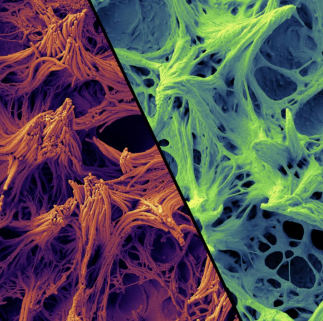
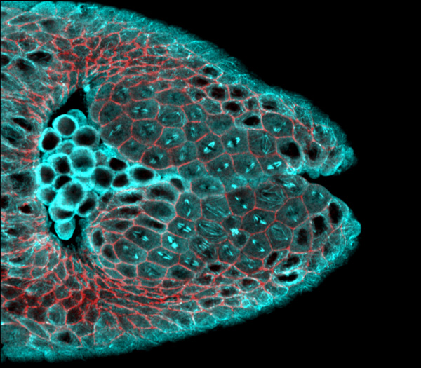
Next they wondered whether the protein might be functioning specifically within one type of airway cell, of which there are many varieties with different roles. To test this, they used a fluorescent protein to mark the cells expressing NEK10, making these cells glow green. When they allowed the cells to differentiate, the brightest glowing cells were those that were covered in cilia. This suggested to the researchers that the woman’s condition was a kind of ciliopathy, or disorder associated with cilia. Nearly all vertebrate cells have some kind of cilia, and mutations that affect their structures can have consequences such as polycystic kidney disease, retinal disease, and conditions such as obesity and cerebral anomalies.

In the lungs, cilia move mucus by wiggling back and forth in tandem with their neighbors. Moreover, previous studies had found that disruption of airway cilia could cause a disease akin to that seen in NEK10 patients. When Chivukula took a closer look at the woman’s airway cilia, he found that they still wiggled at the same speed, but something was off; while normal cilia could transport polystyrene beads on a slithery wave of mucus, her mutated cilia could barely move mucus at all.

Under a microscope, the cilia were strangely clumpy and underdeveloped. The mutation, it turned out, had caused the cilia to be too short to effectively move mucus, leading to a build-up in her airways. This mucus build-up increased her likelihood of respiratory infections and, with each infection, her bronchi grew more enlarged and swollen until she could barely breathe on her own.

A NEW DISEASE

Chivukula, Sabatini, and coauthors published their findings on the new disease in Nature Medicine in February. From what they’ve observed in the seven patients they studied, the condition follows an autosomal recessive inheritance pattern — the gene must be knocked out in both copies for airway cilia to be affected — much like cystic fibrosis and most forms of ciliopathy that affect the lungs or other tissues.

Further research will determine how variable the condition can be depending on the type of mutation in the NEK10 gene. “It’s entirely possible that there are milder or subtler variants of this gene that are not, on their own, causing this sort of end-stage lung disease,” Chivukula says.

That might mean a mutation in the NEK10 gene that led to a protein that was deformed rather than completely unstable, he says, although at this point it is impossible to know for sure. “What we do know is that this double knockout of the gene sort of phenotype is quite rare,” he says. “But like for many genetic diseases, once you understand the severe ones, you can use that information to really dig into the more common forms.”

As for the woman who received the double lung transplant, “She’s doing pretty well,” says Chivukula. “She doesn’t need oxygen and can finally walk around without becoming short of breath. Being sick for 20 years takes its toll like it would for anyone, but she’s in a much better state than she was before her transplant.”

Dr. Armstrong and others at MGH are excited by the potential applications of Chivukula’s findings. “It’s pretty unusual [for a Pathways case] to have quite as beautiful a story as Raghu was able to put together that quickly,” she says.

Maybe in the future, Chivukula says, other patients in the woman’s position will be able to be treated before their condition becomes severe enough to need a transplant in the first place. Although much research remains to be done before the condition could be cured, Chivukula believes the potential is there. Cilia, he points out, have been shown to change slightly due to external causes. For example, smokers can have cilia that are a tiny bit shorter than those of non-smokers.

“We’ve shown that delivering extra active NEK10 protein actually causes cilia function to be improved, so that does suggest that this condition could be druggable in the future,” he says. “We just need to understand the biology a little bit better.”

***

By Eva Frederick

***

Chivukula, R. et al. A human ciliopathy reveals essential functions for NEK10 in airway mucociliary clearance. Nature Medicine. 2020 Feb. doi: 10.1038/s41591-019-0730-x.

Troy Littleton earns Award for Excellence in Undergraduate Advising
April 21, 2020

The Department of Brain and Cognitive Sciences is honored to announce this year’s awards for faculty, graduate students, and undergraduates. These individuals have contributed exceptionally to the academic and intellectual life of our department.

Faculty Awards

BCS Award for Excellence in Undergraduate Advising: Troy Littleton

As one nominator said:

“He is my go-to faculty member for academic and career support, and his door is always open when you need it the most…There have been challenges that I’ve faced here that feel insurmountable, but whenever those times hit, I could talk it through with Professor Littleton. With his guidance, we would work towards a solution… And when I did pull through, when I succeeded beyond my own imaginable expectations, his office was the first place I went to for celebration.”

BCS Award for Excellence in Undergraduate Teaching: Myriam Heiman

This award is based on student evaluations and nominations. Myriam co-teaches 9.09, Cellular and Molecular Neurobiology, and 9.18, Developmental Neurobiology. Some comments from her evaluations show why she is so admired as a teacher:

“Myriam was a great professor. She went through the material at a perfect pace, and really emphasized general understanding of the topics we were learning about. She was also very welcoming to questions.”

“Professor Heiman is thorough, passionate, and insightful. The devil is always in the details and she does an excellent job highlighting the significant points in the context of the course.”

BCS Award for Excellence in Graduate Teaching: Sasha Rakhlin

Sasha teaches 9.521, Mathematical Statistics—an Asymptotic Approach, and co-teaches 9.520, Statistical Learning Theory and Applications. As with the undergraduate teaching award, this recognition is based on both course evaluations and student input. Some comments from Sasha’s evaluations were:

“Very high-quality teaching, with good explanations of difficult ideas and methods.”

“The material by nature requires you to get dirty, and I think he went through the details at the correct level.”

“One of the best lecturers I’ve had at MIT.”

BCS Award for Excellence in Graduate Mentoring: Mark Harnett

This award is based on student nominations. As one of them said of Mark:

“He has been exceptional in supporting the students with their projects and making sure they have all they need to succeed in their experiments, both technically and conceptually. He also always made sure we are prepared for important steps in grad school (qualifying exams and committee meetings) and can deliver excellent presentations. He has always dedicated time to teach fundamental aspects of patch clamp electrophysiology to all rotating students, and is always available, and happy, to answer questions.”

BCS Postdoc Award to an Outstanding Postdoctoral Mentor: Roger Levy

Roger was nominated by one of his postdocs and endorsed by the Building 46 Postdoctoral Association.  His nominator wrote:

“Roger is a brilliant leader and role-model. Roger is compassionate and understanding of the academic and social issues that postdocs face and is someone whose guidance I seek and respect. He has supported me as I try to decide whether or not to pursue an academic career, really proving to me that he has no stake in the outcome besides my wellbeing. He also has [a] tremendously strong moral ethic that reflects science at its best: from issues ranging from conflicts of interest to open access and funding transparency.”

Finally, we have added a special recognition this year:

BCS Award for Excellence in Teaching: Robert Ajemian

Robert is a research scientist in the McGovern Institute who teaches 9.53, Emergent Computations Within Distributed Neural Circuits. Students in this course give exceptionally strong evaluations. For example:

“Robert’s biggest strength is his enthusiasm for the material and for the field in general … The course setup, with Daniel and Karthik sharing some of the instructor responsibility, was a really great feature of the course – the nature of our discussions always benefited from having a variety of expert perspectives.”

“I appreciated the fervor with which the material was presented, which made the class all the more engaging, as well as the emphasis on critical thinking and debate, which is an important but often overlooked aspect of good scientific thinking.”

Robert’s instructor scores support these comments— a 6.4 in his first year, 2018, and a 6.6 in 2019. With scores and comments such as these, it was clear that we should recognize his contributions, and we are pleased to do so.

Graduate Student Awards

Angus MacDonald Award for Excellence in Undergraduate Teaching by a Graduate Student

This award is named for an MIT alum and Corporation Member who was a key supporter of our department and particularly our undergraduate educational mission. This year we are recognizing three graduate students for exemplary teaching of undergrads based on subject evaluations and faculty nominations:

  • Maddie Cusimano
  • Mark Saddler
  • Lupe Cruz

The next two awards named for Walle Nauta, a pioneering neuroanatomist, a founding member of this department, an Institute Professor, and one of the founders of the field of neuroscience.

Walle Nauta Award for Excellence in Graduate Teaching by a Graduate Student, recognizing exemplary teaching of their fellow graduate students based on subject evaluations and faculty nominations.

  • Mahdi Ramadan
  • Victoria Beja-Glasser

Walle Nauta Award for Continuing Dedication to Teaching by a Graduate Student, a special honor for someone who has already received a teaching award from our department and has continued to be exemplary.

  • Mika Braginsky
  • Tobias Kaiser
  • Halie Olson

Undergraduate Awards

Academic Awards (cumulative GPA of 4.9 or greater)

Course 9, Year 4:

  • Katherine Collins
  • Apolonia Gardner
  • Seungweon Pak
  • Ashti Shah
  • Aaditya Singh
  • Yotaro Sueoka
  • Lena Zhu
  • Merryn Daniel
  • Jingxuan Fan
  • Stephanie Hu
  • Ohyoon Kwon
  • Habiba Noamany
  • Raimundo Rodriguez
  • Lauren Schexnayder
  • Sarah Wu
  • Irene Zhou

Course 9, Year 3:

  • Ayesha Ng
  • Albert Gerovitch
  • Kristine Hocker

Course 6-9, Year 4:

  • Alice Zhang

Course 6-9, Year 3:

  • Keith Murray
  • Michelle Yakubek
  • Jasmine Zou

Research Awards (nominated by PI)

  • Keith Skaggs (Course 9, Year 3)
  • Michelle Hung (Course 9, Year 3)
  • Ohyoon Kwon (Course 9, Year 4)

Congratulations once again to all award recipients!

Harnessing the moonseed plant’s chemical know-how
Eva Frederick | Whitehead Institute
April 20, 2020

In overgrown areas from Canada to China, a lush, woody vine with crescent-shaped seeds holds the secret to making a cancer-fighting chemical. Now, Whitehead Institute researchers in Member Jing-Ke Weng’s lab have discovered how the plants do it.

Plants in the family Menispermaceae, from the Greek words “mene” meaning “crescent moon,” and “sperma,” or seed, have been used in the past for a variety of medicinal purposes. Native Americans used the plants to treat skin diseases, and would ingest them as a laxative. Moonseed was also used as an ingredient in curare, a muscle relaxant used on the tips of poison arrows.

But the plants also may have a use in modern-day medicine: a compound called acutumine shown to have anti-cancer properties (although not tested specifically against cancer cells, the chemical has been shown to kill human T-cells, an important quality for leukemia and lymphoma treatments). Acutumine is a halogenated product, which means the molecule is capped on one end by a halogen atom — a group that includes fluorine, chlorine and iodine, among others. In this case, the halogen is chlorine.

Halogenated compounds like acutumine can be useful in medicinal chemistry — their unusual chemical appendages mean they react in interesting ways with other biomolecules, and drug designers can put them to use in creating compounds to complete specific tasks in the body. Today, 20% of pharmaceutical compounds are halogenated. “However, chemists’ ability to efficiently install halogen atoms to desirable positions of starting compounds has been quite limited,” Weng says.

Most natural halogenated products come from microorganisms such as algae or bacteria, and acutumine is one of the only halogenated products made by plants. Chemists finally succeeded in synthesizing the compound in 2009, although the reaction is time-consuming and expensive (10 mg of synthesized acutumine can cost around $2,000).

Colin Kim, a graduate student in the Weng lab at Whitehead Institute, wanted to know how these plants were completing this tricky reaction using only their own genetic material. “We thought, why don’t we ask how the plants make it and then upscale the reaction [to produce it more efficiently]?” Kim says.

“By understanding how living organisms such as the moonseed plant perform chemically challenging halogenation chemistry, we could devise new biochemical approaches to produce novel halogenated compounds for drug discovery,” Weng says.

Kim knew that for every halogenated molecule in an organism, there is an enzyme called a halogenase that catalyzes the reaction that sticks on that halogen. Halogenases are useful in creating pharmaceuticals – a well-placed halogen can help fine-tune the bioactivities of various drugs. So Weng, who is also an associate professor of biology at Massachusetts Institute of Technology, and Kim, who spearheaded the project, began working to identify the helper molecule responsible for creating acutumine in moonseed plants.

First, the scientists obtained three species of Menispermaceae plants. Two of them, common moonseed (Menispermum canadense) and Chinese moonseed (Sinomenium acutum), were known to produce acutumine. They also procured one plant in the same family called snake vine (Stephania japonica) which did not produce the compound.

They began their investigation by using mass spectrometry to look for acutumine in all three plants, and then find out exactly where in the plants it was located. They found the chemical all throughout the first two — and some extra in the roots of common moonseed. As expected, the third plant, snake vine, had none, and could therefore be used as a reference species, since presumably it would not ever express the gene for the halogenase enzyme that could stick on the chlorine molecule.

Next, the researchers started searching for the gene. They began by sequencing the RNA that was being expressed in the plants (RNA serves as a messenger between genomic DNA and functional proteins), and created a huge database of RNA sorted by what tissue it had been identified in.

At this point, the extra acutumine in the roots of common moonseed came in handy. The researchers had some idea of what the enzyme might look like – past research on other halogenases in bacteria suggested that one specific family of enzyme, called Fe(II)/2-oxoglutarate-dependent halogenases, or 2ODHs, for short, was capable of site-specifically adding a halogen in the same way that the moonseed’s mystery enzyme did. Although no 2ODHs had yet been found in plants, the researchers thought this lead was worth a look. So they searched specifically for transcripts similar to 2ODH sequences that were more highly expressed in the roots of common moonseed than in the leaves and stems.

After analyzing the RNA transcripts, Kim and Weng were pretty sure they had found what they were looking for: one gene in particular (which they named McDAH, short for M. canadense dechloroacutumine halogenase) was highly expressed in the roots of common moonseed. Then, in Chinese moonseed, they identified another protein that shared 99.1 percent of McDAH’s sequence, called SaDAH. No similar protein was found in snakevine, suggesting that this protein was likely the enzyme they wanted.

To be sure, the researchers tested the enzyme in the lab, and found that it was indeed the first-ever plant 2ODH, able to stick on the chlorine molecule to the alkaloid molecule dechloroacutumine to form acutumine. Interestingly, the enzyme was pretty picky; when they gave it other alkaloids like codeine and berberine to see if it would install a halogen on those as well, the enzyme ignored them, suggesting it was highly specific toward its preferred substrate, dechloroacutumine, the precursor of acutumine. They compared the enzyme’s activity to other similar enzymes, and found the key to its ability lay in the substitution of one specific amino acid in the active site– aspartic acid — for a glycine.

Now that they had identified the enzyme responsible for the moonseed’s halogenation reactions, Kim and Weng wanted to see what else it could do. A chemical capable of catalyzing such a complex reaction might be useful for chemists trying to synthesize other compounds, they hypothesized.

So they presented the enzyme with some dechloroacutumine and a whole buffet of alternative anions to see whether it might catalyze a reaction with any of these molecules in lieu of chlorine. Of the selection of anions, including bromide, azide, and nitrogen dioxide, the enzyme catalyzed a reaction only with azide, a construct of 3 nitrogen atoms.

“That is super cool, because there isn’t any other naturally occurring azidating enzyme that we know of,” Kim says. The enzyme could be used in click chemistry, a nature-inspired method to create a desired product through a series of simple, easy reactions.

In future studies, Weng and Kim hope to use what they’ve learned about the McDAH and SaDAH enzymes as a starting point to create enzymes that can be used as tools in drug development. They’re also interested in using the enzyme on other plant products to see what happens. “Plant natural products, even without chlorines, are pretty effective and bioactive, so it would be cool to see if you can take those plant natural products and then install chlorines to see what kind of changes and bioactivity it has, whether it develops new-to-nature functions or retain its original bioactivity with enhanced properties,” Kim says. “It expands the biocatalytic toolbox we have for natural product biosynthesis and its derivatization.”

***

Written by Eva Frederick

***

Citation: Kim, Colin Y. et al. The chloroalkaloid (−)-acutumine is biosynthesized via a Fe(II)- and 2-oxoglutarate-dependent halogenase in Menispermaceae plants. Nature Communications. April 20, 2020. DOI: 10.1038/s41467-020-15777-w

Stretch and relax
Lucy Jakub
April 13, 2020

Consider the fruit fly, Drosophila melanogaster. Though it’s only a couple of millimeters long, its body is intricately complex. But it began, as most animals do, as an amorphous blastula—a hollow ball of dividing cells. During embryonic development, the structures of the body emerge as cells multiply and change shape, sculpting tissues into the mature forms dictated by the genetic code. One of the first structural changes is gastrulation, during which the blastula becomes multilayered with an ectoderm, mesoderm, and endoderm. In the developing fly, this occurs through a tissue folding mechanism. The first fold is the invagination of the mesoderm, when cells fated to become muscles contract and curl inward, leaving the cells fated to become skin on the exterior.

Biologists have traditionally focused on how cells generate force to understand cell and tissue shape change. But researchers at MIT have found that there’s another important, though often overlooked, player in tissue folding: cell division, or mitosis. By combining live-imaging with genetic mutations of developing Drosophila embryos, they observed that cell constriction and division can act together to promote folding, and that mitosis interferes with the accumulation of motor proteins that allows cells to generate force.

“What the results tell us is that the cell cycle and cell division might need to be tightly regulated relative to other shape changes that are happening in the tissue,” says Adam Martin, the senior author of the study published on March 13 in Molecular Biology of the Cell. “They present a new paradigm for thinking about how tissue shape might be regulated during development, and provide insight into what might cause birth defects in humans.” Clint Ko PhD ’20, a former graduate student in the Martin lab, was lead author of the study.

In 2000, three different labs identified a genetic mutation that caused premature cell division in developing Drosophila embryos. They found that the gene tribbles, named for the fuzzy, rapidly-reproducing animals in Star Trek, regulates cell division in the mesoderm of the fly, ensuring that cells only divide at the appropriate time. When that gene is deleted, cell division occurs before the mesoderm can properly internalize. What was notable about this mutant was that the blastula never folded, and remained a ball of cells instead of an envelope of tissue with an inside and an outside. This observation led researchers to believe that cell cycle regulation somehow regulates tissue folding. But, at the time, there was no live-imaging technology to visualize how cells changed in the developing embryo.

By using a fluorescent protein to visualize chromosome condensation, which marks the start of mitosis and the cell’s preparation for division, the researchers were able to use live-cell imaging to see how premature division might be interfering with cell constriction. When a cell prepares to divide, it expands and becomes rounded, before elongating—shape changes that exert force on neighboring cells. But something else was going on, too.Specifically, researchers in the Martin lab wanted to see what was happening to networks of the motor protein myosin, which allows cells to contract, in the tribbles mutant. Myosin is the same protein that allows our muscle tissue to contract when we flex. To facilitate tissue folding in the developing fly, myosin is concentrated at the top of the cells in the mesoderm, where they form the surface of the blastula. As this myosin constricts, the outer surface of the tissue shrinks and contracts inward.

“We noticed that when the cells are dividing, the apical myosin networks that are present disappear,” says Ko. Cells that had already begun to contract relaxed when they entered mitosis, indicating that it’s a loss of contractility in the tribbles mutant that prevents folding. The researchers suspect that this reversal occurs because mitosis disrupts signaling from the gene RhoA, which regulates contractility and cell shape changes during development. An undergraduate researcher in the lab, Prateek Kalakuntla, showed that regulation of RhoA changes at the start of mitosis.

“Initially we were just curious about the tribbles mutant,” says Ko. “But then we started exploring other ways of looking at how cell divisions affect myosin accumulation in cells.” They utilized a mutation in which the gene fog, which is located upstream of myosin activation on the genome, was overexpressed. (Fog is short for “folded gastrulation.”) Cells in the Drosophila ectoderm don’t normally contract, but with ectopic fog overexpression, those cells activated myosin, too. With live-cell imaging, the researchers observed furrows develop across the ectoderm.

“It was a bit unexpected to see these tissues folding when they shouldn’t be folding,” says Ko. Specifically, the folds occurred along the boundaries of mitotic domains, regions of spatiotemporally patterned cell divisions that occur in coordinated pulses. “That led to this sort of novel idea that cell divisions—particularly when they’re in this pattern where they’re interspersed between contractile cells—can actually promote tissue folding.”

Understanding the genetic basis for tissue folding, and how our genes control the development of specific bodily features, can help determine how birth defects arise during development. “If cell cycle control is misregulated during development, it could actually alter the shape of that tissue,” says Martin. The study paves the way for further research into how exactly the location of myosin in the cell is regulated, and how it is affected at the molecular level by cell division.

“We observed that when these cells enter mitosis, the localization of myosin activators changes. But we don’t really know how it changes,” says Ko. “That would be a pretty interesting research problem, especially considering that it’s such an integral part of force generation in cells.” Kalakuntla has begun investigating what controls these regulators, which will be an avenue of future research for the lab.

Top image: Myosin networks, in green, contract cell membranes in the mesoderm of a developing Drosophila embryo. Credit: Martin lab.

Citation:
“Apical Constriction Reversal upon Mitotic Entry Underlies Different Morphogenetic Outcomes of Cell Division”
Molecular Biology of the Cell, online March 4, 2020, DOI: 10.1091/mbc.E19-12-0673
Clint S. Ko, Prateek Kalakuntla, and Adam C. Martin

Interested in sharpening your science communication skills?

An internship with MIT Biology can get you on your way.

Raleigh McElvery
April 7, 2020

For the past several years, MIT Biology has been training undergraduates, graduate students, and research associates in the craft of science communication. In an effort to foster professional development and share the exciting research that transpires on campus, our communications team offers science writing and multimedia internships. We develop these positions to align with the interests of our interns, who often help out on a volunteer basis. Assignments range from assisting with videos and podcasts to writing news stories and profiles, aiding with social media, and chronicling the history of the department. After honing their own skills, many of our interns have successfully competed for prestigious communications fellowships, graduate programs in science writing, and communications jobs. Take a look at what they’ve done, and contact us if you’re a member of the department interested in joining our team.

Justin Chen PhD ’18 (Spring 2017 – Spring 2018)

Justin Chen earned his PhD in Hazel Sive’s lab, using frog embryos to model human craniofacial development. As a science writing intern, he composed student profiles for the department website and articles on research papers for MIT News. After graduating from MIT, he earned an AAAS Mass Media and Science and Engineering Fellowship, which he spent at STAT News publishing breaking news and profiles of scientists. He is currently an external affairs associate at OpenBiome, where he drafts press releases, annual reports, academic publications, and patient education materials, while helping to manage the website and social media. In addition to his work at Openbiome, he authors personal essays as a writer-in-residence at Porter Square Books.

Nafisa Syed SB ’19 (Spring 2019)

Nafisa Syed earned her bachelor’s degree in Biology (Course 7), with minors in Science Writing (Course 21W) and Brain and Cognitive Sciences (Course 9). She was an editor at The TechMIT Undergraduate Research Journal (MURJ), and Rune Literary Magazine, while completing a UROP in Evelina Fedorenko’s lab studying the brain’s language regions. As an intern at MIT Biology, Nafisa generated content for the internal newsletter, spearheaded social media campaigns, and analyzed data displaying the distribution of life science funding across the Institute. She is currently earning her master’s degree at MIT’s Graduate Program in Science Writing.

Saima Sidik (Spring 2019 – Spring 2020)

Saima Sidik is a research associate in Sebastian Lourido’s lab, where she studies how the parasite Toxoplasma gondii causes disease. In addition to authoring articles on scientific research for her blog, 10X Objective, Saima composes student profiles for the department website and MIT Newsnews briefs, and archival pieces about the history of biology at MIT. Starting this fall, she will begin her master’s degree at MIT’s Graduate Program in Science Writing.

Lucy Jakub (Fall 2019- Spring 2020)

Lucy Jakub served as the editorial assistant at The New York Review of Books for two years before entering MIT’s Graduate Program in Science Writing in the fall of 2019. As an intern for MIT Biology, she writes news briefs for the department website, student profiles for MIT News, and articles on recent events, in addition to generating the internal newsletter and social media campaigns. Her work has also appeared in Harper’s Magazine and National Geographic.

Sebastian Swanson (Fall 2018 – present)

Sebastian Swanson is a fourth-year graduate student in Amy Keating’s lab, studying the principles of protein-protein interactions in order to develop algorithms for peptide design. As an undergraduate at the University of Minnesota, he served as an officer and co-chair of MinneCinema Studios, which produces a variety of multimedia projects ranging from mock TV episodes to short films. He is currently the department’s primary cinematographer, filming faculty profiles and short videos on research projects.

Are you a member of the MIT Biology community interested in honing your scientific communication skills? Contact biowebmaster@mit.edu to discuss potential internship opportunities.