These genes help explain how malaria parasites survive treatment with common drug
Eva Frederick | Whitehead Institute
September 23, 2020

The essential malaria drug artemisinin acts like a “ticking time bomb” in parasite cells — but in the half a century since the drug was introduced, malaria-causing parasites have slowly grown less and less susceptible to the treatment, threatening attempts at global control over the disease.

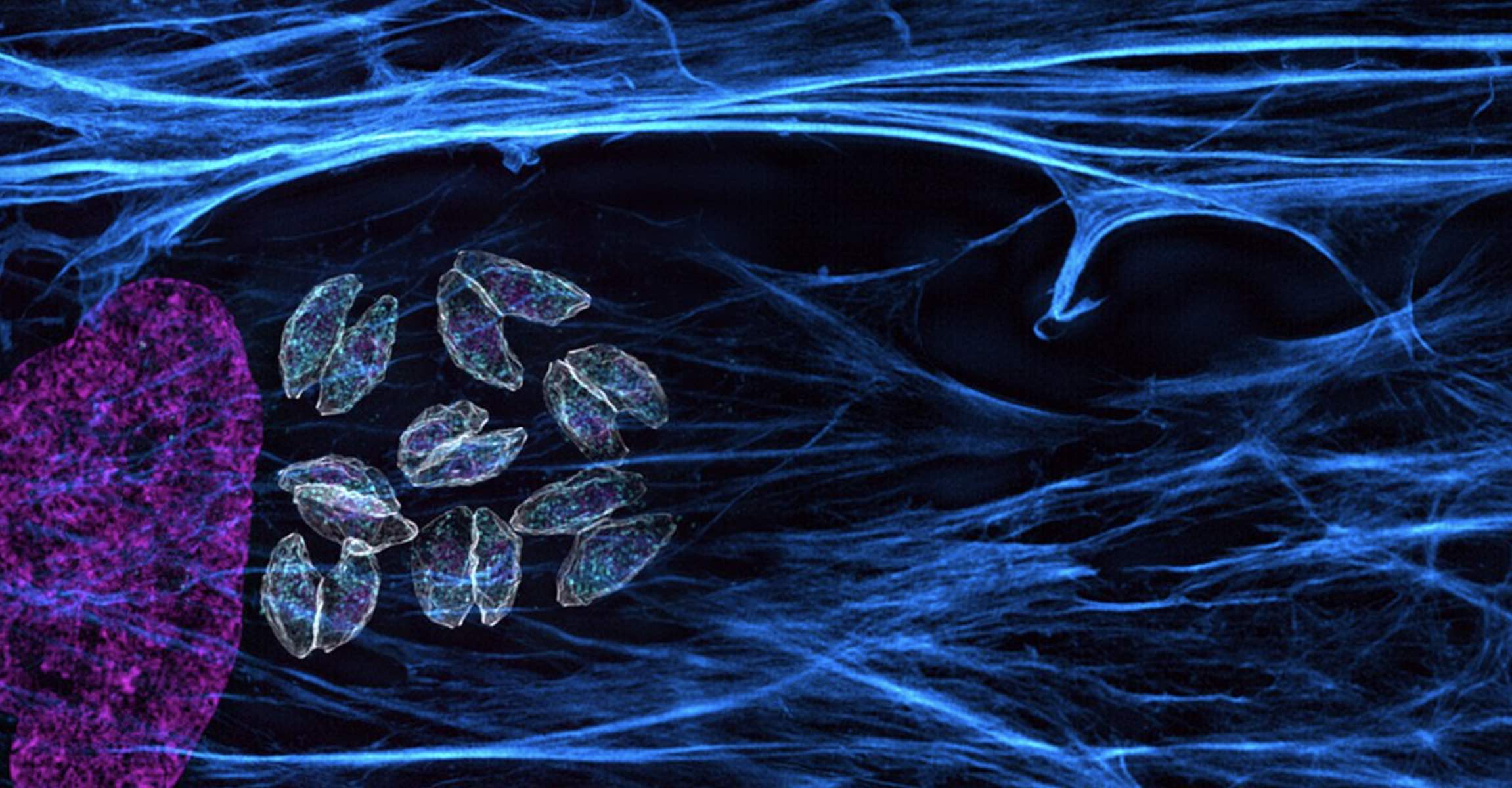
In a paper published September 23 in Nature Communications, Whitehead Institute Member Sebastian Lourido and colleagues use genome screening techniques in the related parasite Toxoplasma gondii (T. gondii) to identify genes that affect the parasites’ susceptibility to artemisinin. Two genes stood out in the screen: one that makes the drug more lethal, and another that helps the parasite survive the treatment.

Artemisinin is derived from the extract of sweet wormwood (Artemisia annua), and is usually used against malaria as part of a combination therapy. “Artemisinin kills malaria-causing parasites super fast—it will wipe out 90 percent of parasites within 24 hours,” says former postdoctoral researcher and co-first author Clare Harding, now a research fellow at the University of Glasgow. Once the fast-acting drug clears out the bulk of the parasites—such as Plasmodium falciparum, the culprit in the deadliest forms of malaria—from the bloodstream, the second drug finishes off the stragglers, curing the infection.

“Artemisinin works differently than most antibiotics,” Lourido said. “You can think of it as a sort of bomb that needs to be turned on in order to work.” The molecule required to light the drug’s fuse is called heme. Heme is a small molecule that facilitates several cellular functions, including electron transport and the delivery of oxygen to tissues as a component of hemoglobin. When heme molecules encounter artemisinin, they activate the drug allowing the creation of small, toxic radicals which react with proteins, lipids and metabolites inside the parasite, leading to its death.

Lourido, Harding, and co-first authors Boryana Petrova and Saima Sidik (“We were the ‘Heme Team,’” Harding said) wanted to understand what mechanisms the less susceptible parasites were using to avoid activating the “bomb”. Previously, Lourido and his lab—which focuses on apicomplexan parasites, a group which includes both Toxoplasma gondii and the malaria-causing Plasmodium falciparum—had developed a method to screen the entire genome of T. gondii to discover beneficial and harmful mutations. For a number of reasons, the screen does not work on Plasmodium parasites, but Lourido hypothesized that the related parasites’ genomes were similar enough that the method could prove helpful.

After running the screen, two genes stood out to the researchers as important factors in the parasites’ susceptibility to artemisinin treatment. One, called Tmem14c, seemed to be protecting the parasites: when the gene was disrupted in the screen, the parasites became more susceptible to treatment with artemisinin. The gene is analogous to one in red blood cells that serves as a transporter for heme and its building blocks, shuttling them in and out of the mitochondrion.

“What could be happening here is that, in the absence of Tmem14c, heme, artemisinin’s activator, collects within the mitochondria where it is being synthesized, thereby rendering the mitochondria better at activating that ticking time bomb,” Lourido said. “Having that high concentration of heme in the mitochondria is like having a flame when there is a gas leak.”

The screen also identified one mutation that led to parasites being less sensitive to artemisinin. The mutation affected a gene called DegP2, the product of which interacts with several mitochondrial proteins and appears to play a role in heme metabolism. When less DegP2 was present, the cells contained a lower amount of heme, which in turn made it less likely that the parasites would be killed by artemisinin.

Both the findings support other research suggesting that heme metabolism is crucial for artemisinin susceptibility. “It is important to consider the role of heme when combining artemisinin with other therapies,” Lourido said. “You would want to avoid combination therapy that might inadvertently suppress the level of heme within the parasite and thereby reduce susceptibility to antiparasitic agents.”

The project also showed the potential of using the Toxoplasma screening method as a model to study other related parasites. The screen confirmed findings in Toxoplasma that had previously been shown in Plasmodium, suggesting that it could be a valuable tool in studying malaria and other diseases caused by apicomplexan parasites.

“Through the amazing screens and molecular biology that you can do in Toxoplasma, we can really learn a lot about the biology of this diverse group of parasites,” Lourido said. “Defeating malaria is going to take a lot of different and creative approaches, and the fundamental research that we can do in Toxoplasma can in fact inform many of the critical clinical questions we need to answer to control this disease.”

***

Written by Eva Frederick

***

Harding, C., Sidik, S, and Petrova, B., et al. “Genetic screens reveal a central role for heme metabolism in artemisinin susceptibility.” Nature Communications. DOI: https://doi.org/10.1038/s41467-020-18624-0

Bringing new energy to mitochondria research
Greta Friar | Whitehead Institute
September 17, 2020

Tiny mitochondria in our cells turn oxygen and nutrients into usable energy in a process called respiration. This process is essential for powering our cells, and yet in spite of its importance many of the finer details of how it happens remain unknown. One long-standing mystery is how a molecule called nicotinamide adenine dinucleotide (NAD), which plays a big part in respiration and metabolism, gets into the mitochondria in humans and other animals. Mitochondria use NAD in order to produce adenosine triphosphate (ATP), the energy supply molecules used throughout the cell. Researchers knew the identities of the molecules that transport NAD from the wider cell into the mitochondria of yeast and plants, but had not found the animal equivalent—in fact, there was some debate over whether one even existed or whether animal cells used other methods altogether.

Now, research from postdoctoral researcher Nora Kory in Whitehead Institute Member David Sabatini’s lab may end the debate. In a paper published in Science Advances on September 9, the researchers show that the missing human NAD transporter is likely the protein MCART1. This discovery not only answers a longstanding question about a vital cellular process, but may contribute to research on aging—during which cells’ NAD levels drop—as well as research on diseases that involve certain mitochondrial dysfunctions, for which cells with broken NAD transporters could be an experimental model.

“I find it striking that mitochondria play such an important role in metabolism in the cell, which in turn plays a huge role in health and disease, but we still don’t understand how all of the molecules involved get in and out of mitochondria. It was exciting to fill in a piece of that puzzle.” Kory says.

AN UNEXPECTED DISCOVERY

Kory did not set out to find the long sought-after transport molecule. Rather, she was trying to better understand mitochondrial respiration by mapping the genes involved. She was comparing gene essentiality profiles, which show how important a gene is to different processes in a cell—the more co-essential two genes are, the more likely they are to be involved in the same cellular process—and one gene stood out: MCART1, also known as SLC25A51. It was highly correlated to other genes involved in mitochondrial respiration, and belonged to a family of genes known to code for transporters, yet its function was unknown. The protein coded for by MCART1 clearly played an important role, so Kory decided to figure out what that was; as her research progressed, she realized she had found the missing NAD transporter.

Kory and colleagues applied a common approach to determine MCART1’s function: inactivate the gene in cells, and see what breaks down in its absence. This approach is like troubleshooting a machine; if you cut a wire in your car and the headlights stop working, but everything else is fine, then that wire was probably linked to the headlights. When the researchers removed MCART1, the cells exhibited much lower oxygen consumption, reduced respiration and ATP production, and reliance on other, far less efficient means of ATP production—exactly what you’d expect to see if the inactivated gene was needed for respiration. Moreover, the biggest change that the researchers observed in cells without MCART1 was reduced levels of NAD in the mitochondria, while NAD levels in the wider cell remained the same, which they quantified using experiments previously developed in the lab. The researchers confirmed that MCART1 is essential for NAD transport into isolated mitochondria and overabundance of MCART1 caused an increased uptake.

“It’s very satisfying when our lab returns to the techniques that we have developed in order to make new findings such as identifying this important protein,” says Sabatini, who is also a professor of biology at Massachusetts Institute of Technology and an investigator with the Howard Hughes Medical Institute.

The evidence supports that the protein MCART1 is itself the transport channel. However, it is possible that the protein may play some other essential contributing role to transportation, or that it combines with other molecules to do its job. To strengthen the case for MCART1 as the transporter, the researchers showed that MCART1 and the known yeast NAD transport could be switched out for each other in both human and yeast cells, suggesting an equivalent function. Still, further experiments are needed to determine the precise mechanism of transport.

A serendipitous case of synchronous discovery reinforces Kory’s findings. A paper by other researchers published on the same day in the journal Nature also put forth that MCART1 is the missing NAD transporter, based on a completely different set of evidence. Combined, the papers provide an even more compelling case.

“It was nice to see how our different approaches complemented each other, and led to the same conclusion,” Kory says.

Understanding how NAD gets into the mitochondria opens up new questions about the details of mitochondrial respiration. Kory will shortly be leaving Sabatini’s lab to open her own lab at the Harvard T.H. Chan School of Public Health, where she intended to continue investigating the role of the mitochondria’s NAD supply in metabolism and signaling.

***

Written by Greta Friar

***

David Sabatini’s primary affiliation is with Whitehead Institute for Biomedical Research, where his laboratory is located and all his research is conducted. He is also a Howard Hughes Medical Institute investigator and a professor of biology at Massachusetts Institute of Technology.

***

Citations:

Kory, N., et al. (2020). MCART1/SLC25A51 is required for mitochondrial NAD transport. Science Advances. doi:10.1126/sciadv.abe5310

Luongo, T. S., et al. (2020). SLC25A51 is a mammalian mitochondrial NAD+ transporter. Nature. doi:10.1038/s41586-020-2741-7

Defining a “new normal” for campus research

Despite the trials and tribulations of the COVID-19 pandemic, Building 68 core facilities have remained open for business.

Raleigh McElvery
September 10, 2020

In mid-March, MIT closed its doors due to the COVID-19 pandemic, and Building 68 temporarily became a ghost town. Home to over 25 life science labs and three core facilities, the Department of Biology’s primary research hub usually teems with activity. But this spring, only a skeleton crew of essential workers came in and out, maintaining the equipment and running select experiments. Since then, the ghost town has gradually come back to life, as scientists are returning to their benches once again while taking safety precautions.

Three of the 24 core facilities affiliated with the life sciences are located in Building 68: the BioMicro Center, Structural Biology Core Facility, and Biophysical Instrumentation Facility. Known fondly as the “BIF,” the latter houses instruments that help researchers elucidate macromolecular structures. Select staff members remained available throughout the research shutdown to help biologists, biological engineers, and chemists run essential protocols.

One floor down, the BioMicro Center also continued to offer limited services — from maintaining multi-year cancer studies to running analyses probing SARS-CoV-2, the virus strain that causes COVID-19. The team there specializes in genomic and transcriptomic technologies, bioinformatics, and research computing.

Stuart Levine SB ’97,  who leads the BioMicro Center, says the facility handled anywhere from six to nine projects a day before the pandemic. But during the research shutdown, requests dwindled to a steady “trickle.”

“We did whatever we could to be helpful,” he adds. “I went to campus a few times early on, and it was eerie to be one of the only people in the building.”

Illustration of virus-like molecule
Illustration of the Bathe lab’s virus-like nanoparticle. Credit: Ella Maru Design Studio

Although the BioMicro Center is situated in Building 68, it serves a wide array of individuals and labs across campus. Chemical engineering graduate student, Grant Knappe, was also among the select few permitted to work in lab during the shutdown, and he relied heavily on the BioMicro Center for a key step in his experimental protocol. Knappe’s advisor, Professor of Biological Engineering Mark Bathe, began shifting his group’s focus to COVID-related projects almost immediately after campus emptied in mid-March.

The Bathe lab studies nanoparticles made from DNA “origami” that’s been folded into tiny geometric shapes. They’ve developed user-friendly algorithms to design these structures, and regularly employ the BioMicro’s oligonucleotide synthesizer to produce their DNA strands. With the help of the facility, Knappe and his colleagues recently created nanoparticles adorned with short DNA strands to mimic the SARS-CoV-2 spike protein — which induces the body’s immune response. They hope these geometric nanoparticles will eventually help develop COVID-19 vaccines.

“The scientific process is usually very collaborative,” Knappe says, “so at the beginning it was difficult to run experiments without other people nearby to bounce ideas back-and-forth.”

Focusing on just one project — rather than several simultaneously — was also a new experience. Knappe is excited to see where his COVID research will go, and what lab instruments will ultimately be key to the process. “You never know what equipment could end up fighting the virus,” he adds.

On June 1, Phase 1 of the research ramp-up began, and labs were permitted to begin operating at 25% capacity. Scientists started working in shifts with reduced hours, conducting their experiments many feet apart, and visiting MIT Medical for regular COVID-19 testing. Levine remembers that the number of requests for BioMicro services surged almost immediately as researchers returned.

At the same time, Robert Grant, the Research Scientist responsible for the Structural Biology Core Facility, started up the core’s state-of-the-art X-ray crystallography equipment. Leaving in March had been a “mad scramble,” and he remembers hastily terminating non-essential experiments and distributing extra resources (like liquid nitrogen tanks) to labs in-need. When he returned in June he already had project requests.

“A big part of my job is interacting with people, which I really enjoy,” he says. “But we’ve had to adapt, and and devise new ways to train people on equipment and data processing that don’t require close contact.”

Grant has recently started socially distant one-on-one trainings, where both parties remain as far apart as possible while wearing masks and gloves. In some cases, he’s processed samples and collected data for users, helping them perform analyses via Zoom. He’s also found ways to revive collaborations with other institutes. He recently sent crystals to Argonne National Laboratory in Chicago. The student who grew the crystals then remotely controlled an X-ray beamline at Argonne’s Advanced Photon Source synchrotron to collect diffraction data from home while Zooming with Grant.

“We’re definitely open for business, although things look a little different than before,” Grant says. “We’ve reached a new normal.”

Building 68's Structural Biology Core Facility
Building 68’s Structural Biology Core Facility
Posted: 9.10.20
Sebastian Lourido earns ASM Award for Early Career Basic Research
August 28, 2020

Washington, D.C. – August 27, 2020 – The 2021 American Society for Microbiology (ASM) awardees in research, education and leadership have now been announced. ASM congratulates all of the award recipients for their achievements. The ASM Awards program is managed by the American Academy of Microbiology, the honorific leadership group within ASM. The mission of the Academy is to recognize microbiologists for outstanding contributions to the microbial sciences and to provide microbiological expertise in the service of science and the public.

The 2021 ASM Award Laureates:

ASM Alice C. Evans Award for Advancement of Women
Recognizes outstanding contributions toward the full participation and advancement of women in the microbial sciences. This award is given in memory of Alice C. Evans, the first woman to be elected ASM president (elected in 1928).
•    Jennifer Glass, Ph.D.

ASM Award for Applied and Biotechnological Research
Recognizes an outstanding scientist with distinguished research achievements in the development of products, processes and technologies that have advanced the microbial sciences.
•    Dennis Hruby, Ph.D.

ASM Award for Early Career Applied and Biotechnological Research
Recognizes an early career investigator with distinguished research achievements in the development of products, processes and technologies that have advanced the microbial sciences.
•    Kizzmekia Corbett, Ph.D.

ASM Award for Basic Research
Recognizes an outstanding scientist whose discoveries have been fundamental to advancing our understanding of the microbial world.
•    Sue Wickner, Ph.D.

ASM Award for Early Career Basic Research
Recognizes an early career investigator with distinguished basic research achievements in the microbial sciences.
•    Sebastian Lourido, Ph.D.

ASM Award for Environmental Research
Recognizes an outstanding scientist with distinguished research achievements that have improved our understanding of microbes in the environment, including aquatic, terrestrial and atmospheric settings.
•    Terry Hazen, Ph.D.

ASM Award for Early Career Environmental Research
Recognizes an early career investigator with distinguished research achievements that have improved our understanding of microbes in the environment, including aquatic, terrestrial and atmospheric settings.
•    A. Murat Eren, Ph.D.

ASM Award for Education
Recognizes general excellence in microbiology education. Education is broadly defined and meant to include any and all activities that inform and motivate students about the discipline of microbiology.
•    Nichole Broderick, Ph.D.

ASM Award for Research and Leadership in Clinical Microbiology
Recognizes an outstanding scientist/clinical microbiologist with distinguished research achievements, and a record of innovation and advancement of the clinical microbiology profession. This award represents the merging of the BD Research and Sonnenwirth Awards given annually since 1978 and 1986, respectively.
•    Melissa Miller, Ph.D.

ASM Award for Service
Recognizes outstanding contributions through service to the microbiological community.
•    Barbara Robinson-Dunn, Ph.D.

ASM Carski Award
Recognizes an educator for outstanding teaching of microbiology to undergraduate students and for encouraging them to subsequent achievement.
•    Jason Tor, Ph.D.

ASM D.C. White Award
Recognizes distinguished accomplishments in interdisciplinary research and mentoring in microbiology. This award honors D.C. White, who was known for his interdisciplinary scientific approach and for being a dedicated and inspiring mentor.
•    Ferran Garcia-Pichel, Ph.D.

ASM Lifetime Achievement Award
ASM’s premier award for sustained contributions to the microbiological sciences.
•    Bernard Moss, MD, Ph.D.

ASM Moselio Schaechter Award in Recognition of a Developing-Country Microbiologist
This award, named in honor of Professor Moselio Schaechter, former ASM president, recognizes a scientist who has shown exemplary leadership and commitment towards the substantial furthering of the profession of microbiology in research, education or technology in the developing world.
•    Gustavo Goldman, Ph.D.

ASM Scherago-Rubin Award for Clinical Microbiology
Recognizes an outstanding bench-level clinical microbiologist involved in routine diagnostic work that has distinguished her- or himself by excellent performance. The award was established by the late Sally Jo Rubin, an active member of ASM’s Clinical Microbiology Division, in honor of her grandfather, Professor Morris Scherago.
•    Brandon Ellis, B.S.

ASM William A. Hinton Award for Advancement of a Diverse Community of Microbiologists
Recognizes outstanding contributions toward fostering the research training of minorities and in increasing diversity in microbiology. It is given in memory of William A. Hinton, a physician-research scientist, and one of the first African-Americans to join ASM.
•    Eric Triplett, Ph.D.

###

The American Society for Microbiology is one of the largest professional societies dedicated to the life sciences and is composed of 30,000 scientists and health practitioners. ASM’s mission is to promote and advance the microbial sciences.

ASM advances the microbial sciences through conferences, publications, certifications and educational opportunities. It enhances laboratory capacity around the globe through training and resources. It provides a network for scientists in academia, industry and clinical settings. Additionally, ASM promotes a deeper understanding of the microbial sciences to diverse audiences.

“Runaway” Transcription

Researchers discover new rules governing bacterial gene expression that overturn fundamental assumptions about basic biological pathways.

Raleigh McElvery
August 26, 2020

On the evolutionary tree, humans diverged from yeast roughly one billion years ago. By comparison, two seemingly similar species of bacteria, Escherichia coli and Bacillus subtilis, have been evolving apart for roughly twice as long. In other words: walking, talking bipeds are closer on the tree of life to single-celled fungus than these two bacteria are to one another. In fact, it’s becoming increasingly clear that what is true of one bacterial type may not be true of another — even when it comes down to life’s most basic biological pathways.

E. coli has served as a model organism in scientific research for over a century, and helped researchers parse many fundamental processes, including gene expression. In these bacteria, as one molecular machine, the RNA polymerase, moves along the DNA transcribing it into RNA, it is followed in close pursuit by a second molecular machine, the ribosome, which translates the RNA into proteins. This “coupled” transcription-translation helps monitor and tune RNA output, and is considered a hallmark of bacteria.

However, an interdisciplinary team of biologists and physicists recently showed that the B. subtilis bacterium employs a different set of rules. Rather than working in tandem with the ribosome, the polymerase in B. subtilis speeds ahead. This system of “runaway” transcription creates alternative rules for RNA quality control, and provides insights into the sheer diversity of bacterial species.

“Generations of researchers, including myself, were taught that coupled transcription-translation is fundamental to bacterial gene expression,” says Gene-Wei Li, an associate professor of biology and senior author of the study. “But our very precise, quantitative measurements have overturned that long-held view, and this study could be just the tip of the iceberg.”

Grace Johnson, a graduate student in the Department of Biology, and Jean-Benoît Lalanne, a graduate student in the Department of Physics, are the lead authors on the paper, which appeared in Nature on Aug. 26.

A curious clue

In 2018, Lalanne developed an experimental technique to measure the boundaries of RNA transcripts. When DNA is transcribed into RNA, the resulting transcripts are generally longer than the DNA coding sequence because they also have to include an extra bit at the end to signal the polymerase to stop. In B. subtilis, Lalanne noticed there simply wasn’t enough space between the ends of the coding sequences and the ends of the RNA transcripts — the extra code was too short for both the polymerase and the ribosome to fit at the same time. In this bacterium, coupled transcription-translation didn’t seem possible.

“It was a pretty weird observation,” Lalanne recalls. “It just didn’t square up with the accepted dogma.”

To delve further into these puzzling observations, Johnson measured the speeds of the RNA polymerase and ribosome in B. subtilis. She was surprised to find that they were moving at very different rates: the polymerase was going roughly twice as fast as the ribosome.

During coupled transcription-translation in E. coli, the ribosome is so closely associated with the RNA polymerase that it can control when transcription terminates. If the RNA encodes a “premature” signal for the polymerase to stop transcribing, the nearby ribosome can mask it and spur the polymerase on. However, if something goes awry and the ribosome is halted too far behind the polymerase, a protein called Rho can intervene to terminate transcription at these premature sites, halting the production of these presumably non-functional transcripts.

However, in B. subtilis, the ribosome is always too far behind the polymerase to exert its masking effect. Instead, Johnson found that Rho recognizes signals encoded in the RNA itself. This allows Rho to prevent production of select RNAs while ensuring it doesn’t suppress all RNAs. However, these specific signals also mean Rho likely has a more limited role in B. subtilis than it does in E. coli.

A family trait

To gauge how common runaway transcription is, Lalanne created algorithms that sifted through genomes from over 1,000 bacterial species to identify the ends of transcripts. In many cases, there was not enough space at the end of the transcripts for both the RNA polymerase and the ribosome to fit, indicating that more than 200 additional bacteria also rely on runaway transcription.

“It was striking to see just how widespread this phenomenon is,” Li says. “It raises the question: How much do we really know about these model organisms we’ve been studying for so many years?”

Carol Gross, a professor in the Department of Microbiology and Immunology at University of California San Francisco who was not involved with the study, refers to the work as a “tour de force.”

“Gene-Wei Li and colleagues show transcription-translation coupling, thought to be a foundational feature of bacterial gene regulation, is not universal,” she says. “Instead, runaway transcription leads to a host of alternative regulatory strategies, thereby opening a new frontier in our study of bacterial strategies to thrive in varied environments.”

As researchers widen their experimental radius to include more types of bacteria, they are learning more about the basic biological processes underlying these microorganisms — with implications for those that take up residence in the human body, from helpful gut microbes to noxious pathogens.

“We’re beginning to realize that bacteria can have distinct ways of regulating gene expression and responding to environmental stress,” Johnson says. “It just shows how much interesting biology is left to uncover as we study increasingly diverse bacteria.”

Citation:
“Functionally uncoupled transcription–translation in Bacillus subtilis
Nature, online August 26, 2020, DOI: 10.1038/s41586-020-2638-5
Grace E. Johnson, Jean-Benoît Lalanne, Michelle L. Peters, and Gene-Wei Li

Top illustration: Researchers discovered a new system of transcription and translation in bacteria, where the polymerase (pink) in B. subtilis “runs away” from the ribosome (blue). Credit: Grace Johnson
Posted: 8.26.20
Antibiotic resistance: How to prevent the next public health emergency
Emma H. Yee, Steven S. Cheng, Grant A. Knappe, and Christine A. Moomau | MIT Science Policy Review
August 25, 2020

Article Summary

Antibiotics are a vital component of global health. By killing or inhibiting the growth of bacteria, antibiotics treat infections like pneumonia, staph, and tuberculosis.By preventing infections, they enable major medical procedures such as surgeries and chemotherapy. However,bacteria are becoming increasingly resistant to current antibiotics, causing an estimated 34,000 deaths annually in the US. Left unchecked, antibiotic resistance will have major public health consequences, causing over 5 million deaths each year by 2050. Major causes of this crisis are the misuse of existing antibiotics and the slow development of new antibiotics. To incentivize responsible use, governments and institutions are initiating education programs, mandating comprehensive hospital antibiotic stewardship programs, and funding the development of rapid diagnostics. To bring new antibiotic drugs to market, the US government and other non-governmental organizations are funding scientific research toward antibiotic development.Additional incentives are being pursued to improve the commercial viability of antibiotic development and protect drug developers from the unique challenges of the antibiotic market. With diligent efforts to improve responsible use and encourage novel antibiotic drug discovery, we can decrease the global disease burden, save money, and save lives.

Antibiotics are drugs that kill or inhibit the growth of bacteria, and we have them to thank for the 25-year increase in American life expectancy in the last century[1,2]. In 1900, the three leading causes of death were bacterial infections: pneumonia, tuberculosis, and diarrhea/enteritis[3]. Penicillin, the first antibiotic, was discovered in 1928. But it was not until World War II, when wounded soldiers were more likely to die from infections than the injuries themselves, that governments realized penicillin’s life-saving potential[4]. The US government began developing and mass-producing penicillin through unprecedented public, private, and international collaborations, prompting a new era of antibiotics. Antibiotics are now used to treat a myriad of common infections like strep throat, meningitis, tuberculosis, tetanus, urinary tract infections, and food poisoning. They also enable medical procedures that otherwise create a high risk of infection, such as invasive surgery, organ transplantation, and chemotherapy[5]. However, antibiotics are not “one size fits all”; certain types of antibiotics are only effective against certain kinds of bacteria, and all antibiotics are ineffective against viruses[6].

Antibiotics kill or inhibit bacterial growth via various mechanisms of action; they might attack the protective bacterial cell wall, interfere with bacterial reproduction, or interrupt production of molecules necessary for the bacteria’s survival[7]. However, bacteria reproduce and evolve rapidly, changing over time to resist an antibiotic’s destructive mechanism of action. In fact, the more we use antibiotics, the faster bacteria evolve to resist those antibiotics. As bacteria reproduce, random DNA mutations will occur. Most random mutations have no effect on the bacteria, but sometimes a mutation will give the bacteria a special ability to resist an antibiotic—for instance, the mutation may change the cellular target of the antibiotic, or allow the bacteria to pump the drug out of the cell. When an antibiotic is used on bacteria, most of the population will die, but if any of the bacteria have one of these resistance-conferring mutations, they will survive and continue to reproduce, until the entire population is resistant[5]. The use of antibiotics therefore creates environments where bacteria with antibiotic resistance mutations are more likely to survive and reproduce, while susceptible bacteria are gradually killed off.

Figure 1: Use of an antibiotic gradually increases the prevalence of resistant bacteria. If any cell has developed characteristics allowing it to resist attack by an antibiotic, it is more likely to survive and multiply.

This means that, over time, the bacteria that cause infections in humans are more and more likely to be resistant to common antibiotics. It is important to note that bacteria develop antibiotic resistance–not people. But when people use lots of antibiotics, they change bacterial populations such that more and more bacteria are resistant to those antibiotic drugs. This illustrates the double-edged sword nature of antibiotic use: antibiotics are immensely valuable for combating countless infections and enabling medical procedures, but the more we use them, the less valuable they become.

Today, antibiotic resistance is accelerating at alarming rates. The Centers for Disease Control and Prevention (CDC) estimates there are 3 million antibiotic resistant infections in the US every year, causing at least 34,000 deaths[5]. Globally, at least 700,000 deaths occur due to resistant infections, most of which are bacterial; the actual number is likely higher due to poor reporting and surveillance[8]. The prospect of widespread antibiotic resistance threatens to bring society into a post-antibiotic age where infections are more expensive and difficult to treat. This is a threat to not only public health but also the economic stability of the healthcare system[9] and national security[10].

Figure 2:Annual global deaths due to different factors. Antimicrobial resistance (AMR) accounts for resistance from bacteria, as well as fungi, viruses, parasites, and other microbes[15].
This review will focus on medical use of antibiotics in humans in the US, but antibiotic use in animals and agriculture are also major contributors to the current crisis[6]. It is also critical to understand that combating antibiotic resistance will require global cooperative action because infection-causing bacteria spread rapidly between cities, countries, and continents. A large part of addressing antibiotic resistance in the US is assisting and coordinating with other governments, especially those in low-income countries which have the highest instances of antibiotic resistance, but the fewest resources to deal with it[11]. It is also vital to understand the causes of antibiotic resistance in the US and effective actions US institutions can take.

Misuse and Overuse of Antibiotics

Overuse of antibiotics is a major contributor to the rapid proliferation of antibiotic resistant infections. It is estimated that US doctors’ offices and emergency departments prescribe about 47 million unnecessary antibiotic courses annually, amounting to 30% of all antibiotic prescriptions[12]. Many studies show that even when illnesses do require antibiotics, prescribed time courses are significantly longer than national guidelines[13, 14].

Rapid Diagnostics and Antibiotic Prescriptions: A major cause of ubiquitous antibiotic overuse is a lack of rapid methods for diagnosing infections. Physicians rely on tests that usually take days to weeks to identify if an infection is bacterial and, if so, which antibiotics will be most effective. Waiting this long can be harmful or even fatal for patients[15]. Therefore, physicians usually prescribe broadly effective antibiotics while knowing little about the nature of the infection[15]. This can save lives, but if the infection is caused by a virus or resistant bacteria, the antibiotics will not treat the illness and will give resistant strains a chance to further multiply, leaving patients susceptible to additional infections.

With growing awareness in the last 5-10 years that appropriate antibiotic use is difficult with current diagnostics, the CDC, the National Institute of Allergy and Infectious Diseases (NIAID), and the Biomedical Advanced Research and Development Authority (BARDA) have collectively awarded hundreds of millions of dollars to state health departments, businesses, and universities to develop rapid diagnostics[16]. BARDA and NIAID also organized a $20 million prize, the Antimicrobial Resistance Diagnostic Challenge[17], and fund the global non-profit, CARB-X, which has invested $82.5 million in 55 projects worldwide for antibiotic resistance research, including diagnostics[18]. This surge in resources and funding has increased rapid diagnostic development. For example, the NIAID funded development of BioFire’s FilmArray[19], which is now an FDA-cleared diagnostic test available for purchase in the US[20]. In just an hour, it tests patient samples for several common types of bacteria, viruses, and yeast, including antibiotic resistant ones[21].

However, new diagnostic technologies have limited effectiveness when they fail to meet practical cost and resource requirements. Cepheid’s GeneXpert MTB/RIF test, for example, can diagnose tuberculosis infection and determine resistance to rifampicin, a common antibiotic for tuberculosis, in 2 hours[22]. Unfortunately, it has not been used as widely as initially expected[23], mainly because the equipment costs $17,000, not counting training and set-up costs[24]. This illustrates another major shortcoming of current diagnostic technologies: high healthcare infrastructure and cost requirements that render them inaccessible to many people.

Widespread access to rapid diagnostics is not just about fairness, it’s a necessity. Antibiotic resistance will remain a problem in the US as long as it is a problem anywhere in the country or the world due to inevitable intra- and international bacterial transmission. Many recently developed rapid diagnostics cost $100-$250 per test[25, 26]. These diagnostic innovations are promising and valuable in filling part of the gap in rapid diagnostics, but their benefits will not be felt by the majority of global hospitals and patients that cannot afford or support high cost, high tech diagnostic investments. Increasing institutional funding in the last 10 years has resulted in new rapid diagnostics for identifying and characterizing infections, a potential step towards reducing antibiotic misuse and subsequent development of antibiotic resistance. However, ensuring accessibility of technological improvements is essential in combating antibiotic resistance.

Prescribing Practices: Updating prescription standards and educating healthcare workers and patients on responsible antibiotic use is another key step in reducing antibiotic overuse. In the US, patients are often prescribed antibiotics for far longer than necessary. Two recent studies found that 70% of patients with sinus infections and 70% of adults hospitalized with pneumonia were given antibiotics for 3 or more days longer than recommended[13, 14]. Oftentimes, this stems from an out-of-date belief that longer is better in terms of preventing the development and spread of resistant bacteria. In fact, the opposite is true. Shorter courses of antibiotics lower the selective pressure for development of resistance. This was illustrated in a study of pediatric antibiotic use[27], where children prescribed 5 days of amoxicillin for the treatment of respiratory infections were less likely to carry antibiotic resistant Streptococcus pneumoniae in their nasal passage than their peers who were treated for 10 days. These children were also found to be less likely to transmit resistant bacteria to others.

In many cases, common antibiotic treatments can be shortened without affecting the outcome. A trial of pneumonia patients found that the standard 8-day course of amoxicillin can be shortened to just 3 days with equal symptom relief and fewer side effects[28]. Similarly, treatment of ventilator-associated pneumonia can be effectively shortened from 14 to 8 days[29]. In some cases, shortened antibiotic courses have actually improved patient outcomes. A reduced course for urinary tract infections from 14 days to 7 days is not only effective, it also prevents post-treatment yeast infections[30].

As scientists and clinicians become more aware of the dangers of resistance, more studies are being conducted to determine the minimum amount of antibiotic required to adequately treat infections. The Infectious Diseases Society of America has also updated their Clinical Practice Guidelines to reflect findings that shorter treatment schedules are often just as effective, are easier to comply with, and reduce development and spread of resistant bacteria[31]. Performing
minimum effective antibiotic treatment trials is costly in the short term, but necessary to safely revise guidelines and save on long-term healthcare costs.

Public misunderstanding and misinformation regarding antibiotics also contribute to their overprescription. In many clinical settings where antibiotics are not necessary, patients may believe antibiotics are the most effective treatment and push their doctors to inappropriately prescribe them. For example, patients often seek antibiotics for viral respiratory illnesses (i.e. cold and flu), despite antibiotics being ineffective against viral infections[5]. It has been demonstrated that patient expectation of antibiotics or physician perception of this desire have a significant influence on antibiotic prescription[32–34].

Table 1: Antibiotic overuse is caused largely by shortcomings in diagnostic technologies and prescribing practices, but there are many possible ways to address these challenges.

Efforts to address this issue include educational initiatives for the public and antibiotic stewardship programs for healthcare providers. One such initiative was France’s national campaign to reduce antibiotic use, launched in 2001[35]. France, Europe’s largest antibiotics consumer, sought to address the problem through physician training and a public health campaign called “Antibiotics are not automatic”. This campaign spread public awareness that overusing antibiotics leads to resistance, and, during the winter flu season, that antibiotics kill bacteria – not the viruses responsible for most respiratory infections. Concurrently with this initiative, antibiotic use in France dropped by over 25% from 2000 to 2007, highlighting the ability of public health education to change clinical outcomes. In recent years, steps have been taken both in the US and internationally to encourage responsible antibiotic use via education, updated prescribing standards, and other courses of action. In 2016, the Joint Commission on Hospital Accreditation, an organization that accredits US healthcare organizations, mandated antibiotic stewardship programs in US hospitals that participate in Medicare and Medicaid. The Joint Commission issued standards cited from the CDC’s Core Elements of Hospital Antibiotic Stewardship Programs[36], including educating staff, healthcare practitioners, patients, and their families on responsible antibiotic use and resistance, appointing a pharmacist leaders to improve hospitals’ antibiotic use, tracking and reporting antibiotic prescribing and resistance patterns, and developing protocols for specific antibiotic use cases, such as pneumonia. The number of hospitals reporting an antibiotic stewardship program that meets all the CDC’s Core Elements doubled between 2014 and 2017[37], and will likely increase further, with stewardship programs now tied to accreditation. On an international scale, the UN and CDC have pushed for global implementation of One Health responses by releasing recommendations for engaging all members of society—governments, businesses, healthcare workers, etc.—in coordinated and strategic efforts to address antibiotic resistance[8]. Comprehensive promotion of responsible antibiotic use is vital to maintaining their usefulness for as long as possible, especially given the difficulty of developing new antibiotics.

Revitalizing the Antibiotic Pipeline

While it is important that existing antibiotics are prescribed cautiously and used responsibly, all antibiotics inevitably encounter resistance[38]. Consequently, continuously developing antibiotics with novel mechanisms of action—the method that an antibiotic uses to kill bacteria—that circumvent existing resistances will remain essential. However, developing these new drugs is costly; it can take well over a decade and cost more than $2 billion, with a 90% failure rate looming over the project[38]. Clinical trials, which require large, diverse populations to demonstrate evidence of drug superiority, account for 65% of the risk-adjusted cost for developing antibiotics[15]. The difficulty of antibiotic drug development is illustrated by the 2019 FDA approval of lefamulin, which marked the first approval of an IV/orally-administered antibiotic with a novel mechanism of action in two decades[39]. Scientific challenges inhibit discovery significantly. The immediately apparent antibiotic candidates have been developed, and discovering antibiotics with new mechanisms of action is challenging. It is now thought that any new, effective antibiotics will need multiple capabilities for killing bacteria, making their discovery more complex[3]. Emerging approaches in antibiotic discovery such as deep learning algorithms are promising technologies to solve these scientific challenges, but are far from bringing new antibiotics to patients[40].

In addition to scientific obstacles, the economics of antibiotic development have reduced innovation and output. The free market is failing to meet society’s antibiotic needs via multiple pathways[41]. Traditional sales-based models, in which revenue is directly proportional to the volume of sales, are antagonistic towards society’s goal of sustainable antibiotic use[2]. Evidence of the current system’s failure is the drastic decrease in antibiotic research programs[3] and the sparse output of new [2]. To address these challenges, policymakers are crucial actors; they can facilitate fertile economic conditions using a combination of 1) “push” policies to galvanize antibiotic discovery and development efforts and 2) “pull” policies to create profitable
economic conditions, incentivizing industry to work in this area. Simultaneously, these policies must be supplemented by sufficient regulations to ensure sustainable and equitable usage, broadly maximizing overall societal benefits.

Push Policies: Push policies drive companies to conduct antibiotic research and clinical trials[42] by providing monetary resources to antibiotic developers. Push policies are realized via grants and pipeline coordinators. Government grants allow both academia and industry to investigate antibiotic candidates and conduct clinical trials. Pipeline coordinators are agencies that ensure governmental funding is distributed efficiently across development stages. Coordinators are essential to ensuring equitable funding distribution across antibiotic candidates and identifying gaps and needs in the antibiotic pipeline from basic research through production. These vehicles have broad precedents and have demonstrated effectiveness at stimulating early stage scientific research. Current estimates show $550 million is spent annually on push spending, though some recommendations show that this number should be $800 million to fully meet the demand for antibiotic research[42]. However, push policies and spending do not completely address the major economic issues.

Figure 3:A combination of push and pull policies are necessary to generate conditions to revitalize the antibiotic pipeline. Currently, only push policies are implemented. Pull policies can de-link an antibiotic’s development from its economic success, which is projected to increase the development rate of antibiotics that society needs.

Pull Policies: The primary goal of push policies is to jump start research and development in antibiotic discovery, but issues remain with the current market structure for antibiotics. This is illustrated by the fact that companies are failing after bringing important antibiotics to market. For instance, the biopharmaceutical company Achaogen successfully developed the antibiotic plazomicin in 2018, but filed for bankruptcy the following year due to insufficient profits from plazomicin[43]. Why would a company that successfully brings a new antibiotic to market fail? Antibiotics are generally prescribed for short periods of time (usually under two weeks), modern health policies support reducing or delaying the use of new antibiotics, and the market lifetime of antibiotics is reduced due to the inevitable development of resistance[44].Overall, these realities minimize sales of the new antibiotic and thus the profits of the developing company. In response, policymakers have proposed pull policies to de-link the sales of the new antibiotic to the economic reward given to the developers, improving the economic viability of developing new antibiotics. These pull policies are supported by the Infectious Diseases Society of America[45]. By de-linking sales from economic reward, the revenue from a new antibiotic is not purely based on the sales volume of that antibiotic. For example, a market entry reward (MER) — a large monetary sum given to developers of novel antibiotics upon successful drug approval — can be used to partially or fully de-link the number of sales from the economic reward. Multiple groups, such as the Boston Consulting Group, have estimated that a $1 billion MER per antibiotic is sufficient, suggesting that this award amount would lead to twenty novel antibiotics for society over the next three decades[42, 46].

An important supplement to any MER policy is the antibiotic susceptibility bonus (ASB)[47]. The ASB rewards companies that develop antibiotics that are effective over long periods of time. As an antibiotic remains effective against target bacteria, companies receive monetary awards. This policy helps better align all stakeholders’ (companies, patients, hospitals, insurance networks) interests towards generating and maintaining effective antibiotics. Companies will no longer have an incentive to oversell antibiotics, as they will receive more money the longer their drug is effective. This supplemental policy could safeguard MERs against abuse, and incentivize the development of antibiotics that act in society’s best interest: to develop effective treatments for long periods of time.

Another potential pull policy is the long-term supply continuity model (LSCM)[42], which addresses how companies respond once market exclusivity for a drug ends due to patent expiration. Suppliers may respond to loss of market exclusivity by either manufacturing fewer units in the case of a modest market or by increasing sales through marketing and promotion. Both actions are detrimental to public health in the case of an antibiotic, either promoting antibiotic overuse or making it harder for people who need the antibiotic to get it. The LSCM addresses this by having a country or group of countries make an agreement with manufacturers to produce a predetermined amount of the respective antibiotic for a specified price. This model to generate a predictable supply of an antibiotic acts as a pull mechanism by making the market for novel, essential antibiotics more sustainable for manufacturers.

Pull policies also have some downsides. For one, pull policies only reward successful antibiotic discovery campaigns; the inherent risk in developing these drugs may still dissuade companies. Also, while push policies have been validated with real world results, pull policies have not been evaluated as extensively. To encourage companies to work in this area, push policies, as well as pull policies, are needed to lower the risk of failed discovery programs. To develop the new drugs that society needs, companies need funding to start research and development and economic incentives to take the drugs to market.

Conclusion Proliferation of antibiotic resistance in bacteria is a major public health problem that is only accelerating. This crisis is caused by overuse of existing antibiotic drugs and lagging development of new ones. To address the former, many US and international institutions are working to improve current diagnostic practices and adopt standards for responsible antibiotic use. Increasing funding for rapid diagnostics R&D, initiating educational programs, and mandating the adoption of comprehensive hospital antibiotic stewardship programs are possible ways to reduce antibiotic overuse. To encourage the development of novel antibiotic drugs, many organizations have also subsidized research and development in this area. Additional incentives are being pursued to improve the commercial viability of antibiotic development and protect drug developers from the risks of the antibiotic market. Antibiotic resistance is a major global health crisis, but with efforts to improve responsible use and end the almost 40-year drought of novel antibiotic drug discovery[48], we can take steps to prevent the next public health emergency. 4“The right of the people to be secure in their persons, houses, papers, and effects, against unreasonable searches and seizures, shall not be violated, and no warrants shall issue, but upon probable cause supported by oath or affirmation, and particularly describing the place to be searched, and the persons or things to be seized.”

Acknowledgements

We thank Erika Madrian for her input in shaping the manuscript.

Citation

Yee, E. H., Cheng, S. S., Knappe, G. A. & Moomau, C. A. Antibiotic resistance: How to prevent the next public health emergency. MIT Science Policy Review 1, 10-17 (2020).