Muscle plays surprising role in tissue regeneration

Whitehead Institute researchers have pinpointed distinct muscle subsets that orchestrate and pattern regrowth.

Nicole Davis | Whitehead Institute
November 22, 2017

Researchers at the Whitehead Institute have illuminated an important role for different subtypes of muscle cells in orchestrating the process of tissue regeneration.

In a paper appearing online today in Nature, they reveal that a subtype of muscle fibers in flatworms is required for triggering the activity of genes that initiate the regeneration program. Notably, in the absence of these muscles, regeneration fails to proceed. Another type of muscle, they report, is required for giving regenerated tissue the proper pattern — for example, forming one head instead of two.

“One of the central mysteries in organ and tissue regeneration is: How do animals initiate all of the cellular and molecular steps that lead to regeneration?” says senior author Peter Reddien, a member of Whitehead Institute, professor of biology at MIT, and investigator with the Howard Hughes Medical Institute. “We’ve helped answer this question by revealing a surprising molecular program that operates within a subgroup of muscle cells that helps establish the molecular information required for proper tissue regeneration after injury.”

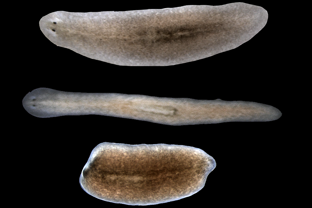
For more than a decade, Reddien and the researchers in his laboratory have studied the biological mechanisms that underlie regeneration in a tiny flatworm called planarians. These worms possess some impressive regenerative capabilities: When sliced in two, each piece of the worm can regrow the body parts needed to form two complete organisms. In previous studies, Reddien’s team identified a set of always-on genes, known as position control genes (PCGs), that provide cells with region-specific instructions, like a set of GPS coordinates, that tell cells where they are in the body, and thus what body part to regenerate. Interestingly, PGCs are active in planarian muscle cells, suggesting muscle may play a major role in the regeneration process.

“This discovery raised a lot of questions about how muscle participates in this process,” Reddien says.

In planarians, there are a handful of muscle cell types. For example, if you imagine the worms as simple cylindrical tubes, there are longitudinal muscle fibers, which run head-to-tail along the tubes’ long axis. There are also circular fibers, which are perpendicular to the longitudinal fibers and hug the tubes’ outer circumference.

To assess the roles of these different muscle cell types in regeneration, first author Lucila Scimone and her colleagues needed a method to selectively remove them. When myoD, a gene found specifically in the longitudinal fibers, is inhibited, those fibers fail to form. Similarly, the nkx1-1 gene marks the circular fibers, and when its function is reduced, they do not develop. Using these genes as molecular scalpels, Scimone and her co-authors could test the effects of ablating these distinct muscle groups on regeneration.

Surprisingly, when the longitudinal fibers were removed, the results were dramatic. The worms live quite normally, but when they are injured (the head removed, for example) they cannot regenerate the missing parts.

“This is an amazing result; it tells us that these longitudinal fibers are essential for orchestrating the regeneration program from the very beginning,” says Scimone, a scientist in Reddien’s lab.

As the researchers dug deeper into the finding, they learned that the functions of two critical genes are disrupted when longitudinal fibers are missing. These genes, called notum and follistatin, are known for their fundamental roles in regeneration, controlling head-versus-tail decisions and sustained cell proliferation, respectively, following tissue injury.

In addition to this essential role for longitudinal fibers, the research team also uncovered a key role for circular fibers. When these muscles are missing, planarians are able to regenerate missing body parts, but what regrows is abnormally patterned. For example, two heads may be regenerated within a single outgrowth, instead of one.

These results underscore an important and previously unappreciated role for muscle, widely known for its contractile properties, in instructing the tissue regeneration program. The Whitehead researchers will continue to probe the role of different muscle cell types in planarian regeneration and also explore whether other animals with regenerative capabilities possess a similar muscle-localized program for conferring positional information.

“It’s hard to understand what limits humans’ abilities to regenerate and repair wounds without first knowing what mechanisms are enabling some animals, like planarians, to do it so amazingly well,” Reddien says.

This work was supported by the National Institutes of Health, Howard Hughes Medical Institute, and the Eleanor Schwartz Charitable Foundation.

A scientific approach to writing fiction

Megan Miranda '02 graduated from MIT intent on pursuing a career in biotechnology. Instead, she became a New York Times best-selling author.

Jay London | MIT Alumni Association
November 15, 2017

Megan Colpitts Miranda ’02, who graduated from MIT with a degree in biology, intended to pursue a career in biotechnology. Instead, she became a successful fiction author whose book, “All the Missing Girls,” is a New York Times best-seller. Both careers share a trial-and-error approach to achieving success, she believes.

“There are a lot of similarities in the process,” Miranda says. “Each book draft is an experiment where I can assess what’s working and what’s not. You start with a blank slate; then each step gets you closer to a solution.”

Miranda worked in biotech in Boston for two years after graduation before moving with her husband, Luis Miranda ’01, to North Carolina, where she spent two years as a high school science teacher.

“Teaching put me back in touch with the elements that made me initially fall in love with science,” she says. “That love of science kind of funneled into writing my first books, which all contained weird scientific elements in their plots.”

Miranda began writing full time after the birth of her two children. After a few years of proposals, rewrites, and revisions, her first book, the young-adult thriller “Fracture,” was released in 2012. Six other books quickly followed, including “Hysteria,” “Vengeance,” “Soulprint,” and “The Safest Lies.” But the one to make the biggest impact has been “All the Missing Girls,” a story about the disappearance of two young women that was named editors’ choice by The New York Times Book Review and one of The Wall Street Journal’s “5 Killer Books for 2016.”

Miranda’s most recent work, “The Perfect Stranger,” was published by Simon and Schuster this year. And her next young-adult book, “Fragments of the Lost,” is due out in early 2018.

Miranda credits the thematic elements of her young-adult books, in part, to her coursework at MIT, where she mixed bioengineering with a steady dose of anthropology and literature.

“My first books combined biology and anthropology,” she says. “They are different sides of the same interests. Biology is the science element, confirmed by process of experiment, while anthropology is the human element.”

MIT’s “fail-forward” mentality also helped lay the groundwork for her literary career. “At MIT, I learned not to fear failure,” Miranda says. “MIT is the type of place where you need self-discipline and a willingness to take risks and try a different approach. Writing is no different.”

Miranda lives near Charlotte with her husband, a senior manager at Accenture, and their 11-year-old daughter and nine-year-old son. She enjoys connecting with readers through school and library visits, and she offers Skype Q&A sessions to book clubs and classes.

This article originally appeared in the September/October 2017 issue of MIT Technology Review.

Mary Clare Beytagh: Finding poetry in medicine

MIT senior and aspiring physician aims to tell stories that humanize the patients behind medical statistics.

Fatima Husain | MIT News correspondent
November 12, 2017

When MIT senior Mary Clare Beytagh isn’t performing research at the Koch Institute for Integrative Cancer Research or writing poetry, she can be found in ballet class at the Harvard Dance Center, continuing her 15 years of intensive dance training.

For Beytagh, ballet provides a reprieve from the hustle and bustle of academics and research. Her twice-a-week classes are “a nice way to de-stress and think about things,” including flashbacks to exciting moments on stage as a preprofessional ballerina, and fond memories with friends.

On days without dance class, Beytagh goes running. The two activities are “sort of antithetical to each other,” she notes. However, she makes it work. Beytagh is majoring in biology and literature at MIT — two fields that, like running and ballet, rarely intersect. But Beytagh aims to change that.

Running start on research

The summer before Beytagh’s senior year in high school, her teachers encouraged her to apply to a research program at the University of Texas Southwestern Medical Center.

The eight-week program took Beytagh out of the the classroom and into to the lab of Kathryn O’Donnell-Mendell, a cancer researcher studying B-cell lymphoma. The program was Beytagh’s first experience with scientific and medical research, and she was hooked.

She continued the research into her senior year of high school and submitted a paper to the prestigious Siemens Competition in Math, Science, and Technology.

While working in the lab, she met an MD-PhD student who opened Beytagh’s eyes to the possibility of pursuing medicine and cancer research simultaneously. When Beytagh applied to college, she looked for schools that emphasized undergraduate research. MIT topped her list.

“MIT rises above everyone else in that aspect,” she says. During an on-campus visit, she took part in a tour that allowed her to learn about the different types of research performed at the Institute. By the end of the tour, Beytagh knew MIT was the right fit. “These are my people,” she recalls thinking.

Upon the advice of her research advisor at UT Southwestern, after Beytagh arrived at MIT she sought out Tyler Jacks, professor of biology and director of the Koch Institute.

Beytagh has worked in the Jacks Lab since her second semester at the Institute. She and the other researchers are developing mouse models for cancer that recapitulate more aspects of the human disease. One goal, for example, is to have the tumors grow in the same locations in the animals as they do in humans.

Last year, Beytagh was invited to speak at the American Association for Cancer Research meeting. There, she presented her research alongside postdocs and early-career cancer biologists.

“That was a cool experience,” she says, “But then, it was back to the lab immediately!”

Documenting experiences

Outside of the lab, Beytagh enjoys expressing herself through her writing as a literature major.

During her sophomore Independent Activities Period (IAP), she traveled to Madrid to study Spanish literature. Her class was taught by MIT professors Stephen Tapscott and Margery Resnick. It examined post-Spanish Civil War novels and poetry — and captivated Beytagh.

After IAP ended, Beytagh continued studying poetry in Tapscott’s course 21L.487 (Modern Poetry). During the class, distinguished American poet Martha Collins visited and performed a poetry reading.

The visit had such an impact on Beytagh that she embarked upon an exercise inspired by one of Collins’ poetry series. The experiment lasted 21 days, during which Beytagh wrote poetic snapshots of each day within a set of predetermined rules.

“I’m a person who likes rules, but within those rules finds creativity,” Beytagh says.

On the 21st day, poetry morphed from hobby to emotional necessity. She found out her good friend had been diagnosed with Hodgkin’s lymphoma. At that moment, her poetry “became catharsis.”

She decided to declare literature as her second major.

“I had been flirting with the idea, but I had never committed,” she says, “Then, at the end of [sophomore] year, I committed.”

“This is it,” she says, recounting her reasoning, “These professors are amazing. I’m having a great time. It’s enriching me as a person.”

Bringing backstories to the forefront

Beytagh often integrates her research and other undergraduate experiences into her writing.

During her junior year IAP, she did an externship in the Yale School of Medicine’s emergency medicine department, with Charles Wira, III. She worked on developing a new risk score system for patients experiencing sepsis, but it was what she witnessed while shadowing in the emergency room that transformed her outlook.

“The most timely and impactful thing I saw there was the nature of the opioid epidemic,” she says, “You can read all you want in The New York Times and look at graphs — but that’s just statistics.”

That winter, she witnessed two to three patients coming into the emergency room for opioid overdoses each day she was there.

“What you don’t get in a graph,” she points out, “are the backstories of all these people.”

After that experience, she began to write about patients she saw and interacted with, in her poetry. In the long term, Beytagh hopes to become a science writer as well as a physician-scientist, telling stories that humanize patients and focus on the social and economic determinants of health.

Though she plans to study cancer biology in an MD-PhD program, she hopes to end up at an institution that allows her to take on other projects such as epidemiological research on opioid addiction.

Facilitating leadership

After a recommendation from her roomates freshman year, Beytagh joined the Leadership Training Institute, an organization which provides leadership training and mentorship to underprivileged Boston area high school students. The institute runs a 12-week program for 50 students each spring.

As the director of the program, Beytagh aims to reach students who are shy but passionate about community service and leadership, and works to provide them with transformative experiences.

“It’s always very gratifying when the students [graduate from the program],” she says. “They say, ‘You guys have made me realize that I not only want to keep service as a part of high school, but as a part of my career and onward.’”

“That gives you chills,” Beytagh says. “If you can spark that in someone and make them realize having an others-focused heart is the way to live life, it can only be good for our world.”

New player in cellular signaling

Researchers have identified a key nutrient sensor in the mTOR pathway that links nutrient availability to cell growth.

Nicole Giese Rura | Whitehead Institute
November 9, 2017

To survive and grow, a cell must properly assess the resources available and couple that with its growth and metabolism — a misstep in that calculus can potentially cause cell death or dysfunction. At the crux of these decisions is the mTOR pathway, a cellular pathway connecting nutrition, metabolism, and disease.

The mTOR pathway incorporates input from multiple factors, such as oxygen levels, nutrient availability, growth factors, and insulin levels to promote or restrict cellular growth and metabolism. But when the pathway runs amok, it can be associated with numerous diseases, including cancer, diabetes, and Alzheimer’s disease. Understanding the various sensors that feed into the mTOR pathway could lead to novel therapies for these diseases and even aging, as dialing down the mTOR pathway is linked to longer lifespans in mice and other organisms.

Although the essential amino acid methionine is one of the key nutrients whose levels cells must carefully sense, researchers did not know how it fed into the mTOR pathway — or if it did at all. Now, Whitehead Institute Member David Sabatini and members of his laboratory have identified a protein, SAMTOR, as a sensor in the mTOR pathway for the methionine derivative SAM (S-adenosyl methionine). Their findings are described in the current issue of the journal Science.

Methionine is essential for protein synthesis, and a metabolite produced from it, SAM, is involved in several critical cellular functions to sustain growth, including DNA methylation, ribosome biogenesis, and phospholipid metabolism. Interestingly, methionine restriction at the organismal level has been linked to increased insulin tolerance and lifespan, similar to the antiaging effects associated with inhibition of mTOR pathway activity. But the connection between mTOR, methionine, and aging remains elusive.

“There are a lot of similarities between the phenotypes of methionine restriction and mTOR inhibition,” says Sabatini, who is also a Howard Hughes Medical Institute investigator and a professor of biology at MIT. “The existence of this protein SAMTOR provides some tantalizing data suggesting that those phenotypes may be mechanistically connected.”

Sabatini identified mTOR as a graduate student and has since elucidated numerous aspects of its namesake pathway. He and his lab recently pinpointed the molecular sensors in the mTOR pathway for two key amino acids: leucine and arginine. In the current line of research, co-first authors of the Science paper Xin Gu and Jose Orozco, both graduate students Sabatini’s lab, identified a previously uncharacterized protein that seemed to interact with components of the mTOR pathway. After further investigation, they determined that the protein binds to SAM and indirectly gauges the pool of available methionine, making this protein — SAMTOR — a specific and unique nutrient sensor that informs the mTOR pathway.

“People have been trying to figure out how methionine was sensed in cells for a really long time,” Orozco says. “I think that this is the first time in mammalian cells a mechanism has been found to describe the way methionine can regulate a major signaling pathway like mTOR.”

The current research indicates that SAMTOR plays a crucial role in methionine sensing. Methionine metabolism is vital for many cellular functions, and the Sabatini lab will further investigate the potential links between SAMTOR and the extended lifespan and increased insulin sensitivity effects that are associated with low methionine levels.

“It is very interesting to consider mechanistically how methionine restriction might be associated in multiple organisms with beneficial effects, and identification of this protein provides us a potential molecular handle to further investigate this question,” Gu says. “The nutrient-sensing pathway upstream of mTOR is a very elegant system in terms of responding to the availability of certain nutrients with specific mechanisms to regulate cell growth. The currently known sensors raise some interesting questions about why cells evolved sensing mechanisms to these specific nutrients and how cells treat these nutrients differently.”

This work was supported by the National Institutes of Health, the Department of Defense, the National Science Foundation, and the Paul Gray UROP Fund.

Department of Biology hosts 2017 Massachusetts Junior Academy of Science Symposium

High school students present research projects to build communication skills while earning membership to the American Junior Academy of Science.

Raleigh McElvery | Department of Biology
October 24, 2017

On Oct. 14, 22 school students from across the state presented their research projects at the annual Massachusetts Junior Academy of Science (MassJAS) Symposium.

The talks were split into two concurrent sessions based on subject: biological and environmental sciences; and engineering, chemistry, mathematics, and physics. Participants were selected based on merit and ranking in this year’s Massachusetts State Science and Engineering Fair.

Judges nominated three students from the biology session and four from physics and engineering as American Junior Academy of Science (AJAS) delegates. Delegates are invited to attend the AJAS Convention, which will be held in Austin, Texas this coming February. The AJAS is a national honor society that meets annually in conjunction with the American Association for the Advancement of Science — the world’s largest science organization and the publisher of Science. All participants were inducted as AJAS fellows.

The sessions took place in adjacent lecture rooms in Building 68. The event was organized by Mandana Sassanfar, director of diversity and science outreach for MIT’s Department of Biology and Department of Brain and Cognitive Sciences, as well as the director of MassJAS. During the event, delegates toured local research institutions, shared their projects with others in the field, and attended conference sessions.

At this year’s MassJAS symposium, the jury for the biological and environmental science session was composed of three graduate students and postdoc from the MIT Department of Biology.

“I really enjoyed hearing how these projects came to be, and what inspired students to ask their respective research questions,” said Sora Kim, a third year graduate student in Tania Baker’s lab and a returning judge. “Some students did these projects at home, while others had collaborations with researchers at local universities. In many cases, these were their first science projects, so being able to understand their own projects and also convey their ideas to a more general audience is really important.”

First-time judge Summer Morrill, a third year graduate student in Angelika Amon’s lab, agreed that learning to present ideas clearly in ways that inspire others is key to the scientific process. “I was excited to hear what people at the high school level think is important in science, because they’re the next generation of scientists,” she said.

Each participant had ten minutes to present, followed by an audience question-and-answer session. The biology-related talks ranged from antimicrobial resistance to gene editing techniques to the effects of wifi router radiation.

Joshua Powers and Natalia Huynh, both juniors at the Everett High School STEM Academy, presented first, describing the results of their summer research project at MIT as part of the LEAH Knox Scholars pilot program. Powers and Huynh pooled their findings, isolating and characterizing bacterial specimens from the Charles River.

“We’re friends and we both go to the same high school, so it was easy to collaborate with both our ideas and our data,” said Powers. “The LEAH Knox Scholars program was intense in that we had the chance to perform more advanced procedures with equipment we’ve never used before in school.”

Huynh also enjoyed tackling larger research questions with more refined tools, adding, “We practiced explaining our results this summer, so today’s presentation was similar to what we’d already done — but a little more intense because it was a competition.”

Nancy Cianchetta, who teaches biotechnology at Everett High School and serves as the coordinator for the STEM Academy, said Powers and Huynh will be part of the very first class to graduate from the Academy. She and many of her students have participated in MIT biology outreach programs over the years.

“I’ve taken my classes here for field trips and career exploration days, and many of my students come for the spring lecture series at the Whitehead Institute,” she said. “The kids get so excited to come to MIT.”

While some participants shared data they’d only just begun to analyze, others had been tackling the same research question for over a year.

Evan Mizerak, a returning MassJAS Symposium winner and senior at Wachusett Regional High School, has spent the past two-and-a-half years collaborating with researchers at the University of Massachusetts Medical School on his project related to heritable infertility in fruit flies.

Mizerak attended last year’s AJAS Convention in Boston, as well as the MIT-sponsored Breakfast with Scientists. This year, delegates met with esteemed faculty, including Institute Professor Phillip Sharp, the winner of the 1993 Nobel Prize in physiology or medicine and a member of the Department of Biology and the Koch Institute for Integrative Cancer Research.

“The AJAS Convention was incredible last year, because we had the chance to meet researchers from around the country — not just in and around Massachusetts,” he said. “At the Breakfast with Scientists, we met with Nobel Prize winners. Being introduced to people I consider celebrities was just amazing.”

“You wouldn’t expect anyone that famous to be interested in our work,” added Emma Kelly, a junior from Newton Country Day School and also a returning presenter. “But these professionals were genuinely curious, and often gave us ideas for new projects and things like that. It was such an incredible opportunity.”

MIT neuroscientists build case for new theory of memory formation

Existence of “silent engrams” suggests that existing models of memory formation should be revised.

Anne Trafton | MIT News Office
October 23, 2017

Learning and memory are generally thought to be composed of three major steps: encoding events into the brain network, storing the encoded information, and later retrieving it for recall.

Two years ago, MIT neuroscientists discovered that under certain types of retrograde amnesia, memories of a particular event could be stored in the brain even though they could not be retrieved through natural recall cues. This phenomenon suggests that existing models of memory formation need to be revised, as the researchers propose in a new paper in which they further detail how these “silent engrams” are formed and re-activated.

The researchers believe their findings offer evidence that memory storage does not rely on the strengthening of connections, or “synapses,” between memory cells, as has long been thought. Instead, a pattern of connections that form between these cells during the first few minutes after an event occurs are sufficient to store a memory.

“One of our main conclusions in this study is that a specific memory is stored in a specific pattern of connectivity between engram cell ensembles that lie along an anatomical pathway. This conclusion is provocative because the dogma has been that a memory is instead stored by synaptic strength,” says Susumu Tonegawa, the Picower Professor of Biology and Neuroscience, the director of the RIKEN-MIT Center for Neural Circuit Genetics at the Picower Institute for Learning and Memory, and the study’s senior author.

The researchers also showed that even though memories held by silent engrams cannot be naturally recalled, the memories persist for at least a week and can be “awakened” days later by treating cells with a protein that stimulates synapse formation.

Dheeraj Roy, a recent MIT PhD recipient, is the lead author of the paper, which appears in the Proceedings of the National Academy of Sciences the week of Oct. 23. Other authors are MIT postdoc Shruti Muralidhar and technical associate Lillian Smith.

Silent memories

Neuroscientists have long believed that memories of events are stored when synaptic connections, which allow neurons to communicate with each other, are strengthened. Previous studies have found that if synthesis of certain cellular proteins is blocked in mice immediately after an event occurs, the mice will have no long-term memory of the event.

However, in a 2015 paper, Tonegawa and his colleagues showed for the first time that memories could be stored even when synthesis of the cellular proteins is blocked. They found that while the mice could not recall those memories in response to natural cues, such as being placed in the cage where a fearful event took place, the memories were still there and could be artificially retrieved using a technique known as optogenetics.

The researchers have dubbed these memory cells “silent engrams,” and they have since found that these engrams can also be formed in other situations. In a study of mice with symptoms that mimic early Alzheimer’s disease, the researchers found that while the mice had trouble recalling memories, those memories still existed and could be optogenetically retrieved.

In a more recent study of a process called systems consolidation of memory, the researchers found engrams in the hippocampus and the prefrontal cortex that encoded the same memory. However, the prefrontal cortex engrams were silent for about two weeks after the memory was initially encoded, while the hippocampal engrams were active right away. Over time, the memory in the prefrontal cortex became active, while the hippocampal engram slowly became silent.

In their new PNAS study, the researchers investigated further how these silent engrams are formed, how long they last, and how they can be re-activated.

Similar to their original 2015 study, they trained mice to fear being placed in a certain cage, by delivering a mild foot shock. After this training, the mice freeze when placed back in that cage. As the mice were trained, their memory cells were labeled with a light-sensitive protein that allows the cells to be re-activated with light. The researchers also inhibited the synthesis of cellular proteins immediately after the training occurred.

They found that after the training, the mice did not react when placed back in the cage where the training took place. However, the mice did freeze when the memory cells were activated with laser light while the animals were in a cage that should not have had any fearful associations. These silent memories could be activated by laser light for up to eight days after the original training.

Making connections

The findings offer support for Tonegawa’s new hypothesis that the strengthening of synaptic connections, while necessary for a memory to be initially encoded, is not necessary for its subsequent long-term storage. Instead, he proposes that memories are stored in the specific pattern of connections formed between engram cell ensembles. These connections, which form very rapidly during encoding, are distinct from the synaptic strengthening that occurs later (within a few hours of the event) with the help of protein synthesis.

“What we are saying is that even without new cellular protein synthesis, once a new connection is made, or a pre-existing connection is strengthened during encoding, that new pattern of connections is maintained,” Tonegawa says. “Even if you cannot induce natural memory recall, the memory information is still there.”

This raised a question about the purpose of the post-encoding protein synthesis. Considering that silent engrams are not retrieved by natural cues, the researchers believe the primary purpose of the protein synthesis is to enable natural recall cues to do their job efficiently.

The researchers also tried to reactivate the silent engrams by treating the mice with a protein called PAK1, which promotes the formation of synapses. They found that this treatment, given two days after the original event took place, was enough to grow new synapses between engram cells. A few days after the treatment, mice whose ability to recall the memory had been blocked initially would freeze after being placed in the cage where the training took place. Furthermore, their reaction was just as strong as that of mice whose memories had been formed with no interference.

Sheena Josselyn, an associate professor of psychology and physiology at the University of Toronto, said the findings run counter to the longstanding idea that memory formation involves strengthening of synapses between neurons and that this process requires protein synthesis.

“They showed that a memory formed during protein-synthesis inhibition may be artificially (but not naturally) recalled. That is, the memory is still retained in the brain without protein synthesis, but this memory cannot be accessed under normal conditions, suggesting that spines may not be the key keepers of information,” says Josselyn, who was not involved in the research. “The findings are controversial, but many paradigm-shifting papers are.”

Along with the researchers’ previous findings on silent engrams in early Alzheimer’s disease, this study suggests that re-activating certain synapses could help restore some memory recall function in patients with early stage Alzheimer’s disease, Roy says.

The research was funded by the RIKEN Brain Science Institute, the Howard Hughes Medical Institute, and the JPB Foundation.

School of Science welcomes new faculty members

This fall brings 14 new professors in the departments of Biology, Chemistry, Mathematics, and Physics.

School of Science
October 10, 2017

This fall, the MIT School of Science has welcomed 14 new professors in the departments of Biology, Chemistry, Mathematics, and Physics.

Ian J. M. Crossfield focuses on the atmospheric characterization of exoplanets through all possible methods — transits, eclipses, phase curves, and direct imaging — from the ground and from space, with an additional interest in the discovery of new exoplanets, especially those whose atmospheres that can be studied in more detail. He joins the MIT Department of Physics as an assistant professor.

Joey Davis, an assistant professor in the Department of Biology, studies the molecular mechanisms underpinning autophagy using biochemical, biophysical, and structural biology techniques such as mass spectrometry and cryo-electron microscopy. This pathway is responsible for protein and organelle degradation and has been linked to a variety of aging associated disorders including neurodegeneration and cancer.

Daniel Harlow works on black holes and cosmology, viewed through the lens of quantum gravity and quantum field theory. He has joined the Department of Physics as assistant professor.

Philip Harris, a new assistant professor in the Department of Physics, searches for dark matter, seeking a deeper understanding of the petabytes of data collected at the Large Hadron Collider. Much of his research exploits new techniques to resolve the structure of quark and gluon decays, known as jet substructure.

Or Hen studies quantum chromodynamics effects in the nuclear medium, and the interplay between partonic and nucleonic degrees of freedom in nuclei, conducting experiments at the Thomas Jefferson and Fermi National Accelerator Laboratories, as well as other accelerators around the world. He has joined the faculty as an assistant professor in the Department of Physics and the Laboratory of Nuclear Science.

Laura Kiessling investigates how carbohydrates are assembled, recognized, and function in living cells, which is crucial to understanding key biological processes such as bacterial cell wall biogenesis, bacteria chemotaxis, enzyme catalysis and inhibition, immunity, and stem cell propagation and differentiation. She is the new Novartis Professor of Chemistry.

Rebecca Lamason investigates how intracellular bacterial pathogens hijack host cell processes to promote infection. In particular, she studies how Rickettsia parkeri and Listeria monocytogenes move through tissues via a process called cell-to-cell spread. She has joined the Department of Biology as an assistant professor.

Sebastian Lourido studies the molecular events that enable parasites in the phylum Apicomplexa to remain widespread and deadly infectious agents. Lourido uses Toxoplasma gondii to model processes conserved throughout the phylum, in order to expand our understanding of eukaryotic diversity and identify specific features that can be targeted to treat parasite infections. He has been welcomed into the Department of Biology as an assistant professor.

Ronald T. Raines, who has joined the faculty as the Firmenich Professor of Chemistry, uses techniques that range from synthetic chemistry to cell biology to illuminate in atomic detail both the chemical basis and the biological purpose for protein structure and protein function. He seeks insights into the relationship between amino-acid sequence and protein function (or dysfunction), as well as to the creation of novel proteins with desirable properties.

Giulia Saccà is an algebraic geometer with a focus on hyperkähler and Calabi-Yau manifolds, K3 surfaces, moduli spaces of sheaves, families of abelian varieties and their degenerations, and symplectic resolutions. She is now an assistant professor in the Department of Mathematics.

Stefani Spranger studies the interactions between cancer and the immune system, with the goal of improving existing immunotherapies or developing novel therapeutic approaches. Spranger seeks to understand how CD8 T cells, otherwise known as killer T cells, are excluded from the tumor microenvironment, with a focus on lung and pancreatic cancers. She has joined the Department of Biology as an assistant professor.

Daniel Suess works at the intersection of inorganic and biological chemistry, studying redox reactions that underpin global biogeochemical cycles, metabolism, and energy conversion. He develops chemical strategies for attaining precise, molecular-level control over the structures of complex active sites. In doing so, his research yields detailed mechanistic insight and enables the preparation of catalysts with improved function. Suess is an assistant professor in the Department of Chemistry.

Wei Zhang is a number theorist who works in arithmetic geometry, with special interest in fundamental objects such as L-functions, which appear in the Riemann hypothesis and its generalizations, and are central to the Langlands program. Zhang has joined the Department of Mathematics as a full professor.

Yufei Zhao, who has joined the Department of Mathematics as an assistant professor, works in combinatorics and graph theory, and is especially interested in problems with extremal, probabilistic, and additive flavors.

Department of Biology hosts its first Science Slam

Eight biology trainees had just three minutes to explain their research and earn favor with the judges and audience in new yearly event.

Raleigh McElvery | Department of Biology
October 5, 2017

Nearly 300 spectators crowded into a lecture hall at the Ray and Maria Stata Center on a recent Tuesday to witness the first annual Science Slam, hosted by MIT’s Department of Biology.

A science slam features a series of short presentations where researchers explain their work in a compelling manner and — as the name suggests — make an impact. The presentations aren’t just talks, they’re performances geared towards a science-literate but non-specialized public audience. In this case, competitors were each given one slide and three minutes to tell their scientific tales and earn votes from audience members and judges.

The jury included Ellen Clegg, editorial page editor of The Boston Globe and co-author of two award-winning books, “ChemoBrain” and “The Alzheimer’s Solution;” Emilie Marcus, CEO of Cell Press and editor-in-chief of the flagship journal, Cell; and Ari Daniel, an independent science reporter who produces digital videos for PBS NOVA and co-produces the Boston branch of Story Collider.

Among the competitors were five graduate students and three postdocs who hailed from labs scattered throughout Building 68, the Whitehead Institute, the Broad Institute, the Koch Institute for Integrative Cancer Research, and the Picower Institute for Learning and Memory. The storytellers were:

  • Sahin Naqvi, from David Page’s lab, who spoke about the evolution of genetic sex differences in mammals, as well as how these differences impact the likelihood of developing certain diseases based on gender;
  • Sudha Kumari, from Darrell Irvine’s lab, who spoke about her work investigating immune cell interactions — specifically how T cells communicate using physical contact;
  • Deniz Atabay, from Peter Reddien’s lab, who spoke about the ways cells in flatworms self-organize during regeneration to re-form organs, tissues, and even neural circuits;
  • Emma Kowal, from Christopher Burge’s lab, who spoke about her goals to demystify the ways in which certain noncoding regions of genetic sequence, known as introns, contribute to protein production;
  • Xin Tang, from Rudolf Jaenisch’s lab, who spoke about a technique to illuminate the seemingly invisible changes in brain cells that trigger disease, using a glowing enzyme from a firefly;
  • Nicole Aponte, from Troy Littleton’s lab, who spoke about her ability to manipulate brain cell activity in the fruit fly, and study how defects in neuronal connections contribute to developmental disorders;
  • Karthik Shekhar, from Aviv Regev’s lab, who spoke about his efforts to identify and manipulate different types of brain cells, understanding how they assemble into complex networks to facilitate learning, memory, and — in some cases — disease; and
  • Monika Avello, from Alan Grossman’s lab, who spoke about “bacterial sexology,” that is, how and why these organisms choose to block unwanted sexual advances from fellow bacteria.

Vivian Siegel, who oversees the department’s communications efforts, moderated the event. Siegel and the Building 68 communications team joined forces with three members of the Building 68 MIT Postdoctoral Association — Ana Fiszbein, Isabel Nocedal, and Peter Sudmant — to publicize the event and to host two pre-slam workshops, as well as one-on-one training sessions with individual participants.

“Participating in a Science Slam seemed like a great way for our trainees to learn how to communicate to a nonspecialized audience, which is something they will need to be able to do throughout their careers,” Siegel said. “We really wanted to develop a camaraderie among the participants, and bring trainees together from across the department to help each other tell compelling stories about their science.”

Kowal — whose talk was titled “Gone but Not Forgotten: How Do Introns Enhance Gene Expression?”  — ultimately took home both the audience and jury cash prizes. Kowal completed her undergraduate degree in chemical and physical biology at Harvard before coming to MIT for graduate school. Her dream is to write science fiction, so she decided she’d better study science so she’d know what to write about.

“I really enjoyed seeing people get stoked about introns, and the fact that they enhance gene expression,” she said. “It’s a great way to get comfortable explaining your project in a compelling way to a broad audience. Since you’ll probably be telling people about your work for a while, I think it’s a very good use of time to practice doing that.”

New target for treating “undruggable” lung cancer

Drug already in clinical trials may be effective on some aggressive adenocarcinomas.

Becky Ham | MIT News correspondent
October 2, 2017

Mutations in the KEAP1 gene could point the way to treating an aggressive form of lung cancer that is driven by “undruggable” mutations in the KRAS gene, according to a new study by MIT researchers.

KEAP1 mutations occur alongside KRAS mutations in about 17 percent of lung adenocarcinoma cases. Tyler Jacks, director of MIT’s Koch Institute for Integrative Cancer Research and co-senior author of the study, and his colleagues found that cancer cells with nonfunctioning KEAP1 genes are hungry for glutamine, an amino acid essential for protein synthesis and energy use. Starving these cells of glutamine may thus offer a way to treat cancers with both KRAS and KEAP1 mutations.

Indeed, small-molecule-based inhibitors of glutaminase, an enzyme crucial to glutamine metabolism, slowed cancer cell growth and led to smaller tumors overall in human lung adenocarcinoma cell lines and in tumors in mice with KEAP1 mutations, the researchers found.

The study offers a way to identify lung cancer patients who might respond well to drugs that block the work of glutaminase, says MIT graduate student Rodrigo Romero, a first author on the paper that appears in the Oct. 2 online edition of Nature Medicine.

“All cell lines that we have currently tested that are KEAP1-mutant — independent of their KRAS status — appear to be exquisitely sensitive to glutaminase inhibitors,” says Romero, a graduate student in Jacks’ lab, who participated in the MIT Summer Research Program (MSRP) as an undergraduate.

Hyperactivating the antioxidant response

Lung adenocarcinoma accounts for about 40 percent of U.S. lung cancers, and 15 to 30 percent of those cases contain a KRAS mutation. KRAS has been “notoriously difficult to inhibit” because the usual ways of blocking the KRAS protein’s interactions or interfering with the protein’s targets have fallen short, says Romero.

Lung cancers containing KRAS mutations often harbor other mutations, including KEAP1, which is the third most frequently mutated gene in lung adenocarcinoma. To find out more about how these co-mutations affect lung cancer progression, the MIT research team created KEAP1 mutations in mouse models of lung adenocarcinoma, using the CRISPR/Cas9 gene-editing system to target the gene.

The KEAP1 protein normally represses another protein called NRF2, which controls the activation of an antioxidant response that removes toxic, reactive oxygen species from cells. When the researchers disabled KEAP1 with loss-of-function mutations, NRF2 was able to accumulate and contribute to a “hyperactivation” of the antioxidant response.

Lung adenocarcinomas bearing the KEAP1 mutation may “take advantage of this [hyper-activation] to promote cellular growth or detoxify intracellular damaging agents,” Romero says.

In fact, when the researchers examined genes targeted by NRF2 across a sample of human lung adenocarcinoma tumors, they concluded that the expression of these genes was greater in advanced stage IV tumors, and that patients with such “up-regulated” NRF2 tumors had significantly worse survival rates than other lung adenocarcinoma patients.

Tumors hungry for glutamine

Romero and colleagues used CRISPR/Cas9 to learn more about other genetic interactions with KEAP1 mutants. Their screening demonstrated that lung cancer cells with KRAS and KEAP1 loss-of function mutations were more dependent than other cells on increased amounts of glutamine.

To learn whether this glutamine hunger could be a therapeutic vulnerability, the researchers tested two glutaminase inhibitors against the cancer cells, including one compound called CB-839 that is in phase I clinical trials for KRAS-mutant lung cancer. CB-839 slowed growth and kept tumors smaller than normal in lung adenocarcinoma with KEAP1 mutations, the researchers found.

Phase I clinical trials that treat KEAP1-mutant lung adenocarcinoma patients with a combination of CB-839 and the cancer immunotherapy drug nivolumab (Opdivo) are also underway, says Romero, who notes the MIT study might help identify patients who would be good candidates for these trials.

“There are also many clinical trials testing the efficacy of glutaminase inhibition in a variety of cancer types, independent of KRAS status. However, the results from these studies are still unclear,” Romero says.

Jacks emphasizes that his laboratory has and will continue to study several mutations beyond KEAP1 that may cooperate with KRAS in their mouse models of human lung adenocarcinoma. “The complexity of human cancer can be quite daunting,” he notes. “The genetic tools that we have assembled allow us to create models of many individual subtypes of the disease and in this way begin to define the exploitable vulnerabilities of each. The observed sensitivity of KEAP1 mutant tumors to glutaminase inhibitors is an important example of this approach. There will be more.”

Co-authors on the Nature Medicine paper include former Koch Institute postdoc Thales Papagiannakopoulos, now at New York University, and MIT professor of biology Matthew Vander Heiden. The research was funded by the Laura and Isaac Perlmutter Cancer Support Grant, the National Institutes of Health, and the Koch Institute Support Grant from the National Cancer Institute.

Michael Rosbash PhD ’71 shares Nobel Prize in physiology or medicine

MIT alumnus and two others honored for discovering the molecular mechanisms of circadian rhythms.

Anne Trafton | MIT News Office
October 2, 2017

Michael Rosbash, who earned his PhD from MIT in 1971, will share the 2017 Nobel Prize in physiology or medicine, the Nobel Committee announced this morning in Stockholm.

Rosbash, now a professor of biology at Brandeis University, shares the prize with Jeffrey C. Hall of the University of Maine and Michael W. Young of Rockefeller University. The scientists were honored for “their discoveries of molecular mechanisms controlling the circadian rhythm.”

Circadian rhythms help living organisms adapt their biological activities to the normal 24-hour cycle of light and darkness. These rhythms influence behavior, sleep, metabolism, body temperature, and many other biological functions.

In 1984, Rosbash, Hall, and Young isolated a gene that regulates these daily rhythms in fruit flies. This gene, known as period, encodes a protein that accumulates during the night and is degraded during the day. Further work revealed that this protein inhibits the gene that encodes it, creating a negative feedback loop that is key to generating continuous oscillations.

Since then, the three scientists have discovered several other genes necessary for maintaining circadian cycles, and similar processes have been found in many other organisms, including humans.

Rosbash was born in Kansas City, Missouri, but grew up in Newton, Massachusetts. He earned his bachelor’s degree at Caltech before coming to MIT to pursue a PhD in biology. He has been on the faculty at Brandeis since 1974, and he is an investigator with the Howard Hughes Medical Institute and a member of the National Academy of Sciences. In 2013, Rosbash, Hall, and Young shared the Shaw Prize in Life Sciences and Medicine for their circadian clock research.

Rosbash is the 35th MIT alumnus to win a Nobel Prize, and the 88th MIT-connected winner of the prize.