New research reveals foundation for bi-directional DNA replication in animal cells
Research from the Bell Lab in the Department of Biology at MIT explores how helicases are loaded in oppositely oriented pairs onto DNA and how the cell regulates that process
By Lillian Eden
The genetic blueprint that directs each of our cells to do its job can be found in our DNA — which in humans is approximately 3 billion bases long. At each cell division, the cells copy each strand of DNA exactly once — under- or over-replicating DNA could lead to disease or cell death.
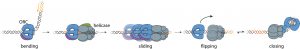
To replicate DNA, cells must first “unzip” the double strands of DNA. In budding yeast, cells do this by loading pairs of proteins called helicases at predetermined points, which are called origins of replication. Eukaryotic cells have many origins of replication. When the pairs of helicases are activated, they separate and travel in opposite directions, allowing replication machinery to access and copy both strands.
Two recent papers from the Bell Lab in the Department of Biology at MIT explore how these helicases are loaded in opposite directions onto DNA and how the cell inhibits that process in all but a specific phase of cell division.
Both helicases are loaded onto the DNA in opposite directions by a single copy of a key protein called the Origin Recognition Complex (ORC). ORC binds to DNA at origins of replication, loads the first helicase, releases from the DNA, then rebinds to the DNA in the reverse orientation to load the second helicase in the opposite direction. The resulting pair of helicases, loaded head-to-head, forms the foundation for bi-directional replication.
Only since 2019 have researchers generally accepted this model, in part because the protein gymnastics — the model requires ORC to ‘flip’ over the first helicase to load the second — was so unexpected. Previously, researchers thought that one ORC would load the first helicase and then a second ORC would load the second helicase.
ORC’s affinity for binding DNA is critical to loading both helicases. In budding yeast, ORC binding sites are found in pairs at each origin of replication: one strong binding site and a weaker, secondary, and oppositely oriented binding site located at various distances from the first. Although it was understood that both sequences are necessary, how ORC releases from the strong binding site and binds the weaker site was not.
First author Annie Zhang, PhD ‘23, found that intermediate steps during helicase loading decrease how tightly ORC binds to DNA, facilitating ORC’s release from the first, strong binding site. ORC remains associated with the first loaded helicase as it flips orientation and binds to the second, weaker binding site. The opposite orientation of the second binding site directs ORC to load the second helicase in the opposite direction. Importantly, once ORC releases from its initial binding site, it allows the complex of ORC and the helicase to slide on the DNA.
“Because the intermediate can slide on DNA, it allows access to many different second binding site locations,” Zhang says. “ORC flipping seems overly complicated, but connecting the two helicase loading events using one molecule is essential to establishing the stoichiometry and the orientation of the helicases. Nature finds a way to solve problems, and it might not be simple, but it’s always very effective.”
To better understand the dynamics and timing of the helicase loading process, Zhang used colocalization single-molecule spectroscopy and a technique called single-molecule FRET (smFRET).
In smFRET, two fluorescent tags are attached to targets of interest—a red dye and a green dye. Red light will be emitted if the tags are close to each other, but only green light will be detected if the tags are far apart. The changes in fluorescence intensity are measured as the reaction progresses, with different smFRET pairs providing clues to parsing out relationships like whether two components interact and, if so, for how long.
Zhang was interested in why cryo-electron microscopy (cryo-EM) images that showed ORC bent the DNA during the helicase loading process. By fluorescently tagging one end of the helicase and the DNA with a smFRET pair, Zhang determined when and how the DNA was unbent and threaded through the ring-shaped helicase.
Even though Zhang anticipated DNA conformational changes, she says it was exciting to see evidence of it occurring in real time. Zhang noted that this approach was only possible because helicase loading is a relatively slow process on the timescale of seconds to minutes. In many biological reactions, conformational changes occur on the timescale of milliseconds to nanoseconds, which these smFRET studies would not have been able to detect.
The helicases are loaded onto the DNA in an open ring shape and later clamp shut around the strands. A second paper from postdoc Audra Amasino, PhD ‘20, explores the ring-closing step.
Helicases are loaded and activated during separate phases of cell division. When loading is undesirable, ORC is deactivated by a process known as phosphorylation–adding a phosphate group. Amasino’s results indicate that phosphorylated ORC can still initiate the process of helicase loading, but there are two critical steps when phosphorylated ORC will inevitably fail.
When ORC is phosphorylated but mixed with otherwise normal proteins in vitro, the first helicase is still recruited and sometimes closes around the DNA–but it won’t stay that way.
Amasino found that the helicase pops off the DNA without closing, or the helicase closes around the DNA and then opens and falls off. She noted that the second failure occurs around the same time in the process when ORC would be flipping to re-orient itself to load the second helicase. Regardless of the presence of otherwise normal proteins, phosphorylated ORC can’t recruit a second helicase.
Amasino notes that when ORC wasn’t phosphorylated, the process often fails at the first helicase ring closing step, indicating that the process isn’t always successful even during the phases of cell division when ORC is meant to be loading helicases.
“Prior to getting this result, we thought that once the ring closes, it’s closed–but we saw that the first helicase is still closing around the DNA when ORC is phosphorylated, but the ring closing is not stable,” Amasino says. “What we found sheds light on what happens when the process is working properly.”
When the process occurs properly, ORC seems to hold the first helicase shut until the second helicase is recruited. The helicases, loaded head-to-head, somehow make the ring closing stable for both. When ORC can’t recruit a second helicase, the process fails.
Amasino used the same approach as Zhang–using structural information from Cryo-EM structures to make educated guesses about where it would be best to attach fluorescent tags for smFRET. By attaching tags to the segments of the first helicase that come together when it closes, Amasino determined that the first helicase clamps shut even when ORC is phosphorylated. Importantly, further experiments showed ORC phosphorylation inhibits the formation of a key intermediate involved in the flipping of ORC; the intermediate is necessary to hold the helicase closed.
MIT Professor of Biology Steve Bell, the senior author of both papers, says that many of the proteins and processes are conserved across all eukaryotic cells. Consequently, both the tools they used and what they discovered in yeast may be broadly applicable.
“The impact here is twofold. We now understand this very fundamental event in more detail, but this approach also illustrates the power of combining structural biology with these single molecule studies to understand how complex assembly events occur in cells,” he says. “This is the next frontier in what you can do in terms of understanding detailed mechanisms.”
The Bell lab is now working to figure out how ORC remains localized to the DNA during helicase loading, which is critical for ORC flipping orientations to load the second helicase.
“We want to understand how ORC retains attachment to the helicase. We know at some level it must do that, but we don’t know how,” Bell says. “We hope to have the answer to that question in the next few months.”