Greta Friar | Whitehead Institute
August 4, 2021
All the buzz in the lab
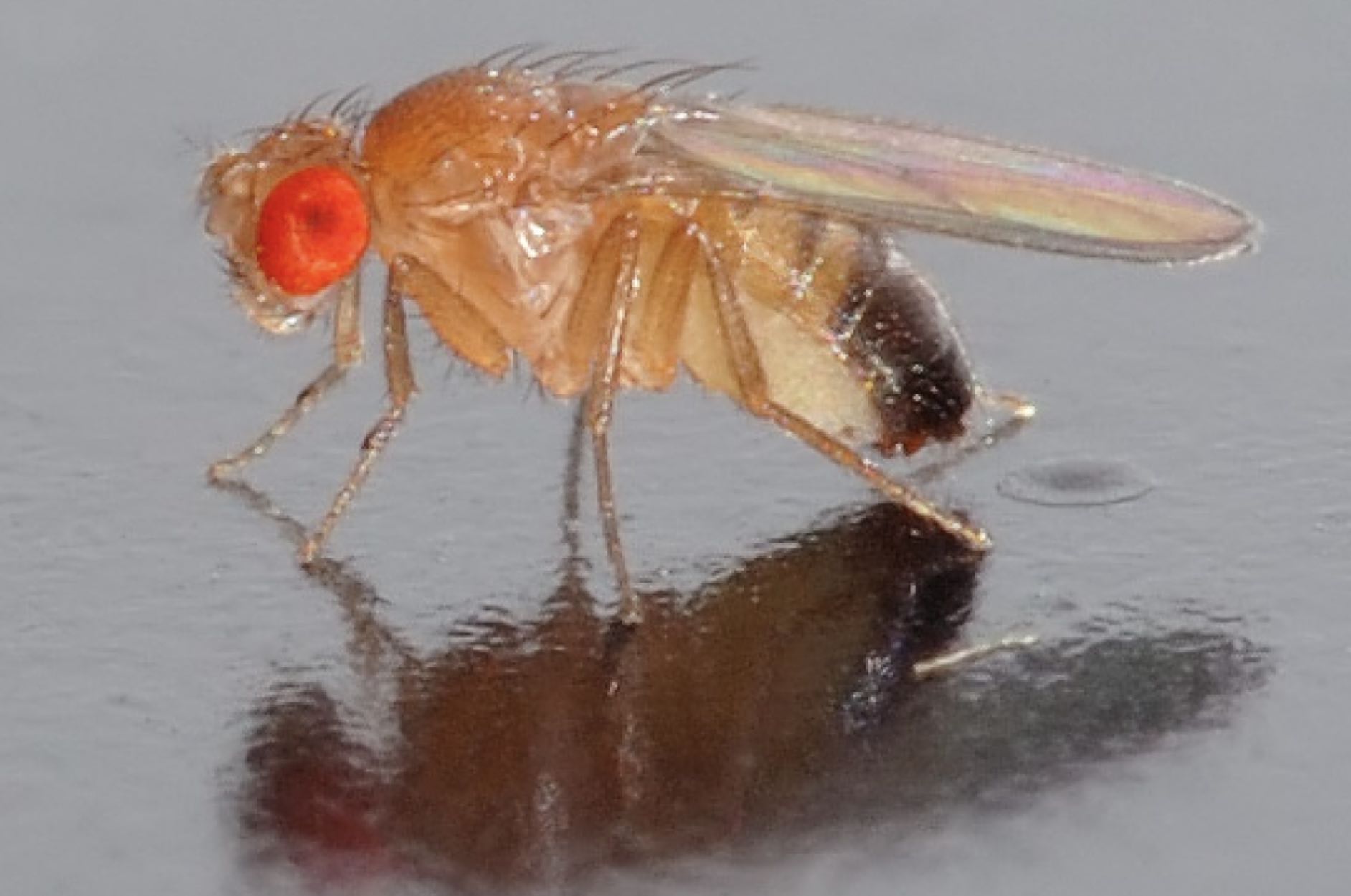
On a sunny summer morning in Cambridge, Massachusetts, Mariyah Saiduddin walked into a room and was met by the sight of thousands of fruit flies. For most people, this would be an emergency: time to call an exterminator, take out the trash, and scrub the room from top to bottom. However, this room full of flies is part of Whitehead Institute Director Ruth Lehmann’s lab, where fruit flies are seen not as pests but as valuable research tools—and are safely contained in vials. Saiduddin is a graduate student researcher in Lehmann’s lab who uses a fraction of the flies in the room in her research.
The flies found in Lehmann’s lab, and in the adjacent lab run by Whitehead Institute Member Yukiko Yamashita, are not exactly like their less-beloved wild counterparts. Fruit flies have been used in research for more than a century, and in that time, they have been engineered to become powerful, malleable models capable of answering questions in many areas of research. The most common species used in research is Drosophila melanogaster, often referred to simply as “Drosophila.” The researchers who use flies call themselves Drosophilists, and their community around the world works together to maintain a rich variety of flies and create new tools with which to manipulate those flies. In the past century, work in fruit flies has led to six Nobel Prizes in Physiology or Medicine, and has shed light on topics from the basics of genetics, to the principles of embryonic development, to circadian rhythms, to the immune system, to a plethora of diseases.
A very fly model organism
Fruit flies became a go-to research tool during the explosion of genetics research around the turn of the 20th century. What makes them such a good model organism? First of all, they are easy and relatively cheap to raise in large numbers. They have short lifespans and quick reproduction times, so researchers can rapidly breed and study multiple generations. Fruit flies are ready to reproduce—growing from embryo to larva to adult—in under two weeks and then can lay hundreds of eggs in a matter of days.
The embryonic days of fly research
The transformation of fruit flies from wild pests into top notch research tools began in the early 1900s. Charles W. Woodworth, at Harvard University at the turn of the 20th century, is credited with being the first researcher to breed Drosophila in large numbers and with suggesting that the species could be used to study genetics, then a new field of research.[2] Thomas Hunt Morgan, at Columbia University, was one of several researchers to follow Woodworth in using fruit flies for his research, and it was Morgan who really established fruit flies as a model organism, through both his own success and that of the students who came out of his lab and the soon-famous Columbia fly room.
For his research, Morgan bred fruit flies until one developed a mutation, white eyes (most fruit flies’ eyes are red), and then continued breeding the mutant and its descendants to track patterns in inheritance of the white-eyes trait. With these experiments, Morgan showed that genes, which had recently been established as the smallest units of inheritance, are organized on chromosomes, cellular structures each one of which contains a certain, consistent set of genes. One of Morgan’s students, Alfred Sturtevant, expanded on this work, showing that the genes on each chromosome can be mapped in a specific linear order. The proof of the chromosome theory of inheritance won Morgan the 1933 Nobel Prize in Physiology or Medicine and the mutation that Morgan identified, white, is still used as a marker in fruit flies today. Morgan and the scientists who came through his lab continued to do groundbreaking research, demonstrating the potency of fruit flies as a model, and soon flies became a popular research tool.
Research in fruit flies has led to five further Nobel Prizes since Morgan’s, including the 1995 prize awarded to Edward B. Lewis, Christiane Nüsslein-Volhard and Eric Wieschaus for their discoveries regarding “the genetic control of early embryonic development.” These three researchers identified and discovered the function of key genes involved in determining and carrying out the blueprint for a fly’s body during development. Nüsslein-Volhard and Wieschaus systematically mutated many flies in order to discover the genes involved in body patterning. After they introduced lots of mutations, they observed what happened to the flies, and then they determined which genes had been mutated to cause the effects to body patterning that they observed. Using this strategy, they identified and characterized many key genes involved in guiding the development of an embryo into a segmented body.
Lewis, meanwhile, identified and determined the function of what would come to be known as homeotic genes, the genes that determine which specific body parts grow in each body segment: these genes essentially determine the blueprint for the fly’s body, and—as Lewis showed—when mutated lead to some very unusual body plans. Collectively, these researchers’ discoveries illuminated both the genetics and the evolution of the body plan in flies—work that was quickly extrapolated to other species, including humans, whose development occurs in a similar fashion.
Lewis, Nüsslein-Volhard and Wieschaus’ work set the stage for future researchers such as Lehmann and Yamashita who study development in flies. In fact, Nüsslein-Volhard had a direct influence on Lehmann: Lehmann trained with her as a graduate student. Nüsslein-Volhard’s later work provided important insights into the morphogen gradients that help guide the developing embryo in assembling itself correctly, and included an in-depth gene screen of zebrafish using the same extensive process of mutation and observation that she and Wieschaus had used in Drosophila.
Researchers at Whitehead Institute are using fruit flies to answer a wide variety of questions. In previous years, former Whitehead Institute Member Terry Orr-Weaver used Drosophila to study the process of cell division during development. She looked at questions such as what determines cell size, what regulates the transition from egg to embryo, and how DNA is accurately replicated and sorted into dividing cells. Whitehead Institute Member David Bartel, also a professor of biology at the Massachusetts Institute of Technology (MIT) and an HHMI investigator, studies RNA and has done some of that research in flies. His work has improved our understanding of how tiny regulatory RNAs called microRNAs target and initiate the destruction of the RNAs that code for proteins in many species, including flies. He used these insights to create TargetScanFly, a database that provides researchers around the world with fly microRNAs’ predicted targets. Bartel’s lab also recently discovered how some microRNAs are rapidly degraded in Drosophila cells and how other types of small regulatory RNAs are protected from this degradation. In other studies of gene regulation in flies, performed in collaboration with Orr-Weaver, the Bartel lab identified the RNAs that are first produced by the developing embryo and determined why RNAs of some genes are much better than those of others at producing proteins in fly oocytes and early embryos.
Lehmann and Yamashita use fruit flies to study germ cells, the cells set aside to make or become eggs and sperm—as did Orr-Weaver. The germ line is set aside from the rest of the body’s cells early on, and has the rare property of being, essentially, immortal: all of the other cell lines in the body will eventually die with it, but germ cells survive to become offspring, which contain new germ cells, and so on through the generations.
Lehmann, who is also a professor of biology at MIT, studies how germ cells are set aside, how they migrate during development, and how the germ line is maintained into adulthood. During development of the gonads, cells must work in perfect sync. They follow cues, many of which are still unknown, to ensure that every cell—including germ cells—ends up in the right place at the right time to form functioning, fertile ovaries or testes. Lehmann’s research has shed light on how these processes occur. Lehmann’s lab also studies how RNA is regulated and organized within germline-specific granules inside of the cells. Another interest of the lab is the inheritance of mitochondria, structures inside of cells that provide energy, which are passed down through the generations exclusively through the female germline. Mitochondria carry their own genomic DNA, and this could accumulate deleterious mutations over time; Lehmann’s lab has helped to determine how the germline manages to selectively inherit mitochondria that are mutation-free or healthy.
Yamashita, who is also a professor of biology at MIT and an investigator with the Howard Hughes Medical Institute, studies many aspects of germ cell biology in the context of the adult Drosophila testis. Germline stem cells divide asymmetrically, so one dividing stem cell produces one differentiated cell that will go on to become sperm and one new stem cell that will replenish the germline. Yamashita’s lab has studied what distinguishes the cellular components that are inherited by the new stem cell versus the differentiating cell, as well as how the stem cell is able to identify and retain those components. If this asymmetrical division goes awry, then the germline could be lost. Yamashita’s lab also studies a type of repetitive DNA, which contains many repeats of the same sequences of nucleotides (DNA building blocks) that do not code for any genes. This genetic material was once considered “junk DNA.” They found that this “junk” actually helps to ensure that each germ cell contains the proper number of chromosomes, which is necessary in order to produce viable offspring. The lab continues to look at how repetitive DNAs are maintained and at their roles in germ cell development.
Fortunately for researchers in Lehmann and Yamshita’s labs, the fly research world is one that promotes resource sharing. As they seek to better understand development and related topics, Whitehead Institute researchers have many rich resources to draw from. FlyBase is an online database of Drosophila genetic and molecular data, which contains the complete annotated Drosophila melanogaster genome. The site also has educational resources, community networking links, images and videos, and more. Fly researchers can order the flies they need from stock centers that maintain thousands of lines with different genetic variations, suitable for different research questions. Two of the main stock centers are in Bloomington, Indiana and Kyoto, Japan.
“The fly community, from the very beginning on, has been an example of sharing before publication and exchanging tools and ideas, which is how the best science happens,” Lehmann says.
Fly researchers regularly gather at meetings such as the Annual Drosophila Research Conference, regional Drosophila meetings, and the biennial Crete meeting for principal investigators—which has been held every other year for more than four decades—to exchange ideas and to foster trust and collaboration in the community.
In the typical communal spirit of Drosophilists, Lehmann and Yamashita have physically joined their labs and share a fly room—a room full of microscopes and tools for examining the fruit flies. In order to pursue their inquiries, researchers in both labs allocate a portion of their time to an important activity: keeping their flies alive and breeding.
Drosophilist: A day in the life
Each researcher in the lab keeps their own stock of flies. The flies live in vials that are partially filled with wet, packed down food—typically a mixture of yeast, cornmeal, agar and a few other ingredients. The tops of the vials are sealed with cotton swabs.
Adult flies rest on the sides of the vial, and lay their eggs in the food. Larva hatch and live in the food mixture as they grow through several molts. Eventually, they crawl out of the food and form pupae on the sides of the vials. In a few days, adult flies emerge from the pupal casings and soon begin to mate, continuing the cycle.
A regular part of work in a fly lab is “flipping flies,” or transferring flies into a new vial with fresh food. This has to be done regularly to keep the vials from overcrowding as the fly population expands. Researchers in fly labs soon become adept at flipping vials.
Researchers also flip flies or clear vials of adults in preparation for breeding specific crosses. If a researcher wants to make sure that a specific line of females flies mates with a specific line of males, then they need to use virgin females. That’s because female flies can store sperm and use it to fertilize their eggs later, so the offspring of a non-virgin female could be from the male she just mated with or from a male she mated with as long as two weeks ago.
The most common way for researchers to ensure that the females they use for breeding are virgins is to clear a vial of all of its adult flies, and then wait for new adults to hatch out of their pupal casings. Adult flies cannot mate for the first few hours of their lives, and if kept in chilly conditions—18 degrees Celsius—then they cannot mate for 18 hours. Therefore, a researcher can clear a vial of adult flies, leave it for up to 18 hours, and then collect all of the adult females from the vial, confident that they are virgins.
Taking a closer look
When the researchers want to get a close-up view of their specimens, they take them out of the vials and examine them under the microscope. In order to do this, they dose the flies with carbon dioxide (CO2), which keeps them asleep on a pad under the microscope.
Researchers use gentle tools to maneuver the unconscious flies. The Lehmann lab uses one type of common fly mover: paintbrushes. The Yamashita lab uses another: feathers.
Each lab member has their own brush or feather that feels best in their hands—the Yamashita lab also selects their feathers based on color.
“You learn everyone’s favorite color quickly,” Fingerhut says—hers is purple. Most of the feathers are of the craft store variety, but Yamashita uses real bird feathers (sanitized for lab use).
Under the microscope, it’s very easy to tell the flies apart by their genetic markers. It’s also possible to view the stages of fruit fly development from start to finish.
“In the stages that we’re studying, the embryo will undergo massive morphological changes that you can watch happen over a few hours using video imaging. There’s just so much of what’s going on during those few hours that we don’t know anything about, but you can see it with your own eyes easily with the microscope,” Saiduddin says.
In the following video, Saiduddin captured germ cell precursors forming in a Drosophila embryo. They appear in the posterior pole, shown on the right. Images were captured every 30 seconds.
“When you look at the testis or the ovary, you can see single cell resolution, so you can see what’s happening in all the different cell types, kind of all at once, and you can get a picture of what genes are important at what stage of germ cell development,” Fingerhut says. “You can see the whole process going on in one snapshot when you look at a single tissue, like you can see the stem cells, and their niche, and then you can see every stage up until a mature sperm that’s ready to go on to fertilization.”
Fruit flies may seem like a nuisance when they suddenly start multiplying in the kitchen trash, but the contributions they have made to science are multitudinous. With the tools to manipulate their genomes, their short generation time, large numbers, and easily observable development, fruit flies make for an excellent model organism, and the success of Drosophila research over the decades bears that out. They have been instrumental in shaping our understanding of genetics, development, health and disease, and more. The many flies inside of Whitehead Institute will help answer important questions about how life begets new life, and how new life develops during its early stages.