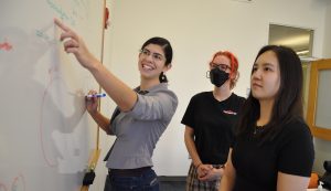
Our advanced undergraduate seminar courses focus on the primary research literature, with the goal of introducing you to the methods of contemporary biological research and the logic of experimental design and interpretation. The seminars are taught by postdoctoral scientists who are practicing researchers. They will expose you to the kind of thinking that is central to contemporary biological research and also impart specific knowledge in particular areas of biology.
Advanced seminars are graded pass/fail, carry six units, and meets for two hours weekly.
Advanced seminars feature:
- Small class size (limit of 8 students)
- High degree of personal contact with the instructor
- Focus on the primary research literature
- Lively discussions about biological problems
Prerequisites:
7.06 Cell Biology or 7.28 Molecular Biology
Subject offering descriptions
Days and class times are flexible and will be determined at the first class meeting.
Fall 2025
7.341 Myo-making and Myo-breaking: How Muscles Develop and Change with Exercise, Aging and Disease, and How CRISPR Gene-editing Can Be Harnessed to Treat Muscle Disorders
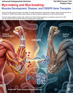
Instructor: Yichi (Tony) Zhang (laboratory of Matt Vander Heiden)
Why do different people excel at different sports? How do differences in genetics and lifestyle affect the development of different muscle groups and muscle fiber types? Do you know someone over the age of 60 or someone suffering from cancer who tells you they are feeling weak and losing muscle? Such muscle loss is real, and is called sarcopenia and cancer cachexia, respectively; there are currently no effective curative therapies for either. These are just some of the questions and problems that will be discussed in this course. Skeletal muscle is the largest tissue in the human body by biomass and is essential to sustain life, both because muscle is necessary for movement and also because, as in severe myopathies like Duchenne Muscular Dystrophy (DMD), perturbed muscle function can lead to respiratory failure and death. We will discuss both classic and recent studies exploring the molecular mechanisms underlying muscle development and dysfunction, including the coordinated transcriptional program that must be activated precisely for muscle cell determination and differentiation and the mechanisms responsible for the subsequent steps of muscle-cell fusion and maturation (i.e. formation of sarcomeres, neuromuscular junctions, etc.). We will learn which aspects of this process are evolutionarily conserved among species. We will discuss how in DMD, cancer, and aging, some of the developmental pathways of muscle are aberrantly activated to stimulate muscle protein breakdown and/or inhibit muscle protein synthesis. We will learn how muscle injury or disease can stimulate muscle stem-cell activation, muscle inflammation, and fibrosis (a process of excessive scar tissue formation). We will end with a critical evaluation of promising therapies involving CRISPR-Cas9 gene editing to correct the underlying monogenic mutations that lead to DMD and other myopathies. We will participate in a field trip to Vertex Pharmaceuticals, which is developing gene-editing therapies to treat diseases such as DMD. The leader of the Vertex DMD gene-editing team will share with us how Vertex views the problem of treating DMD using gene editing and the challenges they have encountered with therapeutic delivery. This course will be discussion-based and focus on the critical reading of the primary literature relevant to muscle biology, gene regulation, gene editing, and therapeutic design and delivery. Students will develop skills for evaluating scientific journal articles, interpreting experimental data, and designing rigorous experiments with appropriate controls. Students will also practice both scientific writing and oral presentation and discussion, with a focus on the critiquing of scientific data. By the end of the course, students will be equipped with a basic knowledge of the field of muscle biology and the critical thinking skills necessary to evaluate the primary research literature in the field of biomedicine more generally.
7.342 The Biology of Regeneration from Wound-Healing to Whole Organism: Molecular, Cellular, Developmental, and Evolutionary Principles and Their Biomedical Applications
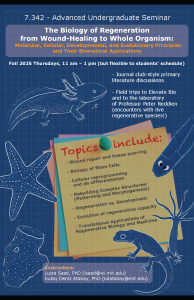 Instructor: Luiza Saad & Kutay Deniz Atabay (laboratory of Peter Reddien)
Many organisms have evolved the capacity to regrow lost tissues and organs to varying degrees. Nonetheless, the underlying mechanisms of regeneration remain one of the most fascinating and unresolved frontiers in biology. This discussion-based course will help students learn to read and critique the primary research literature related to key mechanisms guiding regenerative responses. The instructors invite students from all backgrounds to learn how organisms rebuild themselves. In this course, we will explore the molecular, cellular, and developmental principles that enable tissue regeneration, from simple wound healing to full organ, extremity, and organism regeneration. We will begin by examining the fundamental processes that follow injury: wound closure, cell death, dedifferentiation, and the formation of new cells through proliferation or the activation of stem cells. We will then dive into how the relationship between a cell’s lineage-determined identity and its position, which together specify cell fate, can be reestablished and how newly formed cells and tissues can integrate with pre-existing structures. We will compare and contrast diverse regenerative strategies employed by various organisms, ranging from planarians and axolotls to plants, zebrafish, and mammals, and ask: Why can some species regenerate complex body parts while others cannot? We will discuss the parallels and distinctions between regeneration and embryonic development, including how organisms can repurpose developmental signaling pathways in the adult context. We will also discuss current translational efforts bridging regenerative biology and medicine, with a focus on how fundamental research is being applied to develop new therapies. Examples will include current clinical trials using stem cell-derived pancreatic beta cells to restore insulin production in patients with type 1 diabetes, as well as recent efforts to treat burn wounds using stem cells, either through direct injection or tissue-engineered grafts. Finally, we will compare the distinct mechanisms by which various organisms achieve regeneration, highlighting their similarities and key differences from an evolutionary perspective. The course will explore this phenomenon through the eyes of the fields of developmental biology, evolution, tissue engineering (which uses cells and materials to create functional tissues), and regenerative medicine, a broad field focused on repairing or replacing damaged tissues and organs. We will discuss established and cutting-edge methodologies from the fields of regeneration and broader biomedical science. Topics will include genetic engineering, next-generation DNA and RNA sequencing (high-speed techniques for reading genetic information), high-throughput screening (a method for rapidly testing large numbers of chemical or genetic variables), and gene-expression analysis. During this course, students will have the opportunity to: (1) participate in a field trip to a relevant biotechnology company (for example: Akouos, or Elevate Bio, Merck or Vertex) and (2) observe and manipulate regenerative species (e.g., planarians, axolotls, fish, sea stars, mollusks) during a visit to the laboratory of Professor Peter Reddien at the MIT Whitehead Institute. Throughout the course, students will develop the skills to critically analyze scientific data and communicate their findings effectively in both written (through a short written assignment) and oral formats. Upon completion of this course, students will possess a foundational understanding of the field of regenerative biology and the critical capabilities necessary for the assessment of the primary research literature in the field of biomedicine more generally.
Spring 2026
7.341 Programming Immunity: Bioengineering T Cells to Fight Cancer and Autoimmune Disorders
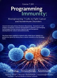 Instructor: Nouran Abdelfattah (laboratory of Stefani Spranger)
How does your body’s immune system distinguish friend from foe? What happens when this system errs? How are scientists “reprogramming” cells of the immune system to fight cancer and autoimmune disorders? In this class, we will explore the immune system with particular emphasis on T cells. T cells and B cells are two classes of lymphocytes, white blood cells in the vertebrate immune system. Unlike B cells, which generate antibodies that recognize pathogens in extracellular spaces, T cells specialize in recognizing fragments of foreign proteins presented on the surface of infected or abnormal cells—an ability that allows them to detect threats such as viruses and cancer. This unique function is matched by the extraordinary diversity of T cell receptors (TCRs): through V(D)J recombination – a random gene rearrangement process that occurs in the thymus – T cells can theoretically generate up to 1015 unique TCRs, vastly exceeding the total number of T cells in the human body. This intricate developmental process equips the T cell repertoire to recognize a wide array of peptides presented by major histocompatibility complex (MHC) molecules – specialized proteins found on the surface of all nucleated cells and that present internal cellular contents for immune surveillance. We will explore the molecular mechanisms underlying T cell antigen recognition, including the structural basis of TCR-peptide-MHC interactions and the cellular processes governing antigen presentation on the surface of both professional antigen-presenting (APCs) cells and all other nucleated cells. While all nucleated cells can display internal proteins using certain types of MHC molecules, professional APCs – such as dendritic cells, macrophages, and B cells – play a specialized role in initiating immune responses by presenting a broader range of antigens and delivering essential activation signals to T cells. We will explore both classical and emerging methodologies for discovering which antigens T cells recognize. Techniques such as peptide–MHC tetramer staining (used to detect antigen-specific T cells) and genetic library screening using cDNA expression systems will be discussed, along with newer high-throughput approaches. These approaches include genome-wide antigen discovery using barcoded peptide libraries, high-throughput T cell receptor (TCR) sequencing to map the diversity of T cell responses, and machine learning-based computational tools to predict antigens from protein sequence data. We will also explore how the principles of T cell biology can be applied to develop novel clinical approaches. We will discuss recent advances in bioengineering that are enabling researchers to harness and redirect T cell specificity for therapeutic applications in oncology and autoimmune disease. One striking example of a clinical approach involves cancer immunotherapy—treatments that redirect the immune system to eliminate tumors. Among these, chimeric antigen receptor (CAR)-engineered T cell therapies have shown remarkable success. For instance, CAR T cells targeting CD19, a molecule expressed on B cell malignancies, have led to the disappearance of all signs of cancer in up to 90% of pediatric patients with relapsed acute lymphoblastic leukemia (ALL)—a disease once considered nearly untreatable. We will also examine how failures in T cell tolerance—the mechanisms that prevent T cells from attacking the body’s own tissues—can lead to autoimmune disease. For example, in Type 1 diabetes, T cells target insulin-producing pancreatic β-cells, leading to their destruction. In multiple sclerosis, T cells mount pathogenic responses against central nervous system (CNS) proteins such as myelin basic protein (MBP), contributing to demyelination and neurodegeneration. Understanding these breakdowns in tolerance provides insight into therapeutic strategies, such as regulatory T cell therapy, which aims to restore immune balance by enhancing the function or number of T cells that suppress inappropriate immune responses. By the end of this course, students will have a foundational understanding of T cell biology, antigen discovery, and immunotherapy development. The course will be discussion-based and center on learning to critically read, evaluate, and debate findings from the primary research literature in immunology and biomedical science more broadly. To connect these insights to real-world applications, we will also examine how companies have translated discoveries from basic research – particularly those involving T cell biology and antigen recognition – into cellular therapeutics for cancer treatment. In this context, we will visit a biotechnology company developing immune-based therapies, to see how research moves from the academic laboratory to drug development and ultimately to the clinic. We will also learn from company scientists about their own career paths and opportunities in the biotechnology and pharmaceutical company world.
Past subject offerings