With new NIH grant, team will learn how neurons build key sites that release neurotransmitters a lot, or a little, to drive nervous system communication
Picower Institute
March 16, 2021
Job descriptions for the thousands of types of neurons in the brain typically include a common function: release chemicals called neurotransmitters to communicate across circuit connections called synapses. In a new study funded by the National Institutes of Health, the lab of MIT Professor Troy Littleton will seek to understand how neurons construct synapses of different strengths, a variety that may be key to the diversity of neural communication.
Littleton, Menicon Professor of Neuroscience in The Picower Institute for Learning and Memory and the Departments of Biology and Brain and Cognitive Sciences at MIT, said the findings could increase scientists’ understanding of how neural circuits develop and change to reflect learning and experience – a phenomenon called plasticity – and might also suggest ways to adjust synaptic strength when it is atypical in disorders such as autism or intellectual disability.
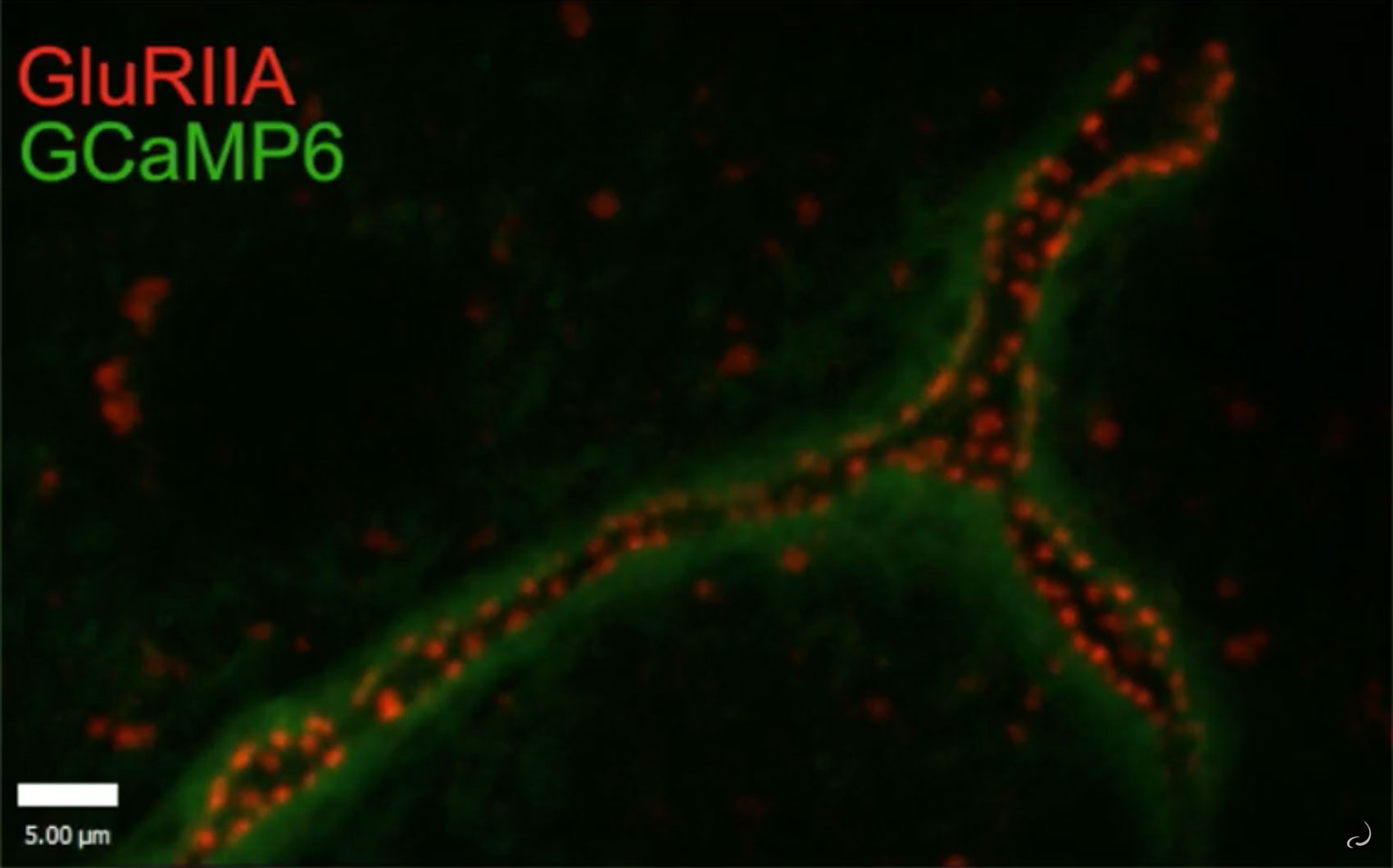
Video from a 2018 Littleton Lab study shows calcium flux (green) indicating the release of glutamate at synapses tagged by the presence of a glutamate receptor (red).
Using neurons that control muscles in the Drosophila fruit fly, the study will focus on “active zones” (AZs), which are tiny neural structures that enable the release of neurotransmitters across each synapse. The flies provide a simple model, Littleton said, that can help elucidate many basic factors affecting AZ strength that are also at play in the neurons of other animals, including mammals.
“Understanding the rules in a simple model like Drosophila that help to define when a synapse is strong or weak allows us to view these principles as fundamental elements of how neurons control synaptic growth and development,” he said. “Depending on which of these factors a neuron modifies or plays around with, it is likely to be able to make synapses stronger or weaker in very different patterns.”
During larval development the neurons build hundreds of AZs. In a 2018 study, Littleton’s lab found that AZs vary widely in their strength: About 10 percent release neurotransmitters as much as 50 times more often than the majority of weaker synapses. The researchers also found that the strongest AZs were typically the ones that had the most time to develop and accumulate their many protein building blocks.
In the new study, which will provide nearly $1.9 million over five years, the team will learn how those active zones get built step by step out of more than a dozen different proteins that arrive at different stages of development. Because some AZs apparently build up bigger and stronger than others, Littleton likens the process to the construction of a variety of houses in a neighborhood—from big four-bedroom homes to little townhomes. The new study, including preliminary work the team has done with the support of the Picower Institute Innovation Fund, will help explain how each kind of structure emerges, in their relative abundance, in the same cell.
In one set of experiments, for instance, his team will study whether the supply of building materials – the various proteins – is a limitation on how many AZs can mature to full strength before development ceases (i.e. maybe they don’t all get enough lumber or nails to fully frame the house in time). The scientists will test that, for instance, with genetic manipulations that change the amount of key proteins produced. By imaging the proteins as they accumulate and by looking in on the same AZs day after day, a technique the lab uses called “intravital imaging,” they can see how changing protein availability changes the construction of AZs in a neuron.
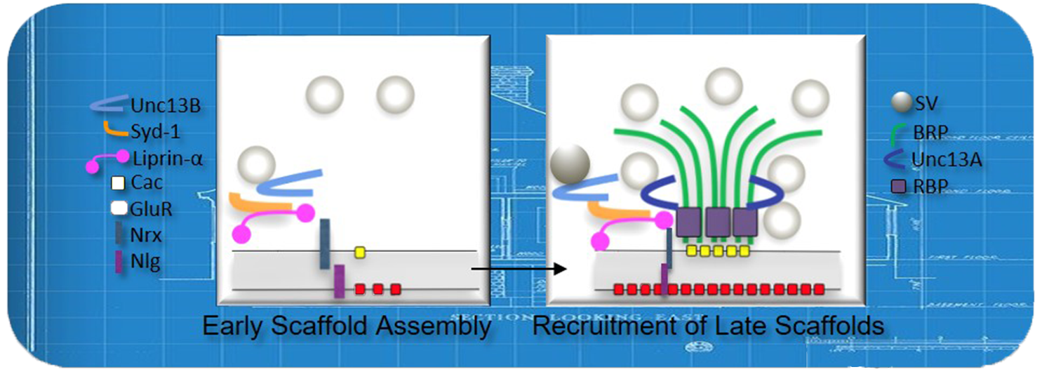
A model of active zone construction: Numerous proteins arrive over time during development to ultimately build a structure for releasing neurotransmitters.
In another set of experiments, the team will test whether some AZs are better than others at acquiring the available material supply and putting it to use (i.e. some may have more carpenters than others to make the best use of the available nails and lumber). And to better understand how the construction process might work in longer-lived animals like mammals, where protein materials not only need to be gathered but also maintained and replaced, they will artificially prolong the flies’ larval stage.
In a third set of tests they will examine the case of two types of neurons that each connect to the same fly muscles but exert control in different ways. Though each type works by releasing the same neurotransmitter, called glutamate, “tonic” neurons feature small but constant glutamate release, while the “phasic” cells release stronger, but more occasional, bursts. The study will examine how AZ development differs, for instance, due to differences in gene expression to promote the different function of these otherwise similar cells.
In all, their goal will be to determine how neurons build their different capacities and styles of connection and communication.
In addition to Littleton the research team includes research scientists Yulia Akbergenova and Suresh Jetti, and graduate students Karen Leopold Cunningham and Andrés Crane.