Study: Fragile X syndrome neurons can be restored

Whitehead Institute researchers are using a modified CRISPR/Cas9-guided activation strategy to investigate the most frequent cause of intellectual disability in males.

Nicole Giese Rura | Whitehead Institute
February 15, 2018

Fragile X syndrome is the most frequent cause of intellectual disability in males, affecting one out of every 3,600 boys born. The syndrome can also cause autistic traits, such as social and communication deficits, as well as attention problems and hyperactivity. Currently, there is no cure for this disorder.

Fragile X syndrome is caused by mutations in the FMR1 gene on the X chromosome, which prevent the gene’s expression. This absence of the FMR1-encoded protein during brain development has been shown to cause the overexcitability in neurons associated with the syndrome. Now, for the first time, researchers at Whitehead Institute have restored activity to the fragile X syndrome gene in affected neurons using a modified CRISPR/Cas9 system they developed that removes the methylation — the molecular tags that keep the mutant gene shut off — suggesting that this method may prove to be a useful paradigm for targeting diseases caused by abnormal methylation.

Research by the lab of Whitehead Institute for Biomedical Research Founding Member Rudolf Jaenisch, which is described online this week in the journal Cell, is the first direct evidence that removing the methylation from a specific segment within the FMR1 locus can reactivate the gene and rescue fragile X syndrome neurons.

The FMR1 gene sequence includes a series of three nucleotide (CGG) repeats, and the length of these repeats determines whether or not a person will develop fragile X syndrome: A normal version of the gene contains anywhere from 5 to 55 CGG repeats, versions with 56 to 200 repeats are considered to be at a higher risk of generating some of the syndrome’s symptoms, and those versions with more than 200 repeats will produce fragile X syndrome.

Until now, the mechanism linking the excessive repeats in FMR1 to fragile X syndrome was not well-understood. But Shawn Liu, a postdoc in Jaenisch’s lab and first author of the Cell study, and others thought that the methylation blanketing those nucleotide repeats might play an important role in shutting down the gene’s expression.

In order to test this hypothesis, Liu removed the methylation tags from the FMR1 repeats using a CRISPR/Cas9-based technique he recently developed with Hao Wu, a postdoc in the Jaenisch lab. This technique can either add or delete methylation tags from specific stretches of DNA. Removal of the tags revived the FMR1 gene’s expression to the level of the normal gene.

“These results are quite surprising — this work produced almost a full restoration of wild type expression levels of the FMR1 gene,” says Jaenisch, whose primary affiliation is with Whitehead Institute, where his laboratory is located and his research is conducted. He is also a professor of biology at MIT. “Often when scientists test therapeutic interventions, they only achieve partial restoration, so these results are substantial,” he says.

The reactivated FMR1 gene rescues neurons derived from fragile X syndrome induced pluripotent stem (iPS) cells, reversing the abnormal electrical activity associated with the syndrome. When rescued neurons were engrafted into the brains of mice, the FMR1 gene remained active in the neurons for at least three months, suggesting that the corrected methylation may be sustainable in the animal.

“We showed that this disorder is reversible at the neuron level,” says Liu. “When we removed methylation of CGG repeats in the neurons derived from fragile X syndrome iPS cells, we achieved full activation of FMR1.”

The CRISPR/Cas-9-based technique may also prove useful for other diseases caused by abnormal methylation including facioscapulohumeral muscular dystrophy and imprinting diseases.

“This work validates the approach of targeting the methylation on genes, and it will be a paradigm for scientists to follow this approach for other diseases,” says Jaenisch.

This work was supported by the National Institutes of Health, the Damon Runyon Cancer Foundation, the Rett Syndrome Research Trust, the Brain and Behavior Research Foundation, and the Helen Hay Whitney Foundation. Jaenisch is co-founder of Fate Therapeutics, Fulcrum Therapeutics, and Omega Therapeutics.

Reading and writing DNA

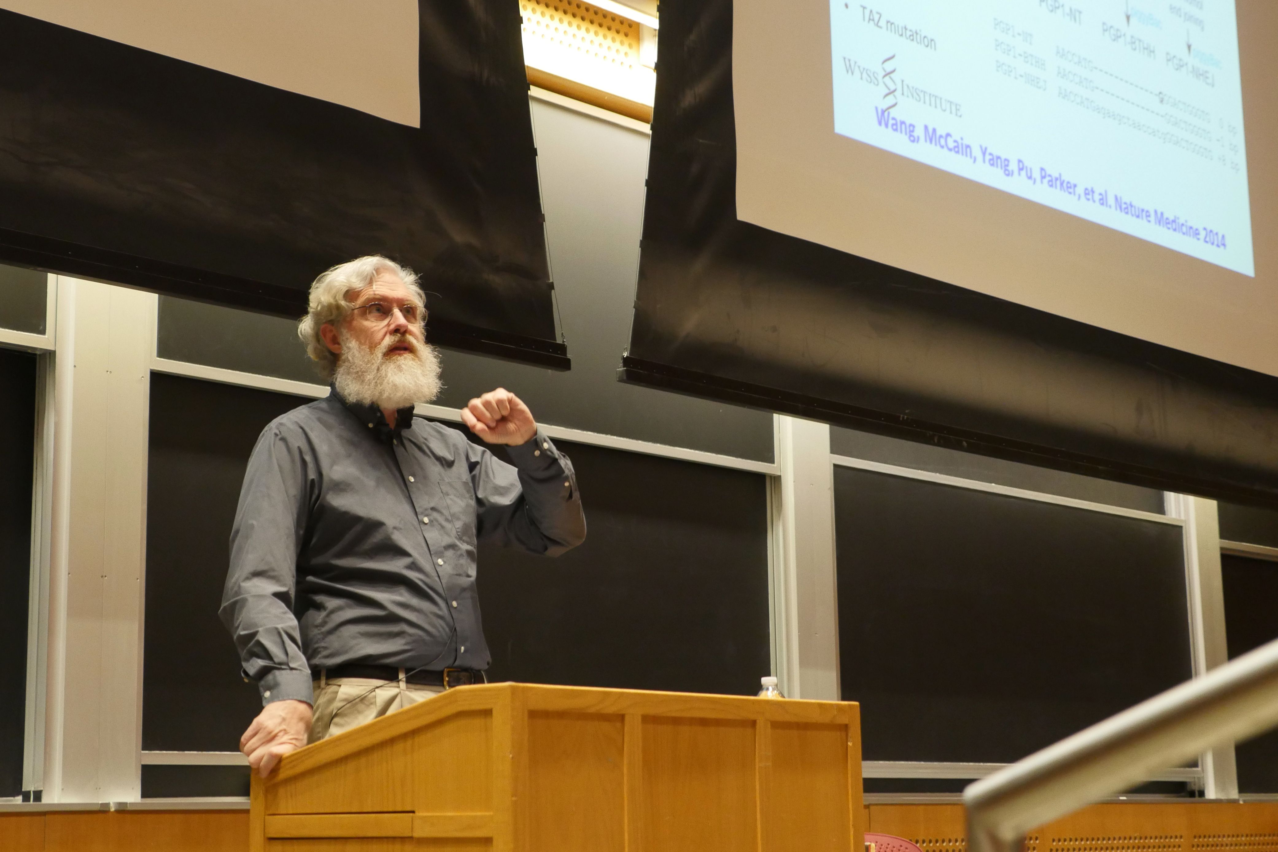
Department of Biology kicks off IAP seminar series with a lecture by synthetic-biology visionary George Church.

Raleigh McElvery | Department of Biology
January 31, 2018

Thanks to the invention of genome sequencing technology more than three decades ago, we can now read the genetic blueprint of virtually any organism. After the ability to read came the ability to edit — adding, subtracting, and eventually altering DNA wherever we saw fit. And yet, for George Church, a professor at Harvard Medical School, associate member of the Broad Institute, and founding core faculty and lead for synthetic biology at the Wyss Institute — who co-pioneered direct genome sequencing in 1984 — the ultimate goal is not just to read and edit, but also to write.

What if you could engineer a cell resistant to all viruses, even the ones it hadn’t yet encountered? What if you could grow your own liver in a pig to replace the faulty one you were born with? What if you could grow an entire brain in a dish? In his lecture on Jan. 24 — which opened the Department of Biology’s Independent Activities Period (IAP) seminar series, Biology at Transformative Frontiers — Church promised all this and more.

“We began by dividing the Biology IAP events into two tracks: one related to careers in academia and another equivalent track for industry,” says Jing-Ke Weng, assistant professor and IAP faculty coordinator for the department. “But then it became clear that George Church, Patrick Brown, and other speakers we hoped to invite blurred the boundaries between those two tracks. The Biology at Transformative Frontiers seminar series became about the interface of these trajectories, and how transferring technologies from lab bench to market is altering society as we know it.”

The seminar series is a staple in the Department of Biology’s IAP program, but during the past several years it has been oriented more toward quantitative biology. Weng recalls these talks as being relegated to the academic sphere, and wanted to show students that the lines between academia, industry, and scientific communication are actually quite porous.

“We chose George Church to kick off the series because he’s been in synthetic biology for a long time, and continues to have a successful academic career even while starting so many companies,” says Weng.

Church’s genomic sequencing methods inspired the Human Genome Project in 1984 and resulted in the first commercial genome sequence (the bacterium Helicobacter pylori) 10 years later. He also serves as the director of the Personal Genome Project, the “Wikipedia” of open-access human genomic data. Beyond these ventures, he’s known for his work on barcoding, DNA assembly from chips, genome editing, and stem cell engineering.

He’s also the same George Church who converted the book he co-authored with Ed Regis, “Regenesis: How Synthetic Biology Will Reinvent Nature and Ourselves,” into a four-letter code based on the four DNA nucleotides (A, T, C, and G), subsisted on nutrient broth from a lab vendor for an entire year, and dreams of eventually resurrecting woolly mammoths. He’s being featured in an upcoming Netflix Original documentary, so when he arrived at the Stata Center to give his lecture last week he was trailed by a camera crew.

According to Church, the transformative technologies that initially allowed us to read and edit DNA have grown exponentially in recent years with the invention of molecular multiplexing and CRISPR-Cas9 (think Moore’s Law but even more exaggerated). But there’s always room for improvement.

“There’s been a little obsession with CRISPR-Cas9s and other CRISPRs,” said Church. “Everybody is saying how great it is, but it’s important to say what’s wrong with it as well, because that tells us where we’re going next and how to improve on it.”

He outlined several of his own collaborations, including those aimed at devising more precise methods of genome editing, one resulting in 321 changes to the Escherichia coli genome — the largest change in any genome yet — rendering the bacterium resistant to all viruses, even those it had not yet come into contact with. The next step? Making similarly widespread changes in plants, animals, and eventually perhaps even human tissue. In fact, Church and his team have set their sights on combatting the global transplantation crisis with humanlike organs grown in animals.

“Since the dawn of transplantation as a medical practice, we’ve had to use either identical twins or rare matches that are very compatible immunologically, because we couldn’t engineer the donor or the recipient,” said Church.

Since it’s clearly unethical to engineer human donors, Church reasoned, why not engineer animals with compatible organs instead? Pigs, to be exact, since most of their organs are comparable in size and function to our own.

“This is an old dream; I didn’t originate it,” said Church. “It started about 20 years ago, and the pioneers of this field worked on it for a while, but dropped it largely because the number of changes to the genome were daunting, and there was a concern that the viruses all pigs make — retroviruses — would be released and infect the immunocompromised organ recipient.”

Church and his team successfully disrupted 62 of these retroviruses in pig cells back in 2015, and in 2017 they used these cells to generate living, healthy pigs. Today, the pigs are thriving and rearing piglets of their own. Church is also considering the prospect of growing augmented organs in pigs for human transplantation, perhaps designing pathogen-, cancer-, and age-resistant organs suitable for cryopreservation.

“Hopefully we’ll be doing nonhuman primate trials within a couple of years, and then almost immediately after that human trials,” he said.

Another possibility, rather than cultivating organs in animals for transplant, is to generate them in a dish. A subset of Church’s team is working on growing from scratch what is arguably the most complicated organ of all, the brain.

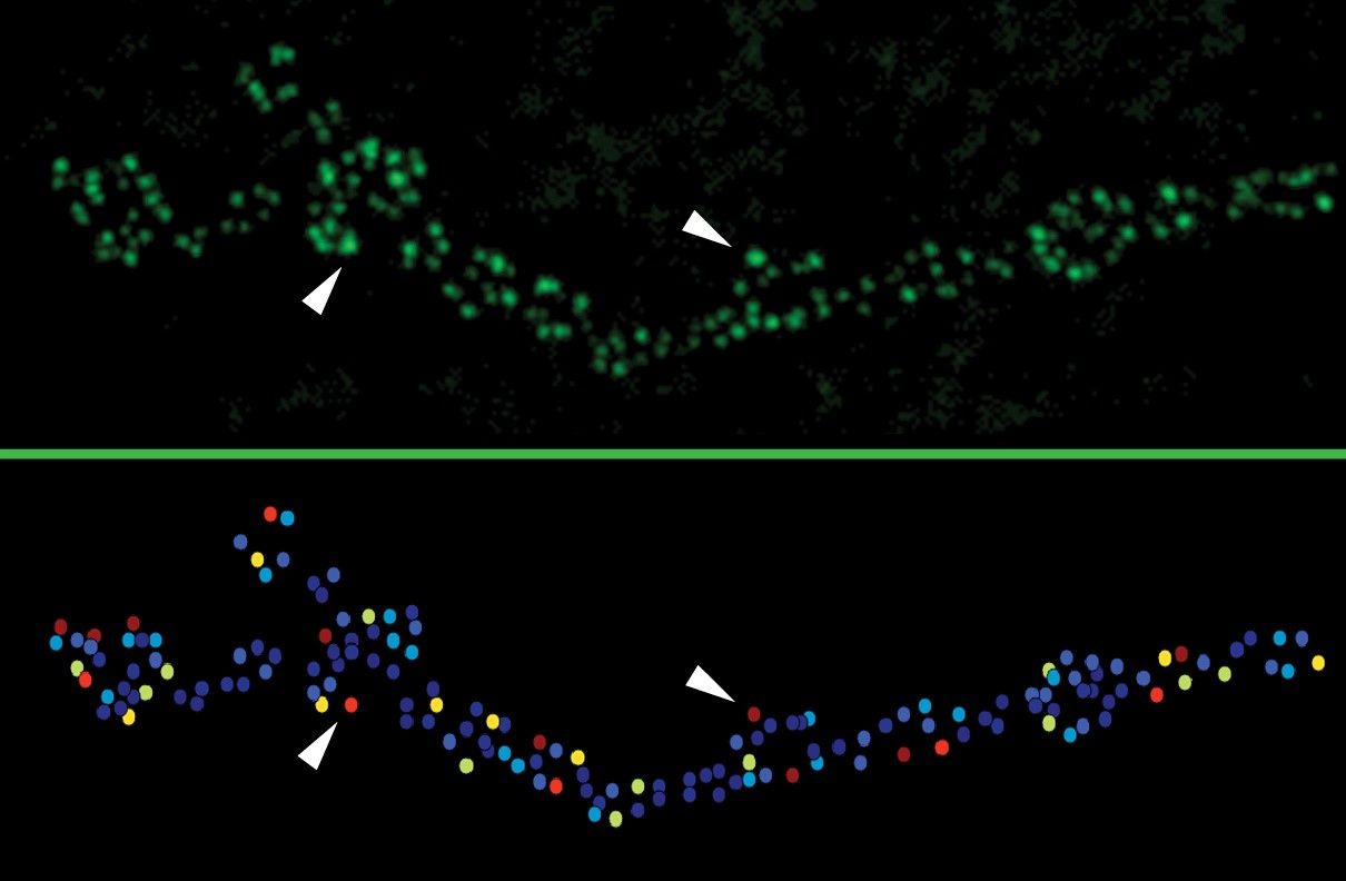
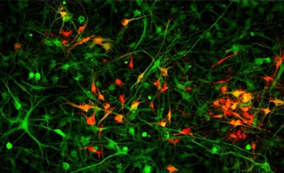
This requires differentiating multiple types of cells in the same dish so they can interact with each other to form the complex systems of communication characteristic of the human brain.

Early attempts at fashioning brain organoids often lacked capillaries to distribute oxygen and nutrients (roughly one capillary for each of the 86 billion neurons in the human brain). However, thanks to their new human transcription factor library, Church and colleagues have begun to generate the cell types necessary to create such capillaries, plus the scaffolding needed to promote the three-dimensional organization of these and additional brain structures. Church and his team have not only successfully integrated the structures with one another, but have also created an algorithm that spits out the list of molecular ingredients required to generate each cell type.

Church noted these de novo organoids are extremely useful in determining which genetic variants are responsible for certain diseases. For instance, you could sequence a patient’s genome and then create an entire organoid with the mutation in question to test whether it was the root cause of the condition.

“I’m still stunned by the breadth of projects and approaches that he’s running simultaneously,” says Emma Kowal, a second-year graduate student, member of Weng’s planning committee, and a former researcher in Church’s lab. “The seminar series is called Biology at Transformative Frontiers, and George is very much a visionary, so we thought it would be a great way to start things off.”

The four-part series also features Melissa Moore, chief scientific officer of the Moderna Therapeutics mRNA Research Platform, Jay Bradner, president of the Novartis Institutes for BioMedical Research, and Patrick Brown, CEO and founder of Impossible Foods.

Decoding the genetics of roundworm mating

Sixth year graduate student Zoë Hilbert investigates how C. elegans react to changes in their environment — and how these changes affect physiology, gene expression, and behavior

Raleigh McElvery
January 30, 2018

Sixth year graduate student Zoë Hilbert is sure of many things. After performing her first dissection in third grade, she was sure she liked science. Before she started college, she was sure she wanted to major in a biology-related discipline. And as she finished her final year at Columbia University, she was sure she would leave the East Coast immediately upon graduation. What she did not anticipate, however, was falling in love with the Cambridge biotechnology hub, applying to MIT for graduate school, and switching fields from biochemistry to genetics.

“I’m incredibly grateful for the MIT first-year program, because dedicating the fall semester solely to taking classes gave me a background in subjects I didn’t take in college,” Hilbert says. “I’d never taken genetics before, and now here I am in Dennis Kim’s lab — a genetics lab.”

Hilbert was enthralled by evolution from an early age, in particular the idea that entire organisms and their proteins change over time in response to internal and external pressures. She recalls becoming “obsessed” with the small and seemingly unremarkable stickleback fish, after she learned that researchers could map the evolution of physical features like additional belly fins or extra armor to variations in specific genes.

“When it came time for the first years to write our National Science Foundation proposals, we had the opportunity to work with a faculty member,” she recalls, “and I chose Dennis because one of the project ideas he’d listed was in a similar vein to the stickleback research. Coming into it, I didn’t know anything about his work or even his model system, but I ended up joining the lab after second semester rotations.”

The Kim lab investigates how the roundworm Caenorhabditis elegans reacts to changes in their environment — and how these changes not only affect physiology and gene expression, but behavior as well.

Today, Hilbert is as enamored by C. elegans as she once was with stickleback fish. With minimal prodding, she’s happy to rattle off their numerous advantages: they’re transparent, so there’s no need to do dissections to look inside; they’re ideal for studying development and the nervous system, because scientists have already charted all the cells in the body and how the neurons communicate; and they’re low-maintenance and easy to keep in lab. The list goes on.

But most pertinent to Hilbert is the fact that — like most species of animals — the two sexes of C. elegans, males and hermaphrodites, often behave differently in similar situations due to differences in gene expression. Take mating, for example.

Hermaphrodites are capable of self-fertilization, and can produce up to several hundred identical progeny over the course of several days. Males are much less common and unable to reproduce on their own, but by mating with hermaphrodites they can introduce some genetic variety into their offspring. Because males must locate a hermaphrodite in order to pass on their genetic material, they’ve developed some specific behaviors to find their mate. And that’s where Hilbert’s work comes in. She makes males choose between the two things they need most: food and mate.

There comes a time in every adult male’s life when finding a mate takes precedence over continuously eating, as younger worms are wont to do. If he is placed in a plate of yummy bacteria by himself, he runs away — not because he’s full, but because he’d rather spend his time searching for a mate. However, if he is placed in a plate of food along with a tempting hermaphrodite, his urge to escape is suppressed and he remains long enough to mate.

That said, C. elegans mating is not always so cut and dry. Researchers understand that a male’s behavior is also food-dependent. If you place a starving male on the plate of food, he no longer prioritizes mating over feeding, and will remain in the food instead of seeking a mate. He is constantly evaluating his priorities, which are heavily influenced by the situation at hand and — as Hilbert discovered — when and where certain genes are expressed.

“We’ve spent a lot of time monitoring how the expression of daf-7 changes in different food and mating situations,” Hilbert says. “When you starve the male, you suppress the gene and as a result you also suppress the fleeing behavior.”Hilbert demonstrated several years ago that this male-specific behavior is controlled by a gene known as daf-7, which encodes a signaling molecule and is expressed in two specific neurons in the male.  (No expression is normally seen in the hermaphrodite.) Curiously, the same gene in the same two neurons is also turned on when any worm — male or hermaphrodite — comes across a pathogen, sending a “WARNING: consume at your own risk” signal, and prompting the worm to avoid the noxious bacteria.

Expression appears to be dependent not only on nutritional state (hungry or full), but also environment (food and/or mate) and sex (since males express daf-7 differently than hermaphrodites).

“All these factors and signals are converging on this one gene,” Hilbert says. “It’s really quite incredible.”

The neurons that express daf-7 are “sensory,” and traditionally viewed as funnels to higher neural centers where information is processed and behaviors are generated. However, Hilbert’s data suggest this information processing is happening right there, directly within these neurons via changes in gene expression without waiting for instructions from on high.

What Hilbert finds particularly intriguing is that the worms rely on just one molecular pathway to dictate behavior in two very different situations: mating and pathogen avoidance. Although the worm flees food in both situations, precisely why one gene is implicated in two distinct settings remains a mystery. Hilbert is still asking herself, For what benefit?

She intends to spend her final semester at MIT tying up loose ends and conducting follow-up experiments to extend the work from her recent paper in the January 2017 issue of eLife, on which she was first author. She’s screening for molecules that could impact whether or not daf-7 is expressed, honing in on chemicals and signaling molecules used by neurons to communicate with one another.

“I’d advise prospective grads to be willing and open to change your mind about what you want to do,” she says. “I was really into protein biochemistry when I first arrived at MIT, and was really surprised when I fell in love with a discipline that was completely different from my initial interests.”

As Hilbert applies to academic postdoctoral positions, she’s still set on fulfilling her longtime dream of heading out West. She’s sure she’d like to end up someplace like California, Washington, or Utah, but only time will tell.

Photo credit: Raleigh McElvery
How the brain selectively remembers new places

Neuroscientists identify a circuit that helps the brain record memories of new locations.

Anne Trafton | MIT News Office
December 25, 2017

When you enter a room, your brain is bombarded with sensory information. If the room is a place you know well, most of this information is already stored in long-term memory. However, if the room is unfamiliar to you, your brain creates a new memory of it almost immediately.

MIT neuroscientists have now discovered how this occurs. A small region of the brainstem, known as the locus coeruleus, is activated in response to novel sensory stimuli, and this activity triggers the release of a flood of dopamine into a certain region of the hippocampus to store a memory of the new location.

“We have the remarkable ability to memorize some specific features of an experience in an entirely new environment, and such ability is crucial for our adaptation to the constantly changing world,” says Susumu Tonegawa, the Picower Professor of Biology and Neuroscience and director of the RIKEN-MIT Center for Neural Circuit Genetics at the Picower Institute for Learning and Memory.

“This study opens an exciting avenue of research into the circuit mechanism by which behaviorally relevant stimuli are specifically encoded into long-term memory, ensuring that important stimuli are stored preferentially over incidental ones,” adds Tonegawa, the senior author of the study.

Akiko Wagatsuma, a former MIT research scientist, is the lead author of the study, which appears in the Proceedings of the National Academy of Sciences the week of Dec. 25.

New places

In a study published about 15 years ago, Tonegawa’s lab found that a part of the hippocampus called the CA3 is responsible for forming memories of novel environments. They hypothesized that the CA3 receives a signal from another part of the brain when a novel place is encountered, stimulating memory formation.

They believed this signal to be carried by chemicals known as neuromodulators, which influence neuronal activity. The CA3 receives neuromodulators from both the locus coeruleus (LC) and a region called the ventral tegmental area (VTA), which is a key part of the brain’s reward circuitry. The researchers decided to focus on the LC because it has been shown to project to the CA3 extensively and to respond to novelty, among many other functions.

The LC responds to an array of sensory input, including visual information as well as sound and odor, then sends information on to other brain areas, including the CA3. To uncover the role of LC-CA3 communication, the researchers genetically engineered mice so that they could block the neuronal activity between those regions by shining light on neurons that form the connection.

To test the mice’s ability to form new memories, the researchers placed the mice in a large open space that they had never seen before. The next day, they placed them in the same space again. Mice whose LC-CA3 connections were not disrupted spent much less time exploring the space on the second day, because the environment was already familiar to them. However, when the researchers interfered with the LC-CA3 connection during the first exposure to the space, the mice explored the area on the second day just as much as they had on the first. This suggests that they were unable to form a memory of the new environment.

The LC appears to exert this effect by releasing the neuromodulator dopamine into the CA3 region, which was surprising because the LC is known to be a major source of norepinephrine to the hippocampus. The researchers believe that this influx of dopamine helps to boost CA3’s ability to strengthen synapses and form a memory of the new location.

They found that this mechanism was not required for other types of memory, such as memories of fearful events, but appears to be specific to memory of new environments. The connections between the LC and CA3 are necessary for long-term spatial memories to form in CA3.

“The selectivity of successful memory formation has long been a puzzle,” says Richard Morris, a professor of neuroscience at the University of Edinburgh, who was not involved in the research. “This study goes a long way toward identifying the brain mechanisms of this process. Activity in the pathway between the locus coeruleus and CA3 occurs most strongly during novelty, and it seems that activity fixes the representations of everyday experience, helping to register and retain what’s been happening and where we’ve been.”

Choosing to remember

This mechanism likely evolved as a way to help animals survive, allowing them to remember new environments without wasting brainpower on recording places that are already familiar, the researchers say.

“When we are exposed to sensory information, we unconsciously choose what to memorize. For an animal’s survival, certain things are necessary to be remembered, and other things, familiar things, probably can be forgotten,” Wagatsuma says.

Still unknown is how the LC recognizes that an environment is new. The researchers hypothesize that some part of the brain is able to compare new environments with stored memories or with expectations of the environment, but more studies are needed to explore how this might happen.

“That’s the next big question,” Tonegawa says. “Hopefully new technology will help to resolve that.”

The research was funded by the RIKEN Brain Science Institute, the Howard Hughes Medical Institute, and the JPB Foundation.

Matthew A. Wilson

Education

  • PhD, 1991, California Institute of Technology
  • BS, 1983, Electrical Engineering, Rensselaer Polytechnic Institute

Research Summary

Our laboratory studies the neural processes within the hippocampus and neocortex that enable memories to form and persist over time. We use a technique that allows us to simultaneously record the activity of hundreds of individual neurons across multiple brain regions in freely behaving animals. When combined with genetic, pharmacological and behavioral manipulations, these recordings allow us to gain a mechanistic understanding of how animals learn and remember.

Awards

  • American Academy of Arts and Sciences, Fellow, 2012
H. Robert Horvitz

Education

  • PhD, 1974, Harvard University
  • BS, 1968, Mathematics and Economics, MIT

Research Summary

Our lab examines how genes control animal development and behavior. We use the experimentally tractable nematode Caenorhabditis elegans to identify and analyze molecular and cellular pathways involved in these important areas of biology. Ultimately, we hope to clarify these fundamental biological mechanisms and provide further insight into human disease.

Awards

  • U.S. National Academy of Inventors, Member, 2015
  • American Association for Cancer Research Academy, Fellow, 2013
  • Royal Society of London, Foreign Member, 2009
  • Genetics Society (U.K.), Mendel Medal, 2007
  • Eli Lilly Lecturer Award, 2007
  • Massachusetts Institute of Technology, James R Killian Jr Faculty Achievement Award, 2006
  • National Academy of Medicine, Member, 2003
  • American Cancer Society, Medal of Honor, 2002
  • The Nobel Foundation, Nobel Prize in Physiology or Medicine, 2002
  • Bristol-Myers Squibb, Award for Distinguished Achievement in Neuroscience, 2001
  • March of Dimes, Developmental Biology, 2000
  • Gairdner Foundation, Gairdner Foundation International Award, 1999
  • National Academy of Sciences, Member, 1991
  • American Academy of Arts and Sciences, Fellow, 1989
  • American Association for the Advancement of Science, Fellow, 1989
  • Howard Hughes Medical Institute, HHMI Investigator, 1988
Elly Nedivi

Education

  • PhD, 1991, Stanford University
  • BSc, 1982, Biology and Biochemistry, Hebrew University, Israel

Research Summary

The property of the brain that allows it to constantly adapt to change is termed plasticity, and is a prominent feature not only of learning and memory in the adult, but also of brain development. Connections between neurons (synapses) that are frequently used become stronger, while those that are unstimulated gradually dwindle away. The Nedivi lab works to identify the cellular mechanisms that underlie the addition and elimination of synaptic connections in response to activity using genetic and in vivo imaging approaches.

Awards

  • Elected Member at Large, AAAS, 2019-2023
  • Elected Member, Dana Alliance, 2019
  • BCS Award for Excellence in Undergraduate Teaching, 2018
  • American Association for the Advancement of Science (AAAS), Fellow, 2016
  • AFAR Julie Martin Mid-Career Award in Aging Research, 2007 – 2011
  • Edgerly Innovation Fund Award, 2006
  • Dean’s Education and Student Advising Award, 2003
  • NSF Powre Award, 1999
  • Alfred P . Sloan Research Fellowship, 1999 – 2001
  • Ellison Medical Foundation New Scholar Award, 1997 – 2002
Rudolf Jaenisch

Education

  • MD, 1967, University of Munich

Research Summary

We aim to understand the epigenetic regulation of gene expression in mammalian development and disease. Embryonic stem cells are important because they have the potential to generate any cell type in the body and, therefore, have great potential for regenerative medicine. We study the way somatic cells reprogram to an embryonic pluripotent state, and use patient specific pluripotent cells to study complex human diseases.

Awards

  • German Society for Biochemistry and Molecular Biology, Otto Warburg Medal, 2014
  • New York Academy, Medicine Medal, 2013
  • Franklin Institute, Benjamin Franklin Medal, 2013
  • National Science Foundation, National Medal of Science, 2011
  • National Science Foundation, National Medal of Science, 2010
  • National Academy of Sciences, Member, 2003
Susumu Tonegawa

Education

  • PhD, 1968, University of California, San Diego
  • BS, 1963, Chemistry, Kyoto University

Research Summary

We are interested in the molecular, cellular and neural circuit mechanisms underlying learning and memory in rodents. We generate genetically engineered mice, and analyze them through multiple methods including molecular and cellular biology, electrophysiology, microscopic imaging, optogenetic engineering, and behavioral studies. Ultimately, we aim to detect the effects of our manipulations at multiple levels in the brain — deducing which behaviors or cognitions are causally linked to specific processes and events taking place at the molecular, cellular, and neuronal circuit levels.

Awards

  • The Nobel Foundation, Nobel Prize in Physiology or Medicine, 1987
  • Albert and Mary Lasker Award in Basic Research, 1987
  • National Academy of Sciences, Member, 1986
Troy Littleton

Education

  • PhD, 1994, Baylor College of Medicine; MD, 1997, Baylor College of Medicine
  • BS, 1989, Biochemistry, Louisiana State University

Research Summary

Using Drosophila, we study how neurons form synaptic connections, as well as how synapses transmit information and change during learning and memory. We also investigate how alterations in neuronal signaling underlie several neurological diseases, including epilepsy, autism, and Huntington’s Disease. We hope to bridge the gap between the molecular components of the synapse and the physiological responses they mediate.