

Neurons are talkers. They each communicate with fellow neurons, muscles, or other cells by releasing neurotransmitter chemicals at “synapse” junctions, ultimately producing functions ranging from emotions to motions. But even neurons of the exact same type can vary in their conversational style. A new open-access study in Cell Reports by neurobiologists at The Picower Institute for Learning and Memory highlights a molecular mechanism that might help account for the nuanced diversity of neural discourse.
The scientists made their findings in neurons that control muscles in Drosophila fruit flies. These cells are models in neuroscience because they exhibit many fundamental properties common to neurons in people and other animals, including communication via the release of the neurotransmitter glutamate. In the lab of Troy Littleton, Menicon Professor in MIT’s departments of Biology and Brain and Cognitive Sciences, which studies how neurons regulate this critical process, researchers frequently see that individual neurons vary in their release patterns. Some “talk” more than others.
In more than a decade of studies, Littleton’s lab has shown that a protein called complexin has the job of restraining spontaneous glutamate chatter. It clamps down on fusion of glutamate-filled vesicles at the synaptic membrane to preserve a supply of the neurotransmitter for when the neuron needs it for a functional reason, for instance to simulate a muscle to move. The lab’s studies have identified two different kinds of complexin in flies (mammals have four) and showed that the clamping effectiveness of the rare but potent 7B splice form is regulated by a molecular process called phosphorylation. How the much more abundant 7A version is regulated was not known, but scientists had shown that the RNA transcribed from DNA that instructs the formation of the protein is sometimes edited in the cell by an enzyme called ADAR.
In the new study from Littleton’s team, led by Elizabeth Brija PhD ’23, the lab investigated whether RNA editing of complexin 7A affects how it regulates glutamate release. What she discovered was surprising. Not only does RNA editing of complexin 7A have a significant impact on how well the protein prevents glutamate release, but also this can vary widely among individual neurons because they can stochastically mix and match up to eight different editions of the protein. Some edits were much more common than others on average, but 96 percent of the 200 neurons the team examined had at least some editing, which affected the structure of an end of the protein called its C-terminus. Experiments to test some of the consequences of this structural variation showed that different complexin 7A edits can dramatically affect the level of electrical current measurable at different synapses. That varying level of activity can also affect the growth of the synapses the neurons make with muscle. RNA editing of the protein might therefore endow each neuron with fine degrees of communication control.
“What this offers the nervous system is that you can take the same transcriptome and by alternatively editing various RNA transcripts, these neurons will behave differently,” Littleton says.
Moreover, Littleton and Brija’s team found that other key proteins involved in synaptic glutamate release, such as synapsin and Syx1A, are also sometimes edited at quite different levels among the same population of neurons. This suggests that other aspects of synaptic communication might also be tunable.
“Such a mechanism would be a robust way to change multiple features of neuronal output,” Brija, Littleton, and colleagues wrote.
The team tracked the different editing levels by meticulously extracting and sequencing RNA from the nuclei and cell bodies of 200 motor neurons. The work yielded a rich enough dataset to show that any of three adenosine nucleotides encoding two amino acids in the C-terminus could be swapped for another, yielding eight different editions of the protein. A slim majority of complexin 7A went unedited in the average neuron, while the seven edited versions composed the rest with widely varying degrees of frequency.
To investigate the functional consequences of some of the different editions, the team knocked out complexin and then “rescued” flies by adding back in unedited or two different edited versions. The experiments showed a stark contrast between the two edited proteins. One, which occurs more commonly, proved to be a less effective clamp than unedited complexin, barely preventing spontaneous glutamate release and upticks in electrical current. The other turned out to be more effective at clamping than the unedited version, keeping a tight lid on glutamate release and synaptic output. And while both of the edited versions showed a tendency to drift away from synapses and into the neuron’s axon, the long branch that extends from the cell body, the edition that clamped well prevented any overgrowth of synapses while the one that clamped poorly provided only a meager curb.
Because multiple editions are often present in neurons, Brija and the team did one more set of experiments in which they “rescued” complexin-less flies with a combination of unedited complexin and the weak-clamping edition. The result was a blend of the two: reduced spontaneous glutamate release than with just the weakly clamping edition alone. The findings suggest that not only does each edition potentially fine-tune glutamate release, but that combinations among them can act in a combinatorial fashion.
In addition to Brija and Littleton the paper’s other authors are Zhuo Guan and Suresh Jetti.
The National Institutes of Health, The JPB Foundation, and The Picower Institute for Learning and Memory supported the research.
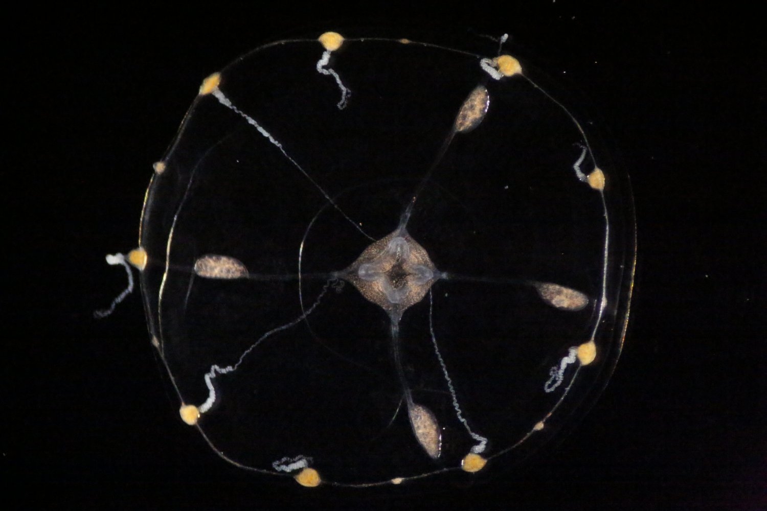
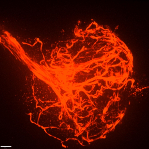
The Clytia hemisphaerica jellyfish is not only a hypnotically graceful swimmer, but also an amazing neuron-manufacturing machine with a remarkable ability to expand and regenerate its nervous system.
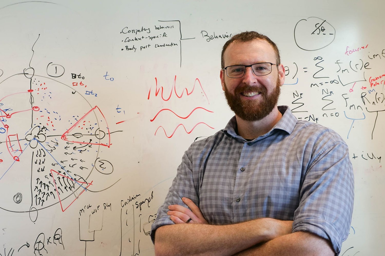
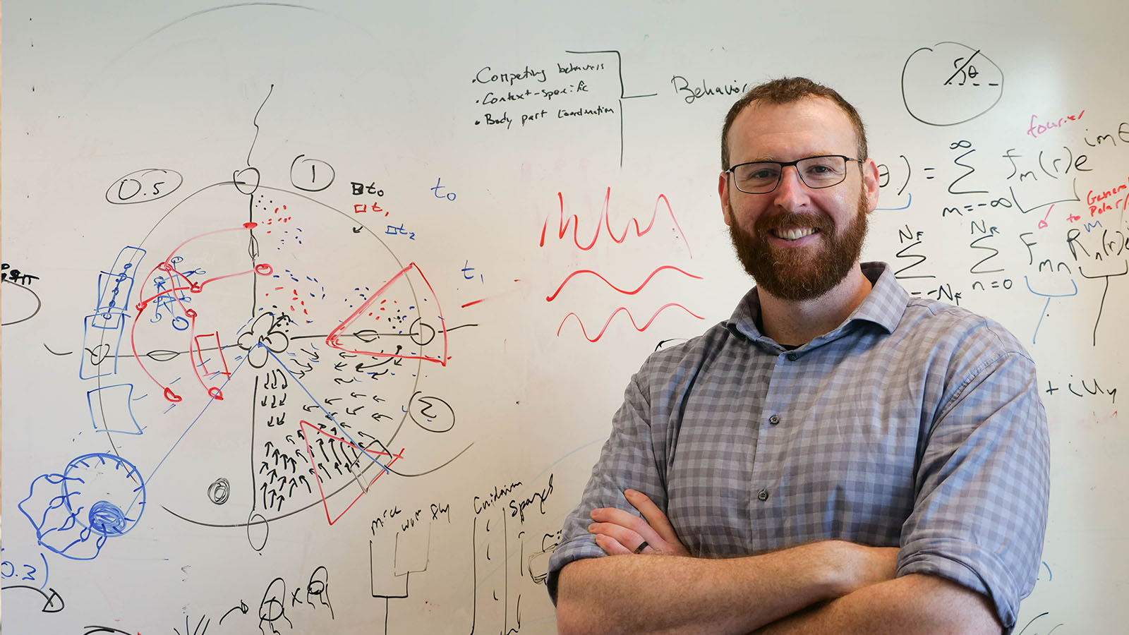
Now, thanks to a prestigious Klingenstein-Simons Fellowship Award in Neuroscience, MIT Assistant Professor Brady Weissbourd will study how the tiny, transparent animals use this ability to build, organize, and rebuild a stable, functional, and robust nervous system throughout their lives.
“As we look more broadly across the animal kingdom it is amazing to see how similar the basic biology is of animals that look completely different — even jellyfish have neurons similar to our own that generate their behavior,” says Weissbourd, a faculty member in MIT’s Department of Biology and The Picower Institute for Learning and Memory, whose work to engineer genetic access to C. hemisphaerica in 2021 established it as a new neuroscience model organism. “At the same time, it could be just as important to examine what is different across species, particularly when it comes to some of the incredible capabilities that have evolved.”
Weissbourd is just one of 13 researchers nationally to be recognized with this fellowship, which provides $300,000 over three years. It will enable Weissbourd’s lab to tackle several questions raised by the jellyfish’s prodigious production of neurons. Where does the constant stream of newborn neurons come from, and what guides them to their eventual places in the jellyfish’s mesh-like neural network? How does the jellyfish organize these ever-changing neural populations — for instance, into functional circuits — to enable its various behaviors?
Another question hails from the surprising results of an experiment in which Weissbourd ablated the entire class of the neurons that the jellyfish uses to fold up its umbrella-shaped body — about 10 percent of the 10,000 or so neurons that it has. He found that within a week enough new neurons had taken their place that the folding behavior was restored. Weissbourd’s studies will also seek to determine how the animal can so readily bounce back from the destruction of a whole major neural network and the behavior it produces.
“We were studying the neural control of a particular behavior and stumbled across this shocking observation that the subnetwork that controls this behavior is constantly changing size and can completely regenerate,” Weissbourd says. “We want to understand the mechanisms that allow this network to be so robust, including the ability to rebuild itself from scratch. I’m very grateful to the Klingenstein Fund and the Simons Foundation for supporting our work.”
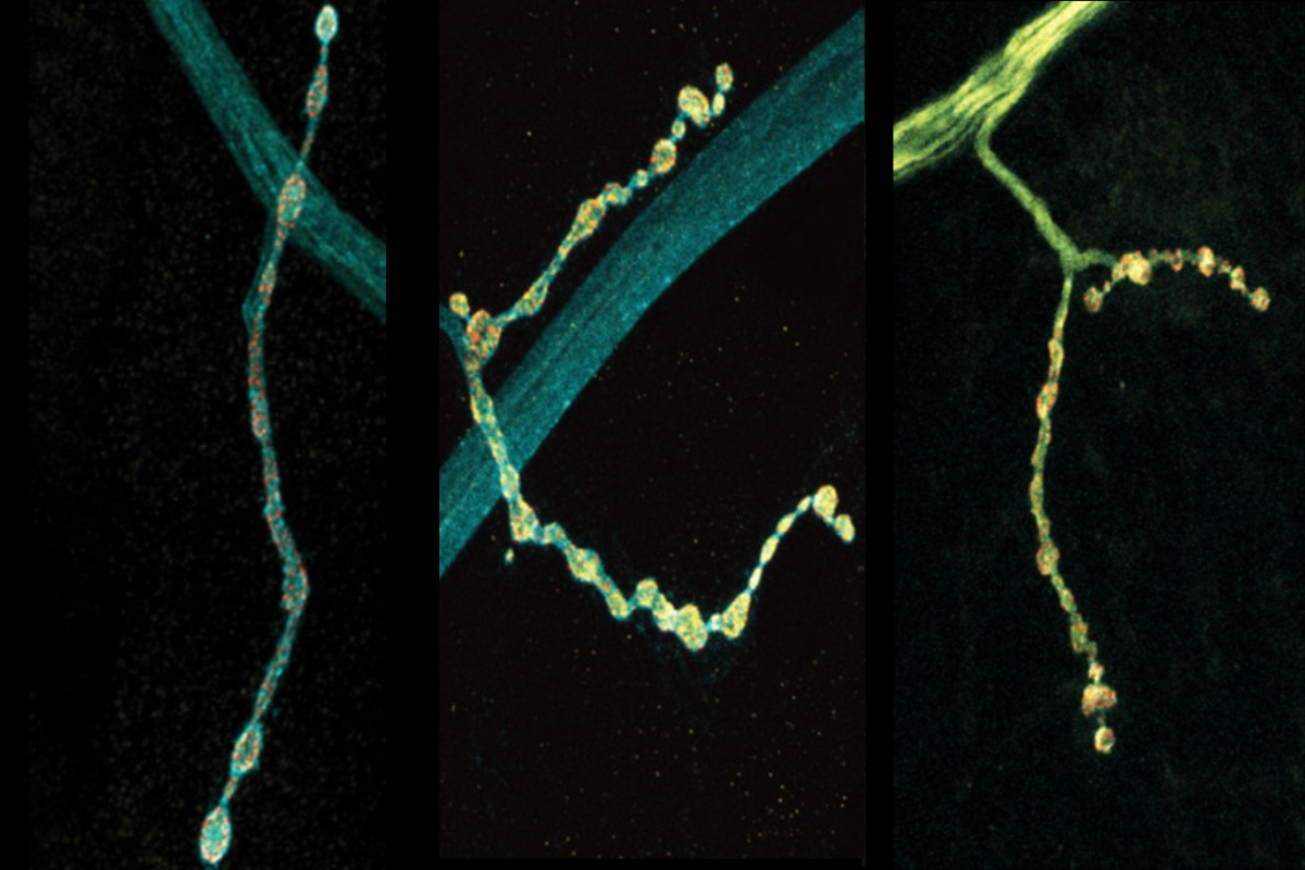
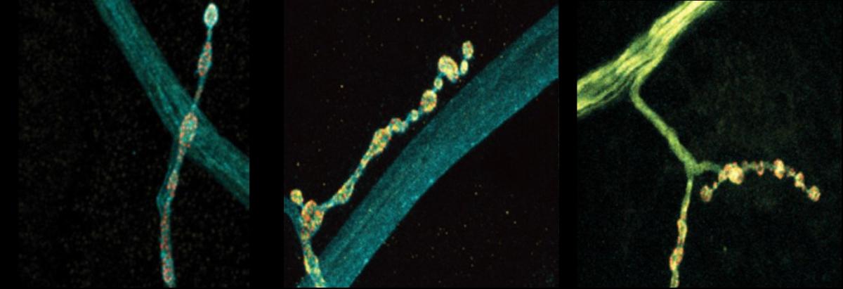
Perhaps the most obvious feature of a neuron is the long branch called an axon that ventures far from the cell body to connect with other neurons or muscles. If that long, thin projection ever seems like it could be vulnerable, a new MIT study shows that its structural integrity may indeed require the support of a surrounding protein called perlecan. Without that protein in Drosophila fruit flies, researchers at The Picower Institute for Learning and Memory found axonal segments can break apart during development and the connections, or synapses, that they form end up dying away.
Perlecan helps make the extracellular matrix, the proteins and other molecules that surround cells, stable and flexible so that cells can develop and function in an environment that is supportive without being rigid.
“What we found was that the extracellular matrix around nerves was being altered and essentially causing the nerves to break completely. Broken nerves eventually led to the synapses retracting,” says study senior author Troy Littleton, the Menicon Professor in MIT’s departments of Biology and Brain and Cognitive Sciences.
Humans need at least some perlecan to survive after birth. Mutations that reduce, but don’t eliminate, perlecan can cause Schwartz-Jampel syndrome, in which patients experience neuromuscular problems and skeletal abnormalities. The new study may help explain how neurons are affected in the condition, Littleton says, and also deepen scientists’ understanding of how the extracellular matrix supports axon and neural circuit development.
Ellen Guss PhD ’23, who recently defended her doctoral thesis on the work, led the research published June 8 in eLife.
At first she and Littleton didn’t expect the study to yield a new discovery about the durability of developing axons. Instead, they were investigating a hypothesis that perlecan might help organize some of the protein components in synapses that fly nerves develop to connect with muscles. But when they knocked out the gene called “trol” that encodes perlecan in flies, they saw that the neurons appeared to “retract” many synapses at a late stage of larval development. Proteins on the muscle side of the synaptic connection remained, but the neuron side of the connection withered away. That suggested that perlecan had a bigger role than they first thought.
Indeed, the authors found that the perlecan wasn’t particularly enriched around synapses. Where it was pronounced was in a structure called the neural lamella, which surrounds axon bundles and acts a bit like the rubbery cladding around a TV cable to keep the structure intact. That suggested that a lack of perlecan might not be a problem at the synapse, but instead causes trouble along axons due to its absence in the extracellular matrix surrounding nerve bundles.
Littleton’s lab had developed a technique for daily imaging of fly neural development called serial intravital imaging. They applied it to watch what happened to the fly axons and synapses over a four-day span. They observed that while fly axons and synapses developed normally at first, not only synapses but also whole segments of axons faded away.
They also saw that the farther an axon segment was from the fly’s brain, the more likely it was to break apart, suggesting that the axon segments became more vulnerable the further out they extended. Looking segment by segment, they found that where axons were breaking down, synapse loss would soon follow, suggesting that axon breakage was the cause of the synapse retraction.
“The breakages were happening in a segment-wide manner,” Littleton says. “In some segments the nerves would break and in some they wouldn’t. Whenever there was a breakage event, you would see all the neuromuscular junctions (synapses) across all the muscles in that segment retract.”
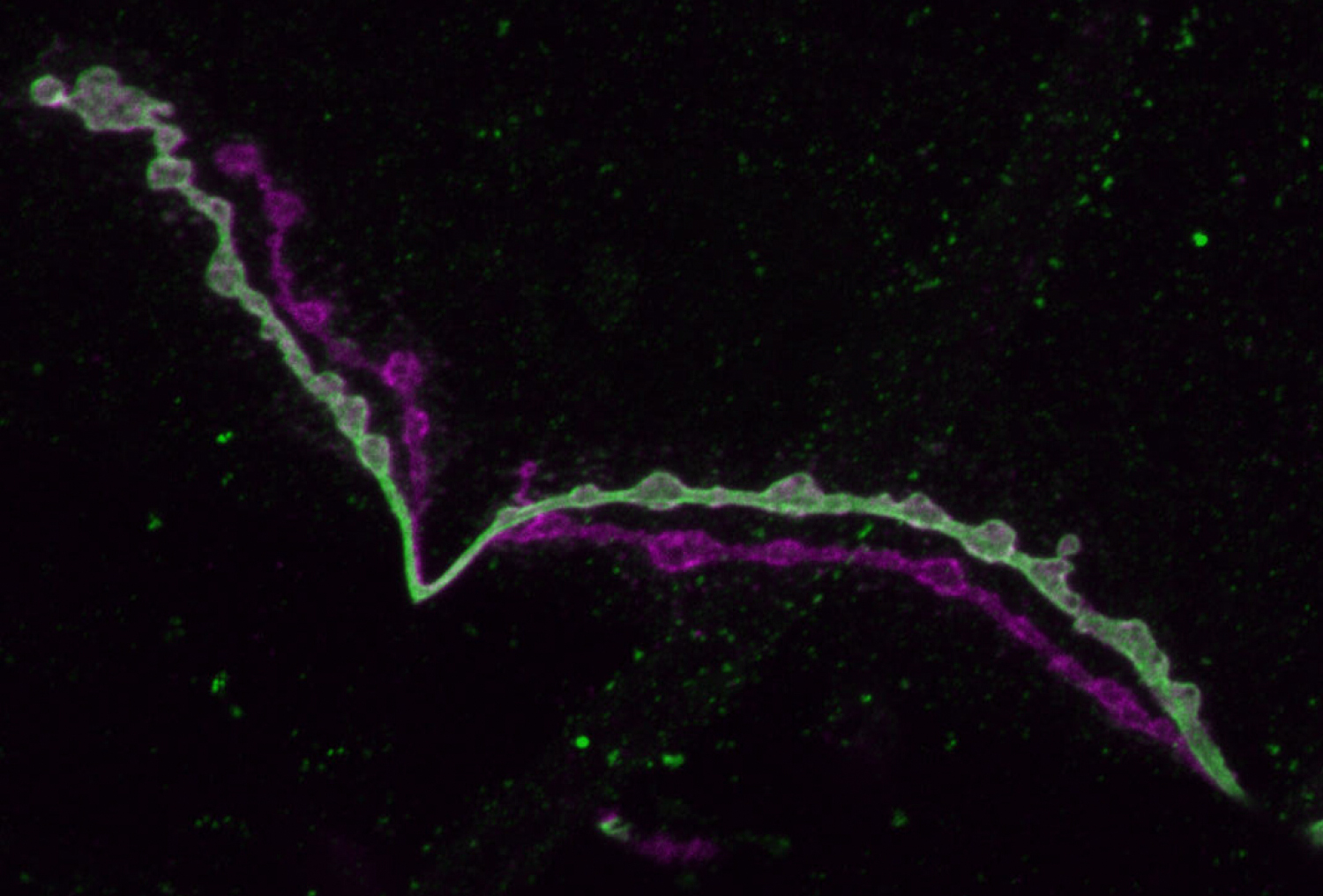
When they compared the structure of the lamella in mutant versus healthy flies, they found that the lamella was thinner and defective in the mutants. Moreover, where the lamella was weakened, axons were prone to break and the microtubule structures that run the length of the axon would become misdirected, protruding outward and becoming tangled up in dramatic bundles at sites of severed axons.
In one other key finding, the team showed that perlecan’s critical role depended on its secretion from many cells, not just neurons. Blocking the protein in just one cell type or another did not cause the problems that total knockdown did, and enhancing secretion from just neurons was not enough to overcome its deficiency from other sources.
Altogether, the evidence pointed to a scenario where lack of perlecan secretion caused the neural lamella to be thin and defective, with the extracellular matrix becoming too rigid. The further from the brain nerve bundles extended, the more likely movement stresses would cause the axons to break where the lamella had broken down. The microtubule structure within the axons then became disorganized. That ultimately led to synapses downstream of those breakages dying away because the disruption of the microtubules means the cells could no longer support the synapses.
“When you don’t have that flexibility, although the extracellular matrix is still there, it becomes very rigid and tight and that basically leads to this breakage as the animal moves and pulls on those nerves over time,” Littleton says. “It argues that the extracellular matrix is functional early on and can support development, but doesn’t have the right properties to sustain some key functions over time as the animal begins to move and navigate around. The loss of flexibility becomes really critical.”
In addition to Littleton and Guss, the paper’s other authors are Yulia Akbergenova and Karen Cunningham.
Support for the study came from the National Institutes of Health. The Littleton Lab is also supported by The Picower Institute for Learning and Memory and The JPB Foundation.
How does animal behavior emerge from networks of connected neurons? How are these incredible nervous systems and behaviors actually generated by evolution? Are there principles shared by all nervous systems or is evolution constantly innovating? What did the first nervous system look like that gave rise to the incredible diversity of life that we see around us?
Combining the study of animal behavior with studies of nervous system form, function, and evolution, Brandon “Brady” Weissbourd, a new faculty member in the Department of Biology and investigator in The Picower Institute for Learning and Memory, uses the tiny, transparent jellyfish Clytia hemisphaerica, a new neuroscience model.
Q: In 2021, you developed a new model organism for neuroscience research, the transparent jellyfish Clytia hemisphaerica. How do these jellyfish answer questions about neuroscience, the nervous system, and evolution in ways that other models cannot?
A: First, I believe in the importance of more broadly understanding the natural world and diversifying the organisms that we deeply study. One reason is to find experimentally tractable organisms to identify generalizable biological principles — for example, we understand the basis of how neurons “fire” from studies of the squid giant axon. Another reason is that transformative breakthroughs have come from identifying evolutionary innovations that already exist in nature — for example, green fluorescent protein (GFP, from jellyfish) or CRISPR (from bacteria). In both ways, this jellyfish is a valuable complement to existing models.
I have always been interested in the intersection of two types of problems: how nervous systems generate our behaviors; and how these incredible systems were actually created by evolution.
On the systems neuroscience side, ever since working on the serotonin system during my PhD I have been fascinated by the problem of how animals control all of their behaviors simultaneously in a flexible and context-dependent manner, and how behavioral choices depend not just on incoming stimuli but on how those stimuli interact with constantly changing states of the nervous system and body. These are extremely complex and difficult problems, with the particular challenge of interactions across scales, from chemical signaling and dynamic cell biology to neural networks and behavior.
To address these questions, I wanted to move into a model organism with exceptional experimental tractability.
There have been exciting breakthroughs in imaging techniques for neuroscience, including these incredible ways in which we can actually watch and manipulate neuronal activity in a living animal. So, the first thing I wanted was a small and transparent organism that would allow for this kind of optical approach. These jellyfish are a few millimeters in diameter and perfectly transparent, with interesting behaviors but relatively compact nervous systems. They have thousands of neurons where we have billions, which also puts them at a nice intermediate complexity compared to other transparent models that are widely used — for example, C. elegans have 302 neurons and larval zebrafish have something like 100,000 in the brain alone. These features will allow us to look at the activity of the whole nervous system in behaving animals to try to understand how that activity gives rise to behaviors and how that activity itself arises from networks of neurons.
On the evolution side of our work, we are interested in the origins of nervous systems, what the first nervous systems looked like, and broadly what the options are for how nervous systems are organized and functioning: to what extent there are principles versus interesting and potentially useful innovations, and if there are principles, whether those are optimal or somehow constrained by evolution. Our last common ancestor with jellyfish and their relatives (the cnidarians) was something similar to the first nervous system, so by comparing what we find in cnidarians with work in other models we can make inferences about the origins and early evolution of nervous systems. As we further explore these highly divergent animals, we are also finding exciting evolutionary innovations: specifically, they have incredible capabilities for regenerating their nervous systems. In the future, it will be exciting to better understand how these neural networks are organized to allow for such robustness.
Q: What work is required to develop a new organism as a model, and why did you choose this particular species of jellyfish?
A: If you’re choosing a new animal model, it’s not just about whether it has the right features for the questions you want to ask, but also whether it technically lets you do the right experiments. The model we’re using was first developed by a research group in France, who spent many years doing the really hard work of figuring out how to culture the whole life cycle in the lab, injecting eggs, and developing other key resources. For me, the big question was whether we’d be able to use the genetic tools that I was describing earlier for looking at neural activity. Working closely with collaborators in France, our first step was figuring out how to insert things into the jellyfish genome. If we couldn’t figure that out, I was going to switch back to working with mice. It took us about two years of troubleshooting, but now we can routinely generate genetically modified jellyfish in the lab.
Switching to a new animal model is tough — I have a mouse neuroscience background and joined a postdoc lab that used mice and flies; I was the only person working with jellyfish, but had no experience. One of my goals is now to optimize and simplify this whole process so that when other labs want to start working with jellyfish we have a simple aquaculture platform to get them started, even if they have no experience.
In addition to the fact that these things are tiny and transparent, the main reason that we chose this particular species is because it has an amazing life cycle that makes it an exciting laboratory animal.
They have separate sexes that spawn daily with the fertilized eggs developing into larvae that then metamorphose into polyps. We grow these polyps on microscope slides, where they form colonies that are thought to be immortal. These colonies are then constantly releasing jellyfish, which are all genetically identical “clones” that can be used for experiments. That means that once you create a genetically modified strain, like a transgenic line or a knockout, you can keep it forever as a polyp colony — and since the animals are so small, we can culture them in large numbers in the lab.
There’s still a huge amount of foundational work to do, like characterizing their behavioral repertoire and nervous system organization. It’s shocking how little we know about the basics of jellyfish biology — particularly considering that they kill more people per year than sharks and stingrays combined — and the more we look into it, the more questions there are.
Q: What drew you to a faculty position at MIT?
A: I wanted to be in a department that does fundamental research, is enthusiastic about basic science, is open-minded, and is very diverse in what people work on and think about. My goal is also to be able to ultimately link mechanisms at the molecular and cellular level to organismal behavior, which is something that [the] MIT [Department of] Biology is particularly strong at doing. It’s been an exciting first few months! MIT Biology is such an amazing place to do science and it’s been wonderful how enthusiastic and supportive everyone in the department has been.
I was additionally drawn to MIT by the broader community and have already found it so easy to start collaborations with people in neuroscience, engineering, and math. I’m also thrilled to have recently become a member of The Picower Institute for Learning and Memory, which further enables these collaborations in a way that I believe will be transformational for the work in my lab.
It’s a new lab. It’s a new organism. There isn’t a huge, well-established field that is taking these approaches. There’s so much we don’t know, and so much that we have to establish from scratch. My goal is for my lab to have a sense of adventure and fun, and I’m really excited to be doing that here in MIT Biology.

Many of our body’s most important functions occur without our conscious knowledge, such as digestion, heartbeat, and breathing. These vital functions depend on the signals generated by the “interoceptive nervous system,” which enables the brain to monitor our internal organs and trigger responses that sometimes save our lives. One second you are breathing normally as you eat your salad and the next, when a vinegar-soaked crouton enters your throat, you are coughing or swallowing to protect and clear your airway. We know our bodies are sensitive to cues like irritants, but we still have a lot to learn about how the interoceptive system works to meet our physiological needs, keep organs safe and healthy, and affect our behavior. We can also learn how chronic insults may lead to organ dysfunction and use what we learn to create therapeutic interventions.
Focusing on the airway, Sara Prescott, a new faculty member in the Department of Biology and investigator in The Picower Institute for Learning and Memory, seeks to understand the ways our nervous systems detect and respond to stimuli in health and disease. Here, she describes her work.
Q: Why is understanding the peripheral nervous system important, and what parts of your background are you drawing on for your current research?
A: The lab focuses on really trying to explore the body-brain connection.
People often think that our mind exists in a vacuum, but in reality, our nervous system is heavily integrated with the rest of the body, and those neural interfaces are important, both for taking information from our body or environment and turning it into an internal representation of the world, and, in reverse, being able to process that information and being able to enact changes throughout the body. That includes things like autonomic reflexes, basic functions of the body like breathing, blood-gas regulation, digestion, and heart rate.
I’ve integrated both my graduate training and postdoctoral training into thinking about biology across multiple scales.
Graduate school for me was quite focused on deep molecular mechanism questions, particularly gene regulation, so I feel like that has been very useful for me in my general approach to neuroscience because I take a very molecular angle to all of this.
It also showed me the power of in vitro models as reductionist tools to explore fundamental aspects of cell biology. During my postdoc, I focused on larger, emergent phenotypes. We were able to manipulate specific circuits and see very impressive behavioral responses in animals. You could stimulate about 100 neurons in a mouse and see that their breathing would just stop until you remove the stimulation, and then the breathing would return to normal.
Both of those experiences inform how we approach a problem in my research. We need to understand how these circuits work, not just their connectivity at the anatomical level but what is driving their changes in sensitivity over time, the receptor expression programs that affect how they sense and signal, how these circuits emerge during development, and their gene expression.
There are still so many foundational questions that haven’t been answered that there’s enough to do in the mouse for quite some time.
Q: How are you specifically looking into interoceptive biology at MIT?
A: Our flagship system is the mammalian airway. We use a mouse model and modern molecular neuroscience tools to manipulate various neural pathways and observe what the effects are on respiratory function and animal health.
Neuroscience and mouse work have a reputation for being a little challenging and intense, but I think this is also where we can ask really important questions that are useful for our everyday lives — and the only place where we can fully recapitulate the complexity of nervous system signaling all the way down to our organs, back to our brain, and back to our organs.
It’s a very fun place to do science with lots of open questions.
One of the core discoveries from my postdoctoral work was focusing on the vagus nerve as a major body-to-brain conduit, as it innervates our lungs, heart, and gastrointestinal tract. We found that there were about 40 different subtypes of sensory neurons within this small nerve, which is really a remarkable amount of diversity and reflects the massive sensory space within the body. About a dozen of those vagal neurons project to the airways.
We identified a rare neuron type specifically responsible for triggering protective responses, like coughing when water or acid entered the airway. We also discovered a separate population of neurons that make us feel and act sick when we get a flu infection. The field now knows what four to five vagal populations of neurons are actually sensing in the airways, but the remaining populations are still a mystery to us; we don’t know what those populations of sensory neurons are detecting, what their anatomy is, and what reflex effects those neurons are evoking.
Looking ahead, there are many exciting directions for the interoceptive biology field. For example, there’s been a lot of focus on characterizing the circuits underlying acute motor reflexes, like rapid responses to visceral stimuli on the timescale of minutes to hours. But we don’t have a lot of information about what happens when these circuits are activated over long periods of time. For example, respiratory tract infections often last for weeks or longer. We know that the airways undergo changes in composition when they’re exposed to different types of infection or stress to better accommodate future threats. One of the hypotheses we’re testing is that chronically activating neural circuits may drive changes in organ composition. We have this idea, which we’re calling reflexive remodeling: neurons may be communicating with stem cells and progenitor cells in the periphery to drive adaptive remodeling responses.
We have the genetic, molecular, and circuit scale tools to explore this phenomenon in mice. In parallel, we’re also setting up some in vitro models of the mouse airway mucosa to expedite receptor screening and to explore basic mechanisms of neuron-epithelium cross-talk. We hope this will inform our understanding of how the airway surface senses and responds to different types of irritants or damage.
Q: This all sounds fascinating. Where does it lead?
A: Human health has been my north star for a long time and I’ve taken a long, wandering path to find particular areas where I can scratch whatever intellectual itch that I have.
I originally thought I would be a doctor and then realized that I felt like I could have a more lasting impact by discovering fundamental truths about how our bodies work. I think there are a number of chronic diseases in which autonomic imbalance is actually a huge clinical component of the disorder.
We have a lot of interest in some of these very common airway remodeling diseases, like chronic obstructive pulmonary disorder — COPD — asthma, and potentially lung cancer. We want to ask questions like how autonomic circuits are altered in disease contexts, and when neurons actually drive features of disease.
Perhaps this research will help us come up with better molecular, cellular, or tissue engineering approaches to improve the outcomes for a variety of autonomic diseases.
It’s very easy for me to imagine how one day, not too far from now, we can turn these findings into something actionable for human health.
How does animal behavior emerge from networks of connected neurons? How are these incredible nervous systems and behaviors actually generated by evolution? Are there principles shared by all nervous systems or is evolution constantly innovating? What did the first nervous system look like that gave rise to the incredible diversity of life that we see around us?
Combining the study of animal behavior with studies of nervous system form, function, and evolution, Brady Weissbourd, a new faculty member in the Department of Biology and investigator in The Picower Institute for Learning and Memory, uses the tiny, transparent jellyfish Clytia hemisphaerica, a new neuroscience model.
Q: In your work, you developed a new model organism for neuroscience research, the transparent jellyfish Clytia hemisphaerica. How do these jellyfish answer questions about neuroscience, the nervous system, and evolution in ways that other models cannot?
A: First, I believe in the importance of more broadly understanding the natural world and diversifying the organisms that we deeply study. One reason is to find experimentally tractable organisms to identify generalizable biological principles – for example, we understand the basis of how neurons “fire” from studies of the squid giant axon. Another reason is that transformative breakthroughs have come from identifying evolutionary innovations that already exist in nature – for example, green fluorescent protein (GFP, from jellyfish) or CRISPR (from bacteria). In both ways, this jellyfish is a valuable complement to existing models.
I have always been interested in the intersection of two types of problems: how nervous systems generate our behaviors; and how these incredible systems were actually created by evolution.
On the systems neuroscience side, ever since working on the serotonin system during my PhD I have been fascinated by the problem of how animals control all of their behaviors simultaneously in a flexible and context-dependent manner, and how behavioral choices depend not just on incoming stimuli but on how those stimuli interact with constantly changing states of the nervous system and body. These are extremely complex and difficult problems, with the particular challenge of interactions across scales, from chemical signaling and dynamic cell biology to neural networks and behavior.
To address these questions, I wanted to move into a model organism with exceptional experimental tractability.
There have been exciting breakthroughs in imaging techniques for neuroscience, including these incredible ways in which we can actually watch and manipulate neuronal activity in a living animal. So, the first thing I wanted was a small and transparent organism that would allow for this kind of optical approach. These jellyfish are a few millimeters in diameter and perfectly transparent, with interesting behaviors but relatively compact nervous systems. They have thousands of neurons where we have billions, which also puts them at a nice intermediate complexity compared to other transparent models that are widely used – for example, C. elegans have 302 neurons and larval zebrafish have something like 100,000 in the brain alone. These features will allow us to look at the activity of the whole nervous system in behaving animals to try to understand how that activity gives rise to behaviors and how that activity itself arises from networks of neurons.
On the evolution side of our work, we are interested in the origins of nervous systems, what the first nervous systems looked like, and broadly what the options are for how nervous systems are organized and functioning: to what extent there are principles versus interesting and potentially useful innovations, and if there are principles, whether those are optimal or somehow constrained by evolution. Our last common ancestor with jellyfish and their relatives (the cnidarians) was something similar to the first nervous system, so by comparing what we find in cnidarians with work in other models we can make inferences about the origins and early evolution of nervous systems. As we further explore these highly divergent animals, we are also finding exciting evolutionary innovations: specifically, they have incredible capabilities for regenerating their nervous systems. In the future, it will be exciting to better understand how these neural networks are organized to allow for such robustness.
Q: What work is required to develop a new organism as a model, and why did you choose this particular species of jellyfish?
A: If you’re choosing a new animal model, it’s not just about whether it has the right features for the questions you want to ask, but also whether it technically lets you do the right experiments. The model we’re using was first developed by a research group in France, who spent many years doing the really hard work of figuring out how to culture the whole life cycle in the lab, injecting eggs, and developing other key resources. For me, the big question was whether we’d be able to use the genetic tools that I was describing earlier for looking at neural activity. Working closely with collaborators in France, our first step was figuring out how to insert things into the jellyfish genome. If we couldn’t figure that out, I was going to switch back to working with mice. It took us about two years of troubleshooting, but now we can routinely generate genetically modified jellyfish in the lab.
Switching to a new animal model is tough – I have a mouse neuroscience background and joined a postdoc lab that used mice and flies; I was the only person working with jellyfish but had no experience. For example, building an aquaculture system and figuring out how to keep jellyfish healthy is not trivial, particularly now that we’re trying to do genetics. One of my goals is now to optimize and simplify this whole process so that when other labs want to start working with jellyfish we have a simple aquaculture platform to get them started, even if they have no experience.
In addition to the fact that these things are tiny and transparent, the main reason that we chose this particular species is because it has an amazing life cycle that makes it an exciting laboratory animal.
They have separate sexes that spawn daily with the fertilized eggs developing into larvae that then metamorphose into polyps. We grow these polyps on microscope slides, where they form colonies that are thought to be immortal. These colonies are then constantly releasing jellyfish, which are all genetically identical “clones” that can be used for experiments. That means that once you create a genetically modified strain, like a transgenic line or a knockout, you can keep it forever as a polyp colony – and since the animals are so small, we can culture them in large numbers in the lab.
There’s still a huge amount of foundational work to do, like characterizing their behavioral repertoire and nervous system organization. It’s shocking how little we know about the basics of jellyfish biology – particularly considering that they kill more people per year than sharks and stingrays combined – and the more we look into it the more questions there are.
Q: What drew you to a faculty position at MIT?
A: I wanted to be in a department that does fundamental research, is enthusiastic about basic science, is open-minded, and is very diverse in what people work on and think about. My goal is also to be able to ultimately link mechanisms at the molecular and cellular level to organismal behavior, which is something that MIT Biology is particularly strong at doing. It’s been an exciting first few months! MIT Biology is such an amazing place to do science and it’s been wonderful how enthusiastic and supportive everyone in the department has been.
I was additionally drawn to MIT by the broader community and have already found it so easy to start collaborations with people in neuroscience, engineering, and math. I’m also thrilled to have recently become a member of The Picower Institute for Learning and Memory, which further enables these collaborations in a way that I believe will be transformational for the work in my lab.
It’s a new lab. It’s a new organism. There isn’t a huge, well-established field that is taking these approaches. There’s so much we don’t know, and so much that we have to establish from scratch. My goal is for my lab to have a sense of adventure and fun, and I’m really excited to be doing that here in MIT Biology.