Biologists’ new peptide could fight many cancers

Drug that targets a key cancer protein could combat leukemia and other types of cancer.

Anne Trafton | MIT News Office
January 15, 2018

MIT biologists have designed a new peptide that can disrupt a key protein that many types of cancers, including some forms of lymphoma, leukemia, and breast cancer, need to survive.

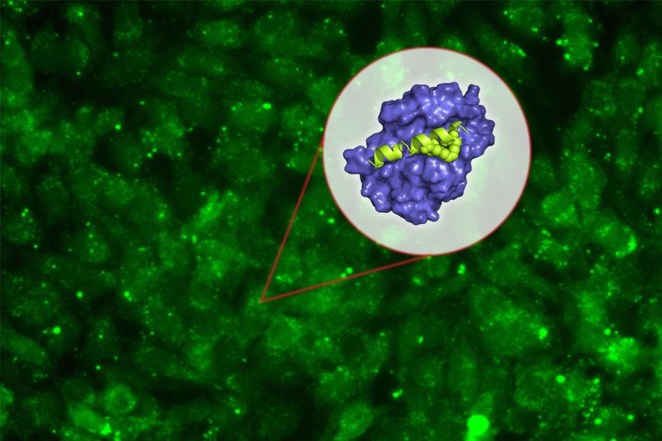
The new peptide targets a protein called Mcl-1, which helps cancer cells avoid the cellular suicide that is usually induced by DNA damage. By blocking Mcl-1, the peptide can force cancer cells to undergo programmed cell death.

“Some cancer cells are very dependent on Mcl-1, which is the last line of defense keeping the cell from dying. It’s a very attractive target,” says Amy Keating, an MIT professor of biology and one of the senior authors of the study.

Peptides, or small protein fragments, are often too unstable to use as drugs, but in this study, the researchers also developed a way to stabilize the molecules and help them get into target cells.

Loren Walensky, a professor of pediatrics at Harvard Medical School and a physician at Dana-Farber Cancer Institute, is also a senior author of the study, which appears in the Proceedings of the National Academy of Sciences the week of Jan. 15. Researchers in the lab of Anthony Letai, an associate professor of medicine at Harvard Medical School and Dana-Farber, were also involved in the study, and the paper’s lead author is MIT postdoc Raheleh Rezaei Araghi.

A promising target

Mcl-1 belongs to a family of five proteins that play roles in controlling programmed cell death, or apoptosis. Each of these proteins has been found to be overactive in different types of cancer. These proteins form what is called an “apoptotic blockade,” meaning that cells cannot undergo apoptosis, even when they experience DNA damage that would normally trigger cell death. This allows cancer cells to survive and proliferate unchecked, and appears to be an important way that cells become resistant to chemotherapy drugs that damage DNA.

“Cancer cells have many strategies to stay alive, and Mcl-1 is an important factor for a lot of acute myeloid leukemias and lymphomas and some solid tissue cancers like breast cancers. Expression of Mcl-1 is upregulated in many cancers, and it was seen to be upregulated as a resistance factor to chemotherapies,” Keating says.

Many pharmaceutical companies have tried to develop drugs that target Mcl-1, but this has been difficult because the interaction between Mcl-1 and its target protein occurs in a long stretch of 20 to 25 amino acids, which is difficult to block with the small molecules typically used as drugs.

Peptide drugs, on the other hand, can be designed to bind tightly with Mcl-1, preventing it from interacting with its natural binding partner in the cell. Keating’s lab spent many years designing peptides that would bind to the section of Mcl-1 involved in this interaction — but not to other members of the protein family.

Once they came up with some promising candidates, they encountered another obstacle, which is the difficulty of getting peptides to enter cells.

“We were exploring ways of developing peptides that bind selectively, and we were very successful at that, but then we confronted the problem that our short, 23-residue peptides are not promising therapeutic candidates primarily because they cannot get into cells,” Keating says.

To try to overcome this, she teamed up with Walensky’s lab, which had previously shown that “stapling” these small peptides can make them more stable and help them get into cells. These staples, which consist of hydrocarbons that form crosslinks within the peptides, can induce normally floppy proteins to assume a more stable helical structure.

Keating and colleagues created about 40 variants of their Mcl-1-blocking peptides, with staples in different positions. By testing all of these, they identified one location in the peptide where putting a staple not only improves the molecule’s stability and helps it get into cells, but also makes it bind even more tightly to Mcl-1.

“The original goal of the staple was to get the peptide into the cell, but it turns out the staple can also enhance the binding and enhance the specificity,” Keating says. “We weren’t expecting that.”

Killing cancer cells

The researchers tested their top two Mcl-1 inhibitors in cancer cells that are dependent on Mcl-1 for survival. They found that the inhibitors were able to kill these cancer cells on their own, without any additional drugs. They also found that the Mcl-1 inhibitors were very selective and did not kill cells that rely on other members of the protein family.

Keating says that more testing is needed to determine how effective the drugs might be in combating specific cancers, whether the drugs would be most effective in combination with others or on their own, and whether they should be used as first-line drugs or when cancers become resistant to other drugs.

“Our goal has been to do enough proof-of-principle that people will accept that stapled peptides can get into cells and act on important targets. The question now is whether there might be any animal studies done with our peptide that would provide further validation,” she says.

Joshua Kritzer, an associate professor of chemistry at Tufts University, says the study offers evidence that the stapled peptide approach is worth pursuing and could lead to new drugs that interfere with specific protein interactions.

“There have been a lot of biologists and biochemists studying essential interactions of proteins, with the justification that with more understanding of them, we would be able to develop drugs that inhibit them. This work now shows a direct line from biochemical and biophysical understanding of protein interactions to an inhibitor,” says Kritzer, who was not involved in the research.

Keating’s lab is also designing peptides that could interfere with other relatives of Mcl-1, including one called Bfl-1, which has been less studied than the other members of the family but is also involved in blocking apoptosis.

The research was funded by the Koch Institute Dana-Farber Bridge Project and the National Institutes of Health.


Harnessing nature’s riches
December 19, 2017

Cambridge, MA – Researchers at Whitehead Institute have reconstructed the full suite of biochemical steps required to make salidroside, a plant-derived compound widely used in traditional medicine to combat depression and fatigue and boost immunity and memory. Their new study, which appears online this week in the journal Molecular Plant, resolves some long-standing questions about how this compound is manufactured by a type of high-altitude plant, known commonly as golden root. This work not only paves a path toward large-scale synthetic efforts—thereby protecting plants already in danger of extinction—but also provides a model for dissecting the biochemical synthesis of a host of natural products, which represent a treasure trove for modern medical discoveries.

“By cracking open the natural synthesis of this compound, known as salidroside, we have helped eliminate a major bottleneck in the broader development of plant-derived natural products into pharmaceuticals,” says Jing-Ke Weng, the senior author of the paper, a Member of Whitehead Institute, and an assistant professor of biology at Massachusetts Institute of Technology. “We simply can’t rely on the native plants as the sole sources of these biologically important molecules.”

Golden root, also called Tibetan ginseng, typically grows in high-altitude, arctic environments, such as Tibet. It is well known in Eastern cultures for its medicinal properties and produces a variety of chemical substances, particularly salidroside, which have garnered interest in the biomedical research community for their potential therapeutic effects.

“People have tried to farm golden root, but the medicinal value is much lower because the plants make much less salidroside when cultivated outside of their normal habitat,” says Weng.

That means collecting enough salidroside to fuel scientific studies is largely impossible, without risking the viability of these plants and their surroundings. So Weng and his team, including first author Michael Torrens-Spence, set out to find a better way. “If we can figure out how plants make these high-value natural products, then we can devise sustainable engineering approaches to recreate such molecules—we won’t have to destroy nature in order to harness its riches,” says Torrens-Spence, a postdoctoral researcher in Weng’s laboratory.

Torrens-Spence and his colleagues used a systematic multi-omics approach to characterize various tissues from a three-month-old, greenhouse-grown golden root plant. By correlating the active genes with the abundance of key metabolites between various tissue types, the researchers created a massive biochemical catalog of the plant’s tissues.

The researchers then mined these data and matched the likely biochemical precursors of salidroside with the candidate genes (and their corresponding enzymes) responsible for those compounds’ synthesis. This approach allowed Weng and his team to create a kind of draft blueprint of how salidroside is made in nature.

To test the validity of this draft blueprint—and the molecular players from the golden root plant that comprise it—the scientists turned to two well-studied laboratory organisms: the baker’s yeast Saccharomyces cerevisiae and the tobacco plant Nicotiana benthamiana. Normally, these organisms do not make salidroside. But if the researchers’ model was correct, by inserting the candidate genes involved in salidroside synthesis Weng and his colleagues should be able to bestow that special property upon them.

That is precisely what the researchers did. Using three key enzymes they identified through their “-omics” approach, including 4HPAAS (4-hydroxyphenylacetaldehyde synthase), 4HPAR (4-hydroxyphenylacetaldehyde reductase), and T8GT (tyrosol:UDP-glucose 8-O-glucosyltransferase), they engineered yeast and tobacco plants with the capacity to make salidroside. Notably, this biochemical pathway for synthesizing salidroside involves three enzymes, rather than four, as had previously been proposed.

“This is an exciting proof-of-principle for how we can systematically unlock the biochemistry behind a range of intriguing plant-derived natural products,” says Weng. “With this capability, we can accelerate biomedical studies of these unique compounds as well as their potential therapeutic development.”

Written by Nicole Davis
* * *
Jing-Ke Weng’s primary affiliation is with Whitehead Institute for Biomedical Research, where his laboratory is located and all his research is conducted. He is also an assistant professor of biology at Massachusetts Institute of Technology.
* * *
Full citation:
“Complete pathway elucidation and heterologous reconstitution of Rhodiola salidroside biosynthesis”
Molecular Plant, online December 19, 2017. DOI: 10.1016/j.molp.2017.12.007
Michael P. Torrens-Spence (1), Tomáš Pluskal (1), Fu-Shuang Li (1), Valentina Carballo (1) and Jing-Ke Weng (1,2).
1. Whitehead Institute for Biomedical Research, Cambridge, Massachusetts, USA
2. Department of Biology, Massachusetts Institute of Technology, Cambridge, Massachusetts, USA
Jacqueline Lees

Education

  • PhD, 1990, University of London
  • BSc, 1986, Biochemistry, University of York

Research Summary

We identify the proteins and pathways involved in tumorigenicity — establishing their mechanism of action in both normal and tumor cells. To do so, we use a combination of molecular and cellular analyses, mutant mouse models and genetic screens in zebrafish.

Michael T. Hemann

Education

  • PhD, 2001, Johns Hopkins University
  • BS, 1993, Molecular Biology and Biochemistry, Wesleyan University

Research Summary

Many human cancers do not respond to chemotherapy, and often times those that initially respond eventually acquire drug resistance. Our lab uses high-throughput screening technology — combined with murine stem reconstitution and tumor transplantation systems — to investigate the genetic basis for this resistance. Our goal is to identify novel cancer drug targets, as well as strategies for tailoring existing cancer therapies to target the vulnerabilities associated with specific malignancies.

Rethinking transcription factors and gene expression

Study shows that, like proteins, genomes must fold appropriately to function properly and that some transcription factors provide the structural support.

Nicole Giese Rura | Whitehead Institute
December 7, 2017

Transcription — the reading of a segment of DNA into an RNA template for protein synthesis — is fundamental for nearly all cellular processes, including growth, responding to stimuli, and reproduction. Now, Whitehead Institute researchers have upended our understanding of how transcription is controlled and the role of transcription factors in the process.

The paradigm shift, described in an article online on Dec. 7 in the journal Cell, hinges on a small protein that plays a key role in genome structure and gives us new insights into how changes in the control of transcription and gene expression can lead to disease.

Transcription has several important players that must all be in the right place at the right time: the transcription machinery, transcription factors, promoters, and enhancers.  According to the existing model, transcription factors are proteins that bind to enhancer regions of the genome and recruit the transcription machinery to the promoter DNA regions, which then initiate the genes’ transcription.

“We’ve always assumed that the role of transcription factors was to recruit the transcription machinery to genes to turn them on or turn them off,” says Richard Young, a Whitehead Insistute member and professor of biology at MIT. “But we never imagined that the transcription factors we’ve studied for three decades actually contribute to the genome’s structure. And as a consequence, they regulate genes. So we now look at genomes like proteins: They have to fold up appropriately in order to control genes.”

Scientists have known that the genome’s structure — how it bends and folds — is essential for efficiently compressing two meters of DNA into each human cell, which is the equivalent of packing a strand ten football fields long into a space the size of a marble. Yet until recently, researchers have not had the tools necessary to appreciate this architecture’s importance in fine control of gene expression or study the genome’s structure at sites ready for transcription.

In 2014, Young and his lab determined that portions of the genome reside in loop-based structures, creating insulated neighborhoods that bring enhancers, promoters, and genes into close proximity. Each loop is tied at the top by a pair of molecules, called CTCF, that are bound together. This structure is essential for proper gene control: If the loop structure is broken, gene expression is altered, and cells can become diseased or die.

In the current research, Young along with co-first authors Abraham Weintraub and Charles Li took a closer look at a protein that is well known but not well understood: Yin Yang 1 (YY1). Hundreds of scientific papers have linked YY1 dysfunction to diseases such as viral infections, cancer, and arthritis, and yet the studies produced seemingly contradictory observations of YY1’s function.

According to Young and colleagues, YY1 is a unique transcription factor that occupies both enhancers and promoters, is essential for cell survival, and is found in almost every cell type in humans and mice. Like CTCF, YY1 can also pair with itself and bind to DNA to form loops that enhance DNA transcription.

“YY1 is expressed broadly, and it is necessary for establishing enhancer-promoter loops in multiple cell types,” says Weintraub. “That’s its job, not recruiting the transcription apparatus. When the structure created by YY1 is removed, the genome is no longer folded properly, gene control is lost and transcription of the affected genes is significantly diminished, which can cause dysfunction.”

This model of YY1’s function could account for its association with a number of disparate diseases. Earlier this year, scientists reported YY1 syndrome — a genetic syndrome causing cognitive disabilities in people with mutations in their YY1 gene.

According to Young, YY1 is probably not the only transcription factor with this loop-forming role, and his lab will be searching for additional factors with similar functions.

“YY1 is most likely just the first one, and there are probably a bunch of collaborators that have similar roles,” says Young. “Instead of the classic function that we thought these transcription factors had — interacting with the transcription apparatus and giving instructions on how much or how little of a gene’s transcript to produce — they are bringing together regulatory elements with the gene. The whole job of these transcription factors is just making structure. We are realizing that the things that form physical structures are much more important than we had appreciated.”

The researchers’ work was supported by the National Institutes of Health, the Ludwig Graduate Fellowship funds, the National Science Foundation, the American Cancer Society, a Margaret and Herman Sokol Postdoctoral Award, the Damon Runyon Cancer Research Foundation, and the Cancer Research Institute. The Whitehead Institute has filed a patent application based on this study.

Laurie A. Boyer

Education

  • PhD, 2001, University of Massachusetts Medical School
  • BS, 1990, Biomedical Science, Framingham State University

Research Summary

We investigate how complex circuits of genes are regulated to produce robust developmental outcomes particularly during heart development. A main focus is to determine how DNA is packaged into chromatin, and how ATP-dependent chromatin remodelers modify this packaging to control lineage commitment. We are now applying these principles to develop methods to stimulate repair of damaged cardiac tissue (e.g., regeneration). Our ability to combine genomic, genetic, biochemical, and cell biological approaches both in vitro and in vivo as well as ongoing efforts to use tissue engineering to model the 3D architecture of the heart will ultimately allow us to gain a systems level and quantitative understanding of the regulatory circuits that promote normal heart development and how faulty regulation can lead to disease.

Awards

  • Medicine by Design Distinguished Lecture, 2017
  • Cardiovascular Rising Star Distinguished Lecture, 2017
  • American Heart Association Innovative Research Award, 2013
  • Irvin and Helen Sizer Career Development Award, 2012
  • Smith Family Award for Excellence in Biomedical Science, 2009
  • Massachusetts Life Sciences Center New Investigator Award, 2008
  • Pew Scholars Award in the Biomedical Sciences, 2008
  • Honorary Doctorate, Framingham State College, 2007
  • The Scientific American World’s 50 Top Leaders in Research, Business or Policy, 2006
Monty Krieger

Education

  • PhD, 1976, California Institute of Technology
  • BS, 1971, Chemistry, Tulane University

Research Summary

We use genetic, biochemical, physiologic, chemical, cellular and molecular biological methods to study cell surface receptor structure and function. We focus on lipoprotein receptors — in particular, the High Density Lipoprotein (HDL) receptor called Scavenger Receptor, Class B, Type I (SR-BI). Our analyses have provided insight into basic biological processes, contributed to our understanding of atherosclerosis and coronary heart disease (CHD) and have uncovered an unexpected connection between cholesterol and mammalian female infertility.

No longer accepting new students.

Awards 

  • Tulane University School of Science and Engineering Outstanding Alumnus Award, 2010
  • National Academy of Sciences, Member, 2009
  • Outstanding Achievement Award for Contributions to Atherosclerosis Research, International Atherosclerosis Society, 2009
  • Margaret MacVicar Faculty Fellow, 1993-2003
Omer H. Yilmaz

Education

  • PhD, 2008, University of Michigan; MD, 2008, University of Michigan Medical School
  • BS, 1999, Biochemistry and Physics, University of Michigan

Research Summary

The adult intestine is maintained by stem cells that require a cellular neighborhood, or niche, consisting in part of Paneth cells. Our laboratory will investigate the molecular mechanisms of how intestinal stem cells and their Paneth cell niche respond to diverse diets to coordinate intestinal regeneration with organismal physiology and its impact on the formation and growth of intestinal cancers.  By better understanding how intestinal stem cells adapt to diverse diets, we hope to identify and develop new strategies that prevent and reduce the growth of cancers involving the intestinal tract that includes the small intestine, colon, and rectum.

Awards

  • AAAS Martin and Rose Wachtel Cancer Research Award, 2018
  • Pew-Stewart Trust Scholar, 2016-2020
  • Sidney Kimmel Scholar, 2016-2020
  • V Foundation Scholar, 2014-2017
  • Harold M. Weintraub Award, 2007
David C. Page

Education

  • MD, 1984, Harvard Medical School
  • BS, 1978, Chemistry, Swarthmore College

Research Summary

We seek to understand the genetic differences between males and females — both within and beyond the reproductive tract. We study the medical ramifications of these differences in a broad context, through comparative biological, evolutionary, developmental and clinically focused analyses. Our three main veins of research relate to sex differences in health and disease, sex chromosome genomics, and germ cell origins and development.

Awards

  • American Academy of Arts and Sciences, Fellow, 2012
  • March of Dimes, Developmental Biology, 2011
  • National Academy of Medicine, Member, 2008
  • National Academy of Sciences, Member, 2005
  • Howard Hughes Medical Institute, HHMI Investigator, 1990
  • MacArthur Foundation, MacArthur Fellowship, 1986
Richard A. Young

Education

  • PhD, 1979, Yale University
  • BS, 1975, Biological Sciences, Indiana University

Research Summary

We use experimental and computational technologies to determine how signaling pathways, transcription factors, chromatin regulators and small RNAs regulate gene expression in healthy and diseased cells. Our interests range from the basic molecular mechanisms behind gene control to drug development for cancer and other diseases caused by gene misregulation.

Awards

  • National Academy of Medicine, Member, 2019
  • National Academy of Sciences, Member, 2012