How Many Evolutionary Events Can It Take To Screw in Nature’s Lightbulb?

Firefly genomics reveals independent evolution of bioluminescence in beetles

Lisa Girard | Whitehead Institute
October 16, 2018

Cambridge, MA — Researchers at Whitehead Institute and collaborators from fourteen other institutions around the world have shed light on the evolutionary origins of luciferase, the key enzyme behind the glow of fireflies and other bioluminescent beetles. By sequencing the genomes of two American and Japanese firefly species that diverged approximately 100 million years ago, along with a more evolutionarily distant bioluminescent Caribbean click beetle, the team discovered that luciferase appears to have arisen independently in fireflies and click beetles. Examining the genes flanking that encoding the luciferase gene, suggests an evolutionary path along which the luciferase gene arose from duplications and divergences of CoA ligase genes involved in fat metabolism. As described online October 16 in the journal eLife, these findings provide fundamental insights into how enzymes can evolve, potentially inform strategies to help protect bioluminescent beetles from a shifting climate and habitat, and could extend the utility of luciferase, which has also been harnessed for biomedical and agricultural research, as a laboratory tool.

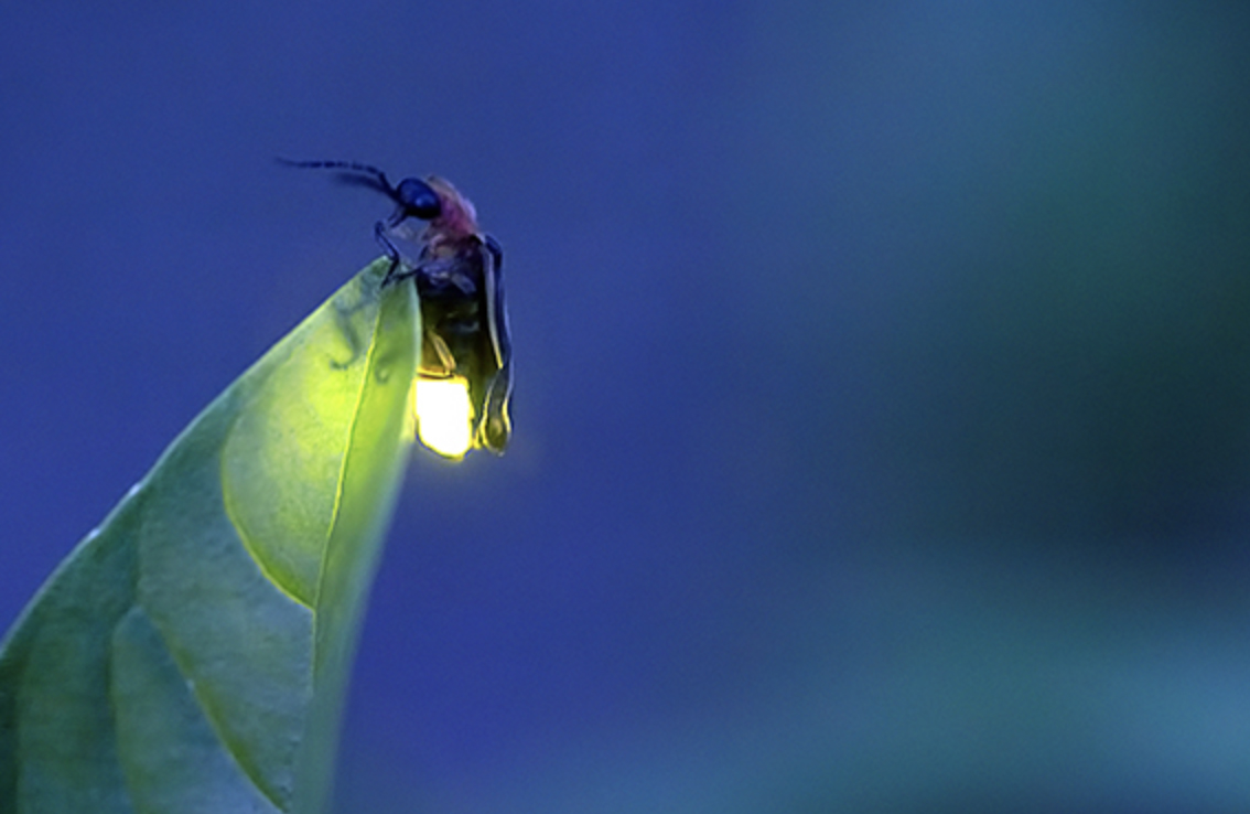
Throughout much of the world, the silent flash of a firefly on a warm evening can only mean one thing-Summer has arrived. But fireflies don’t just signal summer, their glow serves as a mating signal to other fireflies, and is even a warning that they are chemically defended, having a noxious taste capable of repelling the boldest of predators.

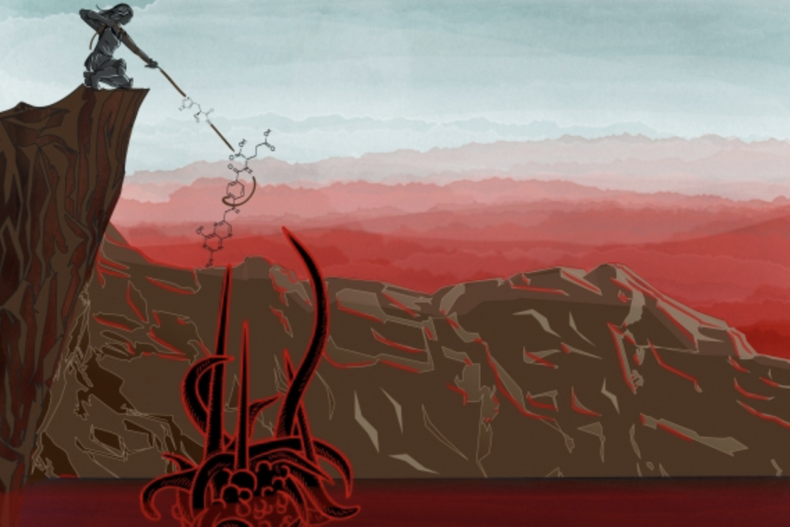
Belying its grandeur, the chemistry of firefly bioluminescence is relatively straightforward. Their light is produced by a specialized firefly enzyme, luciferase, that breaks down a molecule called luciferin, producing light in the process. Luciferase has become a mainstay tool in the laboratory. Scientists can fuse their gene of interest to luciferase and assay for gene expression by measuring the intensity of the glow after luciferin is added.

Beyond fireflies, there are other bioluminescent beetles (despite their name, fireflies are actually beetles), including certain tropical click-beetles. Perplexingly, these diverse bioluminescent beetles use very similar luciferase enzymes and luciferin molecules, but have an unrelated anatomy of their light-producing organs (also known as lanterns), making it unclear if their bioluminescence evolved from a common luminous ancestor, or if their special glow evolved independently.

Since fireflies and bioluminescent click-beetles are not model organisms like mice or fruit flies for which there is a wealth of genetic information, Jing-Ke Weng, Whitehead Institute Member and assistant professor of biology at Massachusetts Institute of Technology (MIT), along with a  graduate student in Weng’s lab, Tim Fallon, and Cornell postdoctoral researcher, Sarah Lower, began their investigations by sequencing the genome of the American Big Dipper firefly, Photinus pyralisNamed for its distinctive swooping “J” flash , this common inhabitant of meadows and suburban lawns has been called the “All-American firefly”. Due to its abundance and ease of identification, it was also the firefly of choice for scientific study, and is the species from which luciferin and luciferase were first characterized. Wanting to start their work quickly and make their progress and data available to others in the firefly community, Whitehead Institute researchers and collaborators crowdsourced funds to sequence the Big Dipper firefly.

The Big Dipper genome sequence, they discovered, revealed interesting insights into the origin of the luciferase gene. Examining the genes flanking that encoding luciferase, they found a cluster, or tandem repeat, of fatty acid CoA ligase genes with the luciferase gene sitting in the middle of this cluster. Sequence similarity and proximity between the luciferase and fatty acid CoA ligase genes suggested an evolutionary path along which the luciferase gene was produced from tandem duplication and divergence of an ancestral fatty acid CoA ligase gene.

“When the luciferase gene was cloned, people knew it was similar to the fatty acid CoA ligase gene in sequence, and hypothesized that it must be related to that ancestry. But what we uncovered from the luciferase gene locus is a tandem repeat of five genes, four are still the fatty acid CoA ligases, but then luciferase evolved right in the middle we believe from divergence of one of these duplications,” says Weng.

The Big Dipper sequencing provided important insights into the origin of luciferase and additional factors involved in bioluminescence, but in order to gain additional insights into the evolution of bioluminescence, the researchers set out to sequence two additional species that they hoped would provide the additional context to help them triangulate on some answers.

The bioluminescent click beetle, Ignelater luminosus, is related to the firefly, but on another branch of the tree of life entirely. Instead of producing light at its tail, it has two lanterns behind its head.

“We thought that sequencing the click beetle would provide insights into the evolution of bioluminescence as well as perhaps into how these animals could acquire very similar traits in terms of their biochemistry, but not in terms of their development,” says Fallon.

The third species they selected to sequence was a Japanese aquatic firefly (Aquatica lateralis), known in Japan as the Heike firefly.  Heike and the Big Dipper diverged from one another over 100 million years ago (to give you a sense of how far this is, it is older than the evolutionary distance between humans and rodents).

The researchers analyzed genomic data from the Japanese aquatic firefly and saw a similar arrangement around the luciferase gene locus as they had in the Big Dipper genome, suggesting that luciferase arose from a common ancestral event in both firefly species. The structure around the luciferase locus in the click beetle, however, was entirely absent, suggesting that luciferase arose through a different event.  Taken together, by sequencing and analyzing data from the genomes of two firefly species that diverged approximately 100 million years ago, along with a more evolutionarily distant bioluminescent click beetle, the team discovered that luciferase appears to have evolved independently in both fireflies and click beetles.

“Having the genome allowed us to understand how the evolution of luciferase happened. Before sequencing, we knew there were five genes cloned, including luciferase, in firefly. By sequencing the genomes we actually uncovered those genomic loci where those initial gene duplication events occured,” says Lower.

In addition to the origins of luciferase, these findings also provided the researchers with insights into the evolution of the light organs.

“Since our findings suggest that luciferase originated independently in both lineages, we can infer that anything that came after luciferase, for example the light organs, or other things dependent on luciferase should also be independent,” says Fallon.

Discovering how bioluminescence arose, as well as other complex traits, can be studied now that genomic information is available. The information can also inform strategies to protect fireflies, whose populations in many parts of the world are diminishing. In addition to adding tools to help reveal a constituent parts list that could allow researchers to optimize bioluminescence as a tool, these findings reveal important insights into the evolution of bioluminescence as well as genomic evolution more broadly.

“Luciferase is a perfect example of how to build a new enzyme, duplication of a related progenitor gene followed by mutation and selection,” says Weng. “And one of the most exciting parts of this study was that by examining the evolutionary scars in the genomes we studied we could actually see it happen.”

* * *
Jing-Ke Weng’s primary affiliation is with Whitehead Institute for Biomedical Research, where his laboratory is located and all his research is conducted. He is also an assistant professor of biology at Massachusetts Institute of Technology.
* * *
Full citation:
“Firefly genomes illuminate parallel origins of bioluminescence in beetles”
eLife, online on October 9, 2018. doi: 10.7554/eLife.36495
Timothy R. Fallon (1,2,*), Sarah E. Lower (3,*), Ching-Ho Chang (4) , Manabu Bessho-Uehara (5,6), Gavin J. Martin (7), Adam J. Bewick (8) , Megan Behringer (9) , Humberto J. Debat (10), Isaac Wong (4) , John C. Day (11), Anton Suvorov (7) , Christian J. Silva (4,12), Kathrin F. Stanger-Hall1 (3), David W. Hall (8) , Robert J. Schmitz (8), David R. Nelson (14), Sara M. Lewis (15), Shuji Shigenobu (16), Seth M. Bybee (7) , Amanda M. Larracuente (4), Yuichi Oba (5), and Jing-Ke Weng (1,2)
1. Whitehead Institute for Biomedical Research, Cambridge, Massachusetts 02142, USA.
2.Department of Biology, Massachusetts Institute of Technology, Cambridge, Massachusetts 02139, USA.
3. Department of Molecular Biology & Genetics, Cornell University, Ithaca, New York 14850, USA.
4. Department of Biology, University of Rochester, Rochester, New York 14627, USA.
5. Department of Environmental Biology, Chubu University, Kasugai, Aichi 487-8501, Japan.
6. Graduate School of Bioagricultural Sciences, Nagoya University, Nagoya, Aichi 464-8601, Japan.
7. Department of Biology, Brigham Young University, Provo, Utah 84602, USA.
8. Department of Genetics, University of Georgia, Athens, Georgia 30602, USA.
9. Biodesign Center for Mechanisms of Evolution, Arizona State University, Tempe, Arizona 85287, USA.
10. Center of Agronomic Research National Institute of Agricultural Technology, Córdoba, Argentina.
11. Centre for Ecology and Hydrology (CEH) Wallingford, Wallingford, Oxfordshire, UK.
12. Department of Plant Sciences, University of California Davis, Davis, California, USA.
13. Department of Plant Biology, University of Georgia, Athens, Georgia 30602, USA.
14. Department of Microbiology Immunology and Biochemistry, University of Tennessee  HSC, Memphis 38163, USA.
15. Department of Biology, Tufts University, Medford, Massachusetts 02155, USA.
16. NIBB Core Research Facilities, National Institute for Basic Biology, Okazaki 444-8585, Japan.
Only in Your Head
Greta Friar | Whitehead Institute
October 9, 2018

Cambridge, Mass. — Brain development is a delicately choreographed dance in which cell division and differentiation into mature cell types must be performed in the right balance for normal growth. In order to better understand factors affecting brain development, Whitehead Institute researchers investigated a genetic mutation that leads to a brain-specific developmental disorder in spite of the gene’s prevalent expression in other cell types.

Kinetochore null protein 1 (KNL1) acts throughout the body during cell division to help ensure the accurate segregation of chromosomes into each daughter cell. A mutation in the KNL1 gene caused by a single change in its DNA sequence leads to microcephaly, a condition in which the brain fails to properly develop, causing babies to be born with small heads, often accompanied by intellectual disabilities and other health problems. In an article published online October 9 in the journal Cell Reports, Whitehead Institute Founding Member Rudolf Jaenisch and colleagues investigated how this KNL1 mutation can lead to microcephaly without affecting other cell types, providing important insights into the underlying basis of microcephaly and the role that KNL1 normally plays in brain development.

“The key question we were interested in was why, if the gene is ubiquitously expressed, is there a brain-specific phenotype,” says Jaenisch, who is also a professor of biology at the Massachusetts Institute of Technology.

Jaenisch lab graduate student Attya Omer Javed, a co-first author on the paper along with past lab members Yun Li and Julien Muffat, used CRISPR-Cas9 to recreate the mutation—a point mutation, or one-letter change in the DNA sequence of the KNL1 gene—in several different cell types derived from human stem cells in the lab. Of the three cell types tested, they found that only the neural progenitor cells, early stage cells that become brain cells, appeared to be affected.

As the brain develops, each neural progenitor can either keep dividing to increase the overall number of cells in the brain, or it can mature into a differentiated brain cell, at which point it is no longer able to divide. For a healthy brain to develop, there needs to be a careful balance between these two processes of proliferation and differentiation. If the progenitors take too long to differentiate, the developing brain won’t have the specific cells it needs to assemble. But if all of the cells differentiate too quickly, before they can divide, there will be a shortage of cells and the brain will be too small.

“Neural progenitors are going through many cell cycles, dividing quickly during brain development. Even a small defect could accumulate to have a huge impact,” Omer Javed says.

The researchers discovered that neural progenitors with the KNL1 mutation differentiated prematurely at the cost of proliferation, resulting in the small brain size that characterizes microcephaly. The brain cells with the mutation also were at a greater risk of cell death, disruption of the cell cycle, ending up with the wrong number of chromosomes, and malfunctions during attempted cell division.

KNL1’s role is in the kinetochore, an assembly of proteins that operate during mitosis to attach chromosomes to the machinery that will pull them apart into the daughter cells. This is why the KNL1 mutation negatively affects cell division. Co-author and Whitehead Member Iain Cheesemanhelped identify KNL1’s role in the kinetochore as a postdoctoral researcher years ago, and his expertise provided an opportunity for collaboration between his lab and Jaenisch’s.

“I have always found it interesting that inherited mutations to the kinetochore seem to lead to microcephaly,” Cheeseman says. “Investigating KNL1 together was an exciting chance to combine our labs’ diverse scientific knowledge.”

In order for the researchers to study the cells in an environment that more closely mimicked a human brain, they used a 3D cell culture technique to grow organoids made up of neural progenitors. Omer Javed found that the neural progenitors were extremely sensitive, as the organoids with the mutation expressed the microcephaly phenotype after as little as two weeks of growth.

Omer Javed then looked for differences between neural progenitors and the other cell types that would explain the brain-specific effects of the mutation. Even with the mutation, the KNL1 gene appeared able to make a functioning protein, explaining its lack of effect on the other cell types. So Omer Javed turned her attention to factors involved in regulating gene expression. For many of our genes to be expressed, first sections called introns must be removed, or spliced out, in order for the correct DNA sequence to then be read into RNA and then translated into a functional protein.

Omer Javed found that the KNL1 mutation created a site for splicing inhibitors to bind and silence the KNL1 gene by preventing it from being read into RNA. She also found a disparity in the level of a protein involved in this process between the cell types: the inhibitory splicing protein hnRNPA1 was much more prevalent in neural progenitors than elsewhere. When hnRNPA1 came across the site caused by the mutation, it prevented the gene from being expressed. The high quantity of hnRNPA1 in neural progenitors appears to be the main factor mediating the brain-specific effects of the mutation.

The work complements and extends previous investigations by the researchers into how neural progenitor proliferation may have contributed to the evolution of large human brains, as well as studies investigating why neural progenitors are so vulnerable to the Zika virus, which has been associated with microcephaly. Given their work suggesting that KNL1 could be a regulator of brain size, Omer Javed hopes that future research will reveal its role in the evolution of the human brain.


This research was supported by Boehringer Ingelheim Fonds, the Simons Foundation, the International Rett Syndrome Foundation, Brain & Behavior Research Foundation, the European Leukodystrophy Association, the National Institutes of Health (NIH grants HD 045022, R37-CA084198 and 1U19AI131135-01). Jaenisch is a cofounder of Fate Therapeutics, Fulcrum Therapeutics, and Omega Therapeutics.


***
Rudolf Jaenisch’s primary affiliation is with Whitehead Institute for Biomedical Research, where his laboratory is located and all his research is conducted. He is also a professor of biology at Massachusetts Institute of Technology.
***
Full citation:
“Microcephaly modeling of kinetochore mutation reveals a brain-specific phenotype”
Cell Reports, online October 9, 2018
Attya Omer (1,2,8), Yun Li (2,3,4,8), Julien Muffat (2,4,5,8), Kuan-Chung Su (2), Malkiel A. Cohen (2), Tenzin Lungjangwa (2), Patrick Aubourg (1,6), Iain M. Cheeseman (2,7), and Rudolf Jaenisch (2,7).
1. Université Paris-Saclay, ED 569, 5 Rue Jean-Baptiste Clément, 92290 Châtenay-Malabry, France
2.  Whitehead Institute for Biomedical Research, 455 Main Street, Cambridge, MA 02142, USA
3. Program in Developmental and Stem Cell Biology, The Hospital for Sick Children, 686 Bay Street, Toronto, ON M4G 0A4, Canada
4. Department of Molecular Genetics, University of Toronto, 1 King’s College Circle, Toronto, ON M5S 1A8, Canada
5. Program in Neurosciences and Mental Health, The Hospital for Sick Children, 686 Bay Street, Toronto, ON M5G 0A4, Canada
6. INSERM U1169, CHU Bicêtre Paris Sud, Le Kremlin-Bicêtre, France.
7. Department of Biology, MIT, 31 Ames Street, Cambridge, MA 02139, USA
8. These authors contributed equally
Immune cell variations contribute to malaria severity

Natural killer cells’ failure to respond to infection may explain why the disease is more grave in some patients.

Anne Trafton | MIT News Office
October 4, 2018

At least 250 million people are infected with malaria every year, and about half a million of those die from the disease. A new study from MIT offers a possible explanation for why some people are more likely to experience a more severe, and potentially fatal, form of the disease.

The researchers found that in some patients, immune cells called natural killer cells (NK cells) fail to turn on the genes necessary to effectively destroy malaria-infected red blood cells.

The researchers also showed that they could stimulate NK cells to do a better job of killing infected red blood cells grown in a lab dish. This suggests a possible approach for developing treatments that could help reduce the severity of malaria infections in some people, especially children, says Jianzhu Chen, one of the study’s senior authors.

“This is one approach to that problem,” says Chen, an MIT professor of biology and a member of MIT’s Koch Institute for Integrative Cancer Research. “Most of the malaria patients who die are children under the age of 5, and their immune system has not completely formed yet.”

Peter Preiser, a professor at Nanyang Technical University (NTU) in Singapore, is also a senior author of the study, which appears in the journal PLOS Pathogens on Oct. 4. The paper’s lead authors are NTU and Singapore-MIT Alliance for Research and Technology (SMART) graduate students Weijian Ye and Marvin Chew.

First line of defense

In 2010, Chen and his colleagues engineered strains of mice that produce several types of human immune cells and red blood cells. These “humanized” mice can be used to study the human immune response to pathogens that don’t normally infect mice, such as Plasmodium falciparum, the parasite that causes malaria.

A few years later, the researchers used those mice to investigate the roles of NK cells and macrophages in malaria infection. These two cell types are key players in the innate immune system, a nonspecific response that acts as the first line of defense against many microbes. Chen and his colleagues found that when they removed human NK cells from the mice and infected them with malaria, the quantity of parasites in the blood was much greater than in mice with NK cells. This did not happen when they removed human macrophages, suggesting that NK cells are the most important first-line defenders against malaria.

A natural killer (NK) cell binds to a malaria-infected red blood cell and destroys it. Credit: Weijian Ye

In that study, the researchers also found that in about 25 percent of the human blood samples they used, the NK cells did not respond to malaria at all. In the new paper, they set out to try to find out why that was the case. To do that, they sequenced the RNA of NK cells before and after they encountered malaria-infected red blood cells. This allowed the researchers to identify a small number of genes that get turned on in malaria-responsive NK cells but not in nonresponsive cells.

Among these genes was one that codes for a protein called MDA5, which was already known to be involved in helping immune cells such as NK cells and macrophages recognize foreign RNA. Further studies revealed that malaria-infected red blood cells secrete tiny bubbles called microvesicles that carry pieces of RNA from the malaria parasite. The studies also showed that NK cells absorb these microvesicles. If MDA5 is present, the NK cell is activated to kill the infected blood cell.

Nonresponsive NK cells, which have lower levels of MDA5, fail to recognize and kill the infected cells. NK cells are also responsible for secreting cytokines that summon T cells and other immune cells, so their failure to activate also hinders other elements of the immune response.

Boosting immunity

Chen and his colleagues also showed that they could activate the nonresponsive NK cells by treating them with a synthetic molecule called poly I:C, which is structurally similar to double-stranded RNA. For poly I:C to be effective, the researchers had to package it into tiny spheres called liposomes, which allow it to enter cells just like the RNA-carrying microvesicles do.

The researchers also found a correlation between the levels of MDA5 in the NK cells and the disease severity experienced by the patients who donated the blood samples. Next, they hope to take cells from human patients and use them to further examine this correlation in humanized mice, and also to explore whether treating the mice with poly I:C would have the same beneficial effect they saw in cells grown in a lab dish.

The research was funded by the National Research Foundation of Singapore through the SMART Interdisciplinary Research Group in Infectious Disease Research Program.

Parasite’s riff on essential enzyme highlights unique biology
Nicole Giese Rura | Whitehead Institute
September 18, 2018

Cambridge, Mass. — The primary currency of energy in cells—adenosine triphosphate (ATP)—is essential for their survival and without it, cellular processes would seize. In the apicomplexan Toxoplasma gondii (T. gondii), a parasite that Whitehead Member Sebastian Lourido studies, key components of the ATP synthase—the enzyme responsible for ATP production—have remained elusive. While investigating indispensable proteins with unknown functions, Lourido and Diego Huet, a postdoctoral researcher in Lourido’s lab, identified a critical component of the enzyme. While highly conserved from yeast to humans, it proved to be considerably different in T. gondii. The findings, published online September 11 in the journal eLife, underscore the unique biology of these parasites and highlight differences between them and their human hosts.

More closely related to plants than to animals, the single-celled apicomplexans are among the most common and deadly human pathogens. According to the World Health Organization, every year these diseases sicken hundreds of millions, kill hundreds of thousands—primarily children—and cost billions of dollars to treat. Species of apicomplexans cause malaria (Plasmodium spp.), cryptosporidiosis (Cryptosporidium spp.), and toxoplasmosis (T. gondii).

Using a CRISPR-based genetic screen that they had adapted to T. gondii, Lourido and Huet had previously identified about 200 genes in T. gondii that are fitness-conferring and specific to apicomplexans. Of that cadre, a few were localized to the mitochondria, where cells manufacture ATP, the cellular currency of energy. Because those genes have not been annotated previously, and the proteins encoded by them have no known function, Huet ran their protein sequences through a database that compared them to protein sequences with known structures.

One of the proteins came back with an interesting hit: it shares structural similarity, but not sequence similarity, with an integral part of the ATP synthase. Most of the protein subunits that compose the apicomplexan ATP synthase have been identified, but key components of the stator—a portion of the enzyme essential for its function—was not yet known.

When Huet experimentally removed the function of the stator subunit in T. gondii, the parasites’ growth stalled, their mitochondria were misshapen and shrunken, and energy production halted—all traits typical of interrupted ATP synthase function.

Because the apicomplexan ATP synthase varies so much from its hosts’ version, those differences, like the unusual stator, could serve as future drug targets. But for Lourido, who is also an assistant professor of biology at Massachusetts Institute of Technology (MIT), the unique stator protein emphasizes how unique and extraordinary apicomplexan organisms are compared to us and their other hosts.

This work was supported by the National Institutes of Health (NIH grants 1DP5OD017892, R21AI123746, and K99AI137218).

* * *

Sebastian Lourido’s primary affiliation is with Whitehead Institute for Biomedical Research, where his laboratory is located and all his research is conducted. He is also an assistant professor of biology at Massachusetts Institute of Technology.

* * *

Full Citation:

“Identification of cryptic subunits from an apicomplexan ATP synthase”

eLife, online September 11, 2018.  DOI: 10.7554/eLife.38097

Diego Huet (1) , Esther Rajendran (2) , Giel G van Dooren (2) , Sebastian Lourido (1,3*).

1. Whitehead Institute for Biomedical Research, Cambridge, United States

2. Research School of Biology, Australian National University, Canberra, Australia

3. Department of Biology, Massachusetts Institute of Technology, Cambridge, Massachusetts, United States

Ankur Jain

Education

  • PhD, 2013, University of Illinois, Urbana-Champaign
  • BTech, 2007,  Biotechnology and Biochemical Engineering, Indian Institute of Technology Kharagpur

Research Summary

We study how biomolecules in a cell self-organize. In particular, we are interested in understanding how membrane-free cellular compartments such as RNA granules form and function. Our lab develops new biochemical and biophysical techniques to investigate these compartments and to understand their dysfunction in human disease.

Awards

  • Young Alumni Achiever’s Award, Indian Institute of Technology Kharagpur, 2019
  • NIH K99/R00 Pathway to Independence Award, 2017
  • Pew Scholar in the Biomedical Sciences, 2022
Study suggests glaucoma may be an autoimmune disease

Unexpected findings show that the body’s own immune system destroys retinal cells.

Anne Trafton | MIT News Office
August 11, 2018

Glaucoma, a disease that afflicts nearly 70 million people worldwide, is something of a mystery despite its prevalence. Little is known about the origins of the disease, which damages the retina and optic nerve and can lead to blindness.

A new study from MIT and Massachusetts Eye and Ear has found that glaucoma may in fact be an autoimmune disorder. In a study of mice, the researchers showed that the body’s own T cells are responsible for the progressive retinal degeneration seen in glaucoma. Furthermore, these T cells appear to be primed to attack retinal neurons as the result of previous interactions with bacteria that normally live in our body.

The discovery suggests that it could be possible to develop new treatments for glaucoma by blocking this autoimmune activity, the researchers say.

“This opens a new approach to prevent and treat glaucoma,” says Jianzhu Chen, an MIT professor of biology, a member of MIT’s Koch Institute for Integrative Cancer Research, and one of the senior authors of the study, which appears in Nature Communications on Aug. 10.

Dong Feng Chen, an associate professor of ophthalmology at Harvard Medical School and the Schepens Eye Research Institute of Massachusetts Eye and Ear, is also a senior author of the study. The paper’s lead authors are Massachusetts Eye and Ear researchers Huihui Chen, Kin-Sang Cho, and T.H. Khanh Vu.

Genesis of glaucoma

One of the biggest risk factors for glaucoma is elevated pressure in the eye, which often occurs as people age and the ducts that allow fluid to drain from the eye become blocked. The disease often goes undetected at first; patients may not realize they have the disease until half of their retinal ganglion cells have been lost.

Most treatments focus on lowering pressure in the eye (also known as intraocular pressure). However, in many patients, the disease worsens even after intraocular pressure returns to normal. In studies in mice, Dong Feng Chen found the same effect.

“That led us to the thought that this pressure change must be triggering something progressive, and the first thing that came to mind is that it has to be an immune response,” she says.

To test that hypothesis, the researchers looked for immune cells in the retinas of these mice and found that indeed, T cells were there. This is unusual because T cells are normally blocked from entering the retina, by a tight layer of cells called the blood-retina barrier, to suppress inflammation of the eye. The researchers found that when intraocular pressure goes up, T cells are somehow able to get through this barrier and into the retina.

The Mass Eye and Ear team then enlisted Jianzhu Chen, an immunologist, to further investigate what role these T cells might be playing in glaucoma. The researchers generated high intraocular pressure in mice that lack T cells and found that while this pressure induced only a small amount of damage to the retina, the disease did not progress any further after eye pressure returned to normal.

Further studies revealed that the glaucoma-linked T cells target proteins called heat shock proteins, which help cells respond to stress or injury. Normally, T cells should not target proteins produced by the host, but the researchers suspected that these T cells had been previously exposed to bacterial heat shock proteins. Because heat shock proteins from different species are very similar, the resulting T cells can cross-react with mouse and human heat shock proteins.

To test this hypothesis, the team brought in James Fox, a professor in MIT’s Department of Biological Engineering and Division of Comparative Medicine, whose team maintains mice with no bacteria. The researchers found that when they tried to induce glaucoma in these germ-free mice, the mice did not develop the disease.

Human connection

The researchers then turned to human patients with glaucoma and found that these patients had five times the normal level of T cells specific to heat shock proteins, suggesting that the same phenomenon may also contribute to the disease in humans. The researchers’ studies thus far suggest that the effect is not specific to a particular strain of bacteria; rather, exposure to a combination of bacteria can generate T cells that target heat shock proteins.

One question the researchers plan to study further is whether other components of the immune system may be involved in the autoimmune process that gives rise to glaucoma. They are also investigating the possibility that this phenomenon may underlie other neurodegenerative disorders, and looking for ways to treat such disorders by blocking the autoimmune response.

“What we learn from the eye can be applied to the brain diseases, and may eventually help develop new methods of treatment and diagnosis,” Dong Feng Chen says.

The research was funded by the National Institutes of Health, the Lion’s Foundation, the Miriam and Sheldon Adelson Medical Research Foundation, the National Nature Science Foundation of China, the Ivan R. Cottrell Professorship and Research Fund, the Koch Institute Support (core) Grant from the National Cancer Institute, and the National Eye Institute Core Grant for Vision Research.

Researchers discover new type of lung cell, critical insights for cystic fibrosis

A comprehensive single-cell analysis of airway cells in mice, validated in human tissue, reveals molecular details critical to understanding lung disease.

Karen Zusi
August 1, 2018

Researchers have identified a rare cell type in airway tissue, previously uncharacterized in the scientific literature, that appears to play a key role in the biology of cystic fibrosis. Using new technologies that enable scientists to study gene expression in thousands of individual cells, the team comprehensively analyzed the airway in mice and validated the results in human tissue.

Led by researchers from the Broad Institute of MIT and Harvard and Massachusetts General Hospital (MGH), the molecular survey also characterized gene expression patterns for other new cell subtypes. The work expands scientific and clinical understanding of lung biology, with broad implications for all diseases of the airway — including asthma, chronic obstructive pulmonary disease, and bronchitis.

Jayaraj Rajagopal, a physician in the Pulmonary and Critical Care Unit at MGH, associate member at the Broad Institute, and a Howard Hughes Medical Institute (HHMI) faculty scholar, and Broad core institute member Aviv Regev, director of the Klarman Cell Observatory at the Broad Institute, professor of biology at MIT, and an HHMI investigator, supervised the research. Daniel Montoro, a graduate student in Rajagopal’s lab, and postdoctoral fellows Adam Haber and Moshe Biton in the Regev lab are co-first authors on the paper published today in Nature.

“We have the framework now for a new cellular narrative of lung disease,” said Rajagopal, who is also a professor at Harvard Medical School and a principal faculty member at the Harvard Stem Cell Institute. “We’ve uncovered a whole distribution of cell types that seem to be functionally relevant. What’s more, genes associated with complex lung diseases can now be linked to specific cells that we’ve characterized. The data are starting to change the way we think about lung diseases like cystic fibrosis and asthma.”

“With single-cell sequencing technology, and dedicated efforts to map cell types in different tissues, we’re making new discoveries — new cells that we didn’t know existed, cell subtypes that are rare or haven’t been noticed before, even in systems that have been studied for decades,” said Regev, who is also co-chair of the international Human Cell Atlas consortium. “And for some of these, understanding and characterizing them sheds new light immediately on what’s happening inside the tissue.”

Using single-cell RNA sequencing, the researchers analyzed tens of thousands of cells from the mouse airway, mapping the physical locations of cell types and creating a cellular “atlas” of the tissue. They also developed a new method called pulse-seq to monitor development of cell types from their progenitors in the mouse airway. The findings were validated in human tissue.

One extremely rare cell type, making up roughly one percent of the cell population in mice and humans, appeared radically different from other known cells in the dataset. The team dubbed this cell the “pulmonary ionocyte” because its gene expression pattern was similar to ionocytes — specialized cells that regulate ion transport and hydration in fish gills and frog skin.

Strikingly, at levels higher than any other cell type, these ionocytes expressed the gene CFTR — which, when mutated, causes cystic fibrosis in humans. CFTR is critical for airway function, and for decades researchers and clinicians have assumed that it is frequently expressed at low levels in ciliated cells, a common cell type spread throughout the entire airway.

But according to the new data, the majority of CFTR expression occurs in only a few cells, which researchers didn’t even know existed until now.

When the researchers disrupted a critical molecular process in pulmonary ionocytes in mice, they observed the onset of key features associated with cystic fibrosis — most notably, the formation of dense mucus. This finding underscores how important these cells are to airway-surface regulation.

“Cystic fibrosis is an amazingly well-studied disease, and we’re still discovering completely new biology that may alter the way we approach it,” said Rajagopal. “At first, we couldn’t believe that the majority of CFTR expression was located in these rare cells, but the graduate students and postdocs on this project really brought us along with their data.”

The results may also have implications for developing targeted cystic fibrosis therapies, according to the team. For example, a gene therapy that corrects for a mutation in CFTRwould need to be delivered to the right cells, and a cell atlas of the tissue could provide a reference map to guide that process.

The study further highlighted where other disease-associated genes are expressed in the airway. For example, asthma development has been previously linked with a gene that encodes a sensor for rhinoviruses, and the data now indicate that this gene is expressed by ciliated cells. Another gene linked with asthma is expressed in tuft cells, which separated into at least two groups — one that senses chemicals in the airway and one that produces inflammation. The results suggest that a whole ensemble of cells may be responsible for different aspects of asthma.

Using the pulse-seq assay, the researchers tracked how the newly characterized cells and subtypes in the mouse airway develop. They demonstrated that mature cells in the airway arise from a common progenitor: the basal cells. The team also discovered a previously undescribed cellular structure in the tissue. These structures, which the researchers called “hillocks,” are unique zones of rapid cell turnover, and their function is not yet understood.

“The atlas that we’ve created is already starting to drastically re-shape our understanding of airway and lung biology,” said Regev. “And, for this and other organ systems being studied at the single-cell level, we’ll have to drape everything we know on top of this new cellular diversity to understand human health and disease.”

Funding for this study was provided in part by the Klarman Cell Observatory at the Broad Institute, Manton Foundation, HHMI, New York Stem Cell Foundation, Harvard Stem Cell Institute, Human Frontiers Science Program, and National Institutes of Health.

Paper(s) cited:

Montoro DT, Haber AL, Biton M et al. A revised airway epithelial hierarchy includes CFTR-expressing ionocytesNature. Online August 1, 2018. DOI: 10.1038/s41586-018-0393-7

Sharpening the edges of cancer chemotherapy
Nicole Davis | Whitehead Institute
July 11, 2018

Cambridge, MA — Tackling unsolved problems is a cornerstone of scientific research, propelled by the power and promise of new technologies. Indeed, one of the shiniest tools in the biomedical toolkit these days is the genome editing system known as CRISPR/Cas9. Whitehead Institute Member David Sabatini and his colleagues pioneered the use of this tool as a foundation for large-scale genetic screens in human cells, turning up a treasure trove of new insights into cellular metabolism, in both normal cells and cancer cells.

When Naama Kanarek, a postdoc in Sabatini’s laboratory, pondered how to apply these state-of-the-art CRISPR/Cas9 screens to her own research, her thoughts turned to a classic cancer chemotherapy drug, methotrexate, which has been in clinical use for nearly seven decades. Often used to treat a form of pediatric leukemia, known as acute lymphoblastic leukemia (ALL), the drug, when deployed as part of a multifaceted treatment plan, can be highly effective. But its power comes at a cost. Because methotrexate can damage not only cancer cells but also healthy tissues, it must be administered with great care. For children who receive high doses of the drug, a mainstay of ALL treatment, that can mean several days spent in the hospital with rigorous clinical monitoring.

In other forms of cancer, methotrexate’s efficacy is more uncertain. For example, in pediatric osteosarcoma, only 65 percent of patients respond. Unfortunately, there is currently no way for doctors to pinpoint who will and who will not.

“From a scientific standpoint, methotrexate is quite special because it was the first metabolic drug to be developed, but much of its biology remains to be discovered — particularly what drives these different responses in patients,” Kanarek says. “So, this is really one of these old, classic questions that has been lingering in the field for some time. We thought we could learn something new.”

And they did. In the July 11 online issue of the journal Nature, Kanarek, Sabatini, and their colleagues report the findings of a CRISPR/Cas9 screen for factors involved in methotrexate sensitivity. The team’s work yielded a surprising set of discoveries that point to the breakdown of histidine — one of several amino acids used by the body to construct proteins — as a critical gatekeeper of cancer cells’ vulnerability to methotrexate. The researchers’ findings not only help illuminate the biology of a well-known cancer chemotherapy, but also suggest a simple dietary supplement that could help broaden its therapeutic window and reduce its toxicity.

 “This study is an example of the power of modern genomic tools to shine a bright light on longstanding questions in human biology,” says senior author David Sabatini, a Member of Whitehead Institute, a professor of biology at Massachusetts Institute of Technology and investigator with the Howard Hughes Medical Institute (HHMI). “While cancer chemotherapies can be quite effective, their biological effects are often poorly understood. By laying bare their biology, we may be able to devise ways to utilize them more wisely.”

ATTACK THE CANCER, NOT THE PATIENT

The history of methotrexate stretches back to the 1940s, a time when strikingly little was known about the origins of cancer much less how best to treat it. The birth of methotrexate as a chemotherapeutic agent was sparked by the astute observations of Sidney Farber, a pediatric pathologist at Boston Children’s Hospital who cared for children with a variety of maladies, including ALL. In the course of caring for patients with ALL, Farber recognized that cancer cells depended on the nutrient folic acid for their own proliferation. That gave him the idea of using folate antagonists to treat ALL. Methotrexate was developed in 1949 precisely for this purpose and was subsequently shown to induce remission in children with ALL. Fast forward to today, and the drug has evolved into a significant tool in oncologists’ toolkit.

“Methotrexate is a major part of the backbone of chemotherapy treatment across many human cancers,” says Loren Walensky, a pediatric hematologist/oncologist at the Dana-Farber Cancer Institute who is not a study co-author but served as an early adviser on the project and will also play a deeper role in planning future follow-up studies. “It is also used outside of the cancer field for the treatment of several autoimmune diseases.”

He added, “But as with all chemotherapy, the critical issue is how to best use it to inflict maximal damage on the cancer without irreparably harming the patient.”

Kanarek explains how new genetic tools are allowing insights into the sensitivity of cancer cells to methotrexate.

The basic mechanics of methotrexate are fairly well known. The drug inhibits dihydrofolate reductase (DHFR), an enzyme that generates the functional form of folate, known as tetrahydrofolate (THF). THF is essential for preparing the raw materials needed to make nucleic acids, such as DNA, which carries cells’ genetic information, and RNA, a close chemical relative involved in making proteins. “Proliferating cells must duplicate their DNA, so they need a lot of THF,” Kanarek explains. “But even cells that are not dividing need to make RNA, and that requires THF, too.”

The results of Kanarek’s CRISPR/Cas9 screen now bring greater clarity to this molecular picture. She and her colleagues uncovered another enzyme, called FTCD, which is involved in the breakdown of histidine. Interestingly, FTCD also requires THF for its function — though not nearly as much as the main target of methotrexate, DHFR. Despite the differential demands of the two enzymes, they both draw from the same, shared pool of THF.

“Under normal conditions, this pool is sufficiently full, so there is no competition for resources, even in rapidly dividing cells,” Kanarek says.

But when the amount of THF becomes limiting — as it does in cells that are treated with methotrexate — the story is quite different, the Whitehead Institute team discovered. In that case, the activity of FTCD poses serious problems, because there isn’t enough THF in the pool to support both cell proliferation and histidine breakdown. When that happens, the cells die.

That got Kanarek thinking more about histidine: Could the nutrient provide a way to tinker with FTCD activity and, by virtue of the cancer cells’ own metabolism, make them more vulnerable to methotrexate?

To explore this question, the researchers used mouse models of leukemia, engineered by transplanting human leukemia cells under the skin of immunocompromised mice. A subset of the mice received injections of methotrexate together with histidine. This one-two punch, Kanarek hypothesized, should ramp up the function of FTCD and more rapidly drain the THF pool, thereby making the cells more sensitive to the cancer-killing effects of methotrexate.

That is precisely what the team observed. Notably, these experiments involved lower than normal doses of methotrexate, suggesting the cells had indeed been made more sensitive to the cancer drug. Moreover, the studies included a human leukemia cell line, called SEM, which harbors a specific genetic mutation that is associated with a particularly poor prognosis in patients — further underscoring the power of the histidine degradation pathway to weaken cells’ defenses.

Now, Kanarek and her colleagues are working to extend these initial findings with additional preclinical studies and, together with Walensky, determine how to best evaluate the potential benefits of histidine supplementation in cancer patients. Their ultimate goal: to pursue clinical trials that will assess histidine’s ability to improve the effectiveness of methotrexate in humans.

In addition to making cancer cells more vulnerable to methotrexate, the Whitehead Institute team’s research also holds promise for another therapeutic challenge: identifying which patients will or will not respond to the drug.

Two other enzymes cooperate with FTCD in breaking down histidine. The levels of one of the enzymes, known as HAL, appears to correlate with cells’ sensitivity to methotrexate: That is, cancer cells with high levels of HAL tend to be more sensitive to the drug. More work is needed to determine whether this correlation extends to a broader swath of patient samples and if it has predictive value in the clinic. Nevertheless, Kanarek and her colleagues are already beginning work on this front. Together with Abner Louissaint, Jr., a hematopathologist at Massachusetts General Hospital who also served as an early adviser on the Nature study, the Whitehead Institute team will launch a second clinical study to examine whether HAL levels can predict methotrexate response in patients with lymphoma.

“Being able to understand who is going to respond to methotrexate and who is not, and how to achieve a therapeutic benefit while mitigating the drug’s potential side effects, could have a profound impact on patient care,” Walensky says. “The insights from this study bring an entirely new dimension to our understanding of a decades-old and critically important cancer medicine. And as a physician and a scientist, that’s truly exciting.”

Written by Nicole Davis

* * *

David Sabatini’s primary affiliation is with Whitehead Institute for Biomedical Research, where his laboratory is located and all his research is conducted. He is also a Howard Hughes Medical Institute investigator and a professor of biology at Massachusetts Institute of Technology.

* * *

Full citation:

“Histidine catabolism is a major determinant of methotrexate sensitivity”

Nature, online on July 11, 2018.

Naama Kanarek (1,2,3,4), Heather R. Keys (1), Jason R. Cantor (1,2,3,4), Caroline A. Lewis (1), Sze Ham Chan (1), Tenzin Kunchok (1), Monther Abu-Remaileh (1,2,3,4), Elizaveta Freinkman (1), Lawrence D. Schweitzer (4), and David M. Sabatini (1,2,3,4).

  1. Whitehead Institute for Biomedical Research and Massachusetts Institute of Technology, Department of Biology, 455 main Street, Cambridge, Massachusetts 02142, USA
  2. Howard Hughes Medical Institute, Department of Biology, Massachusetts Institute of Technology, Cambridge, Massachusetts 02139, USA
  3. Koch Institute for Integrative Cancer Research and Massachusetts Institute of Technology, Department of Biology, 77 Massachusetts Avenue, Cambridge, Massachusetts 02139, USA
  4. Broad Institute of Harvard and Massachusetts Institute of Technology, 415 main Street, Cambridge, Massachusetts 02142, USA
Restricting a key cellular nutrient could slow tumor growth

Researchers identify the amino acid aspartate as a metabolic limitation in certain cancers.

Raleigh McElvery | Department of Biology
June 29, 2018

Remove tumor cells from a living organism and place them in a dish, and they will multiply even faster than before. The mystery of why this is has long stumped cancer researchers, though many have simply focused on the mutations and chains of molecular reactions that could prompt such a disparity. Now, a group of MIT researchers suggests that the growth limitations in live organisms may stem from a different source: the cell’s environment. More specifically, they found that the amino acid aspartate serves as a key nutrient needed for the “proliferation” or rapid duplication of cancer cells when oxygen is not freely available.

The biologists took cancer cells from various tissue types and engineered them to convert another, more abundant substrate into aspartate using the gene encoding an enzyme from guinea pigs. This had no effect on the cells sitting in a dish, but the same cells implanted into mice engendered tumors that grew faster than ever before. The researchers had increased the cells’ aspartate supply, and in doing so successfully sped up proliferation in a living entity.

“There hasn’t been a lot of thought into what slows tumor growth in terms of the cellular environment, including the sort of food cancer cells need,” says Matthew Vander Heiden, associate professor of biology, associate director of the Koch Institute for Integrative Cancer Research, and senior author of the study. “For instance, if you’re trying to get to a given destination and I want to slow you down, my best bet is to set up a roadblock at a place on your route where you’d experience a slow-down anyways, like a long traffic light. That’s essentially what we’re interested in here — understanding what nutrients the cell is already lacking that put the brakes on proliferation, and then further limiting those nutrients to inhibit growth even more.”

Lucas Sullivan, a postdoc in Vander Heiden’s lab, is the lead author of the study, which appeared in Nature Cell Biology on June 25.

Building the case for aspartate

Isolating a single factor that could impact tumor growth within an organism is tricky business. One potential candidate came to Sullivan via a paper he co-authored with graduate student Dan Gui in 2015, which asked a somewhat controversial question: Why is it that cells need to consume oxygen through cellular respiration in order to proliferate?

It’s a rather counter-intuitive question, because some scientific literature suggests just the opposite: Cancer cells in an organism (“in vivo”) do not enjoy the same access to oxygen as they would in a dish, and therefore don’t depend on oxygen to produce enough energy to divide. Instead, they switch to a different process, fermentation, that doesn’t require oxygen. But Sullivan and Gui noted that cancer cells do rely on oxygen for another reason: to produce aspartate as a byproduct.

Aspartate, they soon confirmed, does, in fact, play a crucial role in controlling the rate of cancer cell proliferation. In another study one year later, Sullivan and Gui noted that the antidiabetic drug metformin, known to inhibit mitochondria, slowed tumor growth and decreased aspartate levels in cells in vivo. Since mitochondria are key to cellular respiration, Sullivan reasoned that blocking their function in an already oxygen-constrained environment (the tumor) might make cancer cells vulnerable to further suppression of respiration — and aspartate — explaining why metformin seems to have such a strong effect on tumor growth.

Despite being potentially required for certain amino acids and the synthesis of all four DNA nucleotides, aspartate is already hard to come by, even in oxygen-rich environments. It’s among the lowest concentration amino acids in our blood, and has no way to enter our cells unless a rare protein transporter is present. Precisely why aspartate import is so inefficient remains an evolutionary mystery; one possibility is that its scarcity serves as a “failsafe,” preventing cells from multiplying until they have all the resources to properly do so.

Regardless, the easiest way for cells to get aspartate is not to import it from outside, but rather to make it directly inside, breaking down another amino acid called asparagine to generate it. However, there are very few known mammals that have an enzyme capable of producing aspartate from asparagine — among them, the guinea pig.

Channeling the guinea pig

In the 1950s, a researcher named John Kidd made an accidental discovery. He injected cancer-ridden rats with sera from various animals — rabbits, horses, guinea pigs, and the like — and discovered that guinea pig serum alone shrunk the rats’ tumors. It wasn’t until years later that scientists learned it was an enzyme in the guinea pig blood called guinea pig asparaginase 1 (gpASNase1) that was responsible for this antitumorigenic effect. Today, we know about a host of simpler organisms with similar enzymes, including bacteria and zebrafish. In fact, bacterial asparaginase is approved as a medicine to treat acute lymphocytic leukemia.

Because guinea pigs are mammals and thus have similar metabolisms to our own, the MIT researchers decided to use gpASNase1 to increase aspartate levels in tumors in four different tumor types and ask whether the tumors would grow faster. This was the case for three of the four types: The colon cancer cells, osteosarcoma cells, and mouse pancreatic cancer cells divided more rapidly than before, but the human pancreatic cancer cells continued to proliferate at their normal pace.

“This is a relatively small sample, but you could take this to mean that not every cell in the body is as sensitive to loss of aspartate production as others,” Sullivan says. “Acquiring aspartate may be a metabolic limitation for only a subset of cancers, since aspartate can be produced via a number of different pathways, not just through asparagine conversion.”

When the researchers tried to slow tumor growth using the antidiabetic metformin, the cells expressing gpASNase1 remained unaffected — confirming Sullivan’s prior suspicion that metformin slows tumor growth specifically by impeding cellular respiration and suppressing aspartate production.

“Our initial finding connecting metformin and proliferation was very serendipitous,” he says, “but these most recent results are a clear proof of concept. They show that decreasing aspartate levels also decreases tumor growth, at least in some tumors. The next step is to determine if there are other ways to more intentionally target aspartate synthesis in certain tissues and improve our current therapeutic approaches.”

Although the efficacy of using metformin to treat cancer remains controversial, these findings indicate that one means to target tumors would be to prevent them from accessing or producing nutrients like aspartate to make new cells.

“Although there are many limitations to cancer cell proliferation, which metabolites become limiting for tumor growth has been poorly understood,” says Kivanc Birsoy, the Chapman-Perelman Assistant Professor at Rockefeller University. “This study identifies aspartate as one such limiting metabolite, and suggests that its availability could be targeted for anti-cancer therapies.”

Birsoy is a former postdoc in professor of biology David Sabatini’s lab, who authored a paper published in the same issue of Nature Cell Biology, identifying aspartate as a major growth limitation in oxygen-deprived tumors.

“These companion papers demonstrate that some tumors in vivo are really limited by the chemical processes that require oxygen to get the aspartate they need to grow, which can affect their sensitivity to drugs like metformin,” Vander Heiden says. “We’re beginning to realize that understanding which cancer patients will respond to which treatments may be determined by factors besides genetic mutations. To really get the full picture, we need to take into account where the tumor is located, its nutrient availability, and the environment in which it lives.”

The research was funded by an NIH Pathway to Independence Award, the American Cancer Society, Ludwig Center for Molecular Oncology Fund, the National Science Foundation, a National Institutes of Health Ruth Kirschstein Fellowship, Alex’s Lemonade Stand Undergraduate Research Fellowship, Damon Runyon Cancer Research Foundation, Howard Hughes Medical Institute Faculty Scholar Award, Stand Up to Cancer, Lustgarten Foundation, Ludwig Center at MIT, the National Institutes of Health, and the Koch Institute’s Center for Precision Cancer Medicine.

Stem cell-derived zika model suggests mechanisms underlying microcephaly
Nicole Giese Rura | Whitehead Institute
June 21, 2018

Cambridge, MA  – Scientists turn to model organisms, like mice and yeast, to investigate the biology underlying emerging diseases. But for the Zika virus, the lack of a good model hampered this type of research. Now, a team of researchers in the laboratory of Whitehead Institute Founding Member Rudolf Jaenisch has devised a way to model Zika and other neural diseases in a dish. Their work is described this week in the journal PNAS.

The Zika virus was identified in 1947 in Uganda, but a 2013 epidemic in French Guinea first brought it to the public’s attention. As the disease spread throughout the Americas and the Caribbean in 2014, abnormalities, such as microcephaly in newborns, were increasingly reported when mothers were infected during their first trimester. Scientists’ efforts to better understand the virus and its mechanisms quickly hit a snag: mice, which are often used to model disease pathology, are not vulnerable to the Zika virus unless their innate immune defenses are knocked out. Additionally, neural diseases, such as those that cause microcephaly, affect cells that reside deep in the brain, and they cannot be easily accessed for observation and manipulation.

In order to circumvent these challenges and to model Zika in the lab, the researchers turned to induced pluripotent stem cells (iPSCs)–adult cells that have been pushed back to a embryonic stem cell-like state. iPSCs can in turn be nudged to mature into almost any cell type in the body. In previous work, Julien Muffat and Yun Li, former postdoctoral researchers in the Jaenisch lab, were the first to use iPSCs to create microglia, the specialized immune cells that maintain the brain and spinal cord and care for them after injury.

In the current work, Muffat and Li teamed up with Attya Omer, also a graduate student in the Jaenisch lab, and Lee Gehrke’s lab at MIT to study the effect of the Zika virus on iPSC-derived versions of three neural cell types critical during human fetal brain development: microglia, neural progenitors, and astrocytes. Whether the Zika virus can infect these cells and how well the cells can clear the virus could provide insight into why the virus can cause birth defects like microcephaly. Using their model, the team determined that after being infected with a strain derived from the initial Ugandan Zika virus, microglia can survive and can continue to harbor the virus. This is important because in a developing embryo, microglia move from the yolk sac to the developing brain very early in gestation. The study shows that, like their in vivo counterparts, iPSC-derived microglia could invade the immature neural tissue of a brain organoid, and pre-infected microglia could transfer the virus to the organoids. According to Muffat, this suggests that if microglial precursors are infected before their journey, they could shuttle the Zika virus to the developing brain and infect the neural progenitors residing there.

Neural progenitor cells, which during gestation produce the neurons and glia that constitute the majority of the human brain, are particularly vulnerable to the Zika virus and die when infected. To better understand why these cells are so susceptible, the team compared how the Zika virus and the closely related dengue virus affect the neural progenitor cells. Dengue, which does not cause birth defects like microcephaly, triggers a strong cellular immune response, called interferon, in the neural progenitors, which enables the progenitor cells to efficiently fight and clear the dengue virus. In sharp contrast, when exposed to the Zika virus, neural progenitors mount little if any interferon immune defense. Pretreating the neural progenitor cells with interferon before exposure to the Zika virus impedes the virus’s progression and proliferation, and reduces cell death. These results suggest that therapeutically altering interferon levels could prevent some of the more dire effects of Zika infection on the neural progenitor cells.

According to the team, using iPSC-derived cells has great potential for modeling Zika virus as well as many other diseases that affect the central nervous system.

This work was supported by the European Leukodystrophy Association, the Brain & Behavior Research Foundation, the Simons Foundation (SFARI 204106), the International Rett Syndrome Foundation, Howard Hughes Medical Institute, the National Institutes of Health (NIH grants HD 045022, R37-CA084198, AI100190), the ELA Foundation, the Emerald Foundation, and Biogen. Jaenisch is a cofounder of Fate Therapeutics, Fulcrum Therapeutics, and Omega Therapeutics.

Written by Nicole Giese Rura
***
Rudolf Jaenisch’s primary affiliation is with Whitehead Institute for Biomedical Research, where his laboratory is located and all his research is conducted. He is also a professor of biology at Massachusetts Institute of Technology.
 ***
Full citation:
“Human Induced Pluripotent Stem Cell-derived Glial Cells and Neural Progenitors Display Divergent Responses to Zika and Dengue Infections”
PNAS, online June 18, 2018.
Julien Muffat (1,8), Yun Li (1,8), Attya Omer (1,8), Ann Durbin (3,4,5), Irene Bosch (3,4,5), Grisilda Bakiasi (6), Edward Richards (7), Aaron Meyer (7), Lee Gehrke (3,4,5), Rudolf Jaenisch (1,2).
1. Whitehead Institute for Biomedical Research, 9 Cambridge Center, Cambridge, MA 02142, USA
2. Department of Biology, Massachusetts Institute of Technology, 31 Ames Street, Cambridge, MA 02139, USA
3. IMES, Massachusetts Institute of Technology, Cambridge MA 02139, USA
4. Department of Microbiology and Immunobiology, Harvard Medical School, Boston 02115, USA
5. Harvard-MIT Program in Health Sciences and Technology, Cambridge, MA 02139, USA
6. Bryn Mawr College, Bryn Mawr, PA
7. Koch Institute for Integrative Cancer Research at MIT, Cambridge, MA 02139, USA
8. These authors contributed equally