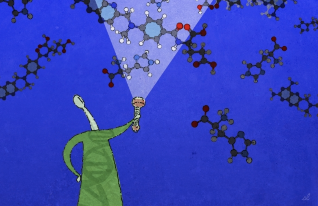
Research Area: Biochemistry, Biophysics, and Structural Biology
Education
PhD, 2016, MIT
BS, 2008, Chemistry, University of Puerto Rico-Mayagüez
Research Summary
We study chromatin — the complex of DNA and proteins that make up our chromosomes. We aim to understand how post-translational modifications to these building-blocks, as well as the factors that regulate these events, play essential roles in maintaining the integrity of cells, tissues, and ultimately entire organisms. We implement a combination of functional genomics, biochemical, genetic, and epigenomic approaches to study how chromatin and epigenetic factors decode the chemical language of chromatin, and how these are dysregulated in diseases such as cancer.
Awards
AACR Gertrude B. Elion Cancer Research Award, 2023
V Foundation Award, 2022
NIH MOSAIC K99/R00 Postdoctoral Career Transition Award, 2021
Eddie Méndez Scholar Award, Fred Hutchinson Cancer Research Center, 2020
Damon Runyon-Sohn Pediatric Cancer Fellowship, Damon Runyon Cancer Research Foundation, 2017
How biologists and mathematicians reached across departmental lines to solve a long-standing problem in electron microscopy
Saima Sidik | Department of Biology
April 19, 2021
MIT’s Hockfield Court is bordered on the west by the ultra-modern Stata Center, with its reflective, silver alcoves that jut off at odd angles, and on the east by Building 68, which is a simple, window-lined, cement rectangle. At first glance, Bonnie Berger’s mathematics lab in the Stata Center and Joey Davis’s biology lab in Building 68 are as different as the buildings that house them. And yet, a recent collaboration between these two labs shows how their disciplines complement each other. The partnership started when Ellen Zhong, a graduate student from the Computational and Systems Biology (CSB) Program, decided to use a computational pattern-recognition tool called a neural network to study the shapes of molecular machines. Three years later, Zhong’s project is letting scientists see patterns that run beneath the surface of their data, and deepening their understanding of the molecules that shape life.
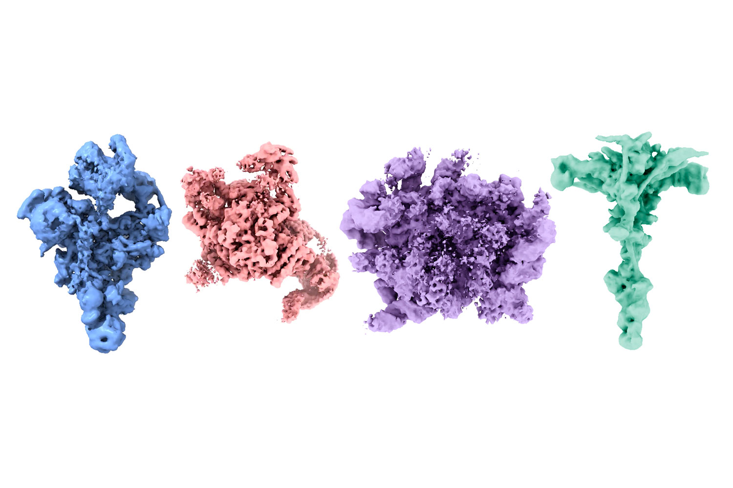
Zhong’s work builds on a technique from the 1970s called cryo-electron microscopy (cryo-EM), which lets researchers take high-resolution images of frozen protein complexes. Over the last decade, better microscopes and cameras have led to a “resolution revolution” in cryo-EM that’s allowed scientists to see individual atoms within proteins. But, as good as these images are, they’re still only static snapshots. In reality, many of these molecular machines are constantly changing shape and composition as cells carry out their normal functions and adjust to new situations.
Along with former Berger lab member Tristan Belper, Zhong devised software called cryoDRGN. The tool uses neural nets to combine hundreds of thousands of cryo-EM images, and shows scientists the full range of three-dimensional conformations that protein complexes can take, letting them reconstruct the proteins’ motion as they carry out cellular functions. Understanding the range of shapes that protein complexes can take helps scientists develop drugs that block viruses from entering cells, study how pests kill crops, and even design custom proteins that can cure disease. COVID-19 vaccines, for example, work partly because they include a mutated version of the virus’s spike protein that’s stuck in its active conformation, so vaccinated people produce antibodies that block the virus from entering human cells. Scientists needed to understand the variety of shapes that spike proteins can take in order to figure out how to force spike into its active conformation.
Graduate student Ellen Zhong (right), and her co-advisor, Professor of Mathematics Bonnie Berger (left)
Getting off the computer and into the lab
Zhong’s interest in computational biology goes back to 2011 when, as a chemical engineering undergrad at the University of Virginia, she worked with Professor Michael Shirts to simulate how proteins fold and unfold. After college, Zhong took her skills to a company called D. E. Shaw Research, where, as a Scientific Programmer, she took a computational approach to studying how proteins interact with small molecule drugs.
“The research was very exciting,” Zhong says, “but all based on computer simulations. To really understand biological systems, you need to do experiments.”
This goal of combining computation with experimentation motivated Zhong to join MIT’s CSB PhD program, where students often work with multiple supervisors to blend computational work with bench work. Zhong “rotated” in both the Davis and Berger labs, then decided to combine the Davis lab’s goal of understanding how protein complexes form with the Berger lab’s expertise in machine learning and algorithms. Davis was interested in building up the computational side of his lab, so he welcomed the opportunity to co-supervise a student with Berger, who has a long history of collaborating with biologists.
Davis himself holds a dual bachelor’s degree in computer science and biological engineering, so he’s long believed in the power of combining complementary disciplines. “There are a lot of things you can learn about biology by looking in a microscope,” he says. “But as we start to ask more complicated questions about entire systems, we’re going to require computation to manage the high-dimensional data that come back.”
Before rotating in the Davis lab, Zhong had never performed bench work before — or even touched a pipette. She was fascinated to find how streamlined some very powerful molecular biology techniques can be. Still, Zhong realized that physical limitations mean that biology is much slower when it’s done at the bench instead of on a computer. “With computational research, you can automate experiments and run them super quickly, whereas in the wet lab, you only have two hands, so you can only do one experiment at a time,” she says.
Zhong says that synergizing the two different cultures of the Davis and Berger labs is helping her become a well-rounded, adaptable scientist. Working around experimentalists in the Davis lab has shown her how much labor goes into experimental results, and also helped her to understand the hurdles that scientists face at the bench. In the Berger lab, she enjoys having coworkers who understand the challenges of computer programming.
“The key challenge in collaborating across disciplines is understanding each other’s ‘languages’,” Berger says. “Students like Ellen are fortunate to be learning both biology and computing dialects simultaneously.”
Bringing in the community
Zhong’s second co-advisor, Professor Joey Davis
Last spring revealed another reason for biologists to learn computational skills: these tools can be used anywhere there’s a computer and an internet connection. When the COVID-19 pandemic hit, Zhong’s colleagues in the Davis lab had to wind down their bench work for a few months, and many of them filled their time at home by using cryo-EM data that’s freely available online to help Zhong test her cryoDRGN software. The difficulty of understanding another discipline’s language quickly became apparent, and Zhong spent a lot of time teaching her colleagues to be programmers. Seeing the problems that non-programmers ran into when they used cryoDRGN was very informative, Zhong says, and helped her create a more user-friendly interface.
Although the paper announcing cryoDRGN was only recently published, the tool created a stir as soon as Zhong posted her code online, many months prior. The cryoDRGN team thinks this is because leveraging knowledge from two disciplines let them visualize the full range of structures that protein complexes can have, and that’s something researchers have wanted to do for a long time. For example, the cryoDRGN team recently collaborated with researchers from Harvard and Washington University to study locomotion of the single-celled organism Chlamydomonas reinhardtii. The mechanisms they uncovered could shed light on human health conditions, like male infertility, that arise when cells lose the ability to move. The team is also using cryoDRGN to study the structure of the SARS-CoV-2 spike protein, which could help scientists design treatments and vaccines to fight coronaviruses.
Zhong, Berger, and Davis say they’re excited to continue using neural nets to improve cryo-EM analysis, and to extend their computational work to other aspects of biology. Davis cited mass spectrometry as “a ripe area to apply computation.” This technique can complement cryo-EM by showing researchers the identities of proteins, how many of them are bound together, and how cells have modified them.
“Collaborations between disciplines are the future,” Berger says. “Researchers focused on a single discipline can take it only so far with existing techniques. Shining a different lens on the problem is how advances can be made.”
Zhong says it’s not a bad way to spend a PhD, either. Asked what she’d say to incoming graduate students considering interdisciplinary projects, she says: “definitely do it.”
Raleigh McElvery
March 26, 2021
Humans breathe oxygen, but many microbes deep within in our gut don’t have access to this precious resource. Instead, they breathe sulfur compounds, releasing hydrogen sulfide in the process. This colorless gas is best-known for its rotten stench, but inside the human colon it has been linked to a thinner mucus barrier, and ailments such as inflammatory bowel disease, Crohn’s disease, ulcerative colitis, and colorectal cancer. In order to develop potential treatments, researchers are probing how microbes create hydrogen sulfide and which molecules they use.
To help further these efforts, Catherine Drennan’s lab and Heather Kulik’s lab at MIT collaborated with Emily Balskus’ lab at Harvard University to investigate the structure and mechanism of an enzyme that’s critical for hydrogen sulfide production: isethionate sulfite-lyase (IslA). The team examined IslA while it was bound to a metabolite that’s readily available in the gut — and revealed how the bacterium Bilophila wadsworthia uses this interaction to help generate the hydrogen sulfide precursor called sulfite. The researchers then compared IslA’s binding behavior to other enzymes in the same family, in order to better understand how these enzymes have evolved to perform challenging chemistry on a wide variety of molecules. Their findings were published on Mar. 26 in the journal Cell Chemical Biology.
“Although abundant, sulfide-producing bacteria are not well understood,” says Drennan, a professor of biology and chemistry and a Howard Hughes Medical Institute investigator. “By characterizing the enzymes in these bacteria that are responsible for sulfur metabolism, we can develop therapeutic strategies to limit production of hydrogen sulfide that can lead to disease.”
Although researchers have been studying bacterial sulfur respiration for decades, IslA was only recently identified. This enzyme breaks the bond between a carbon atom and a sulfur atom in a compound called isethionate, which is a prevalent metabolite in the human body. In doing so, IslA releases the sulfite that bacteria such as B. wadsworthia use to produce hydrogen sulfide.
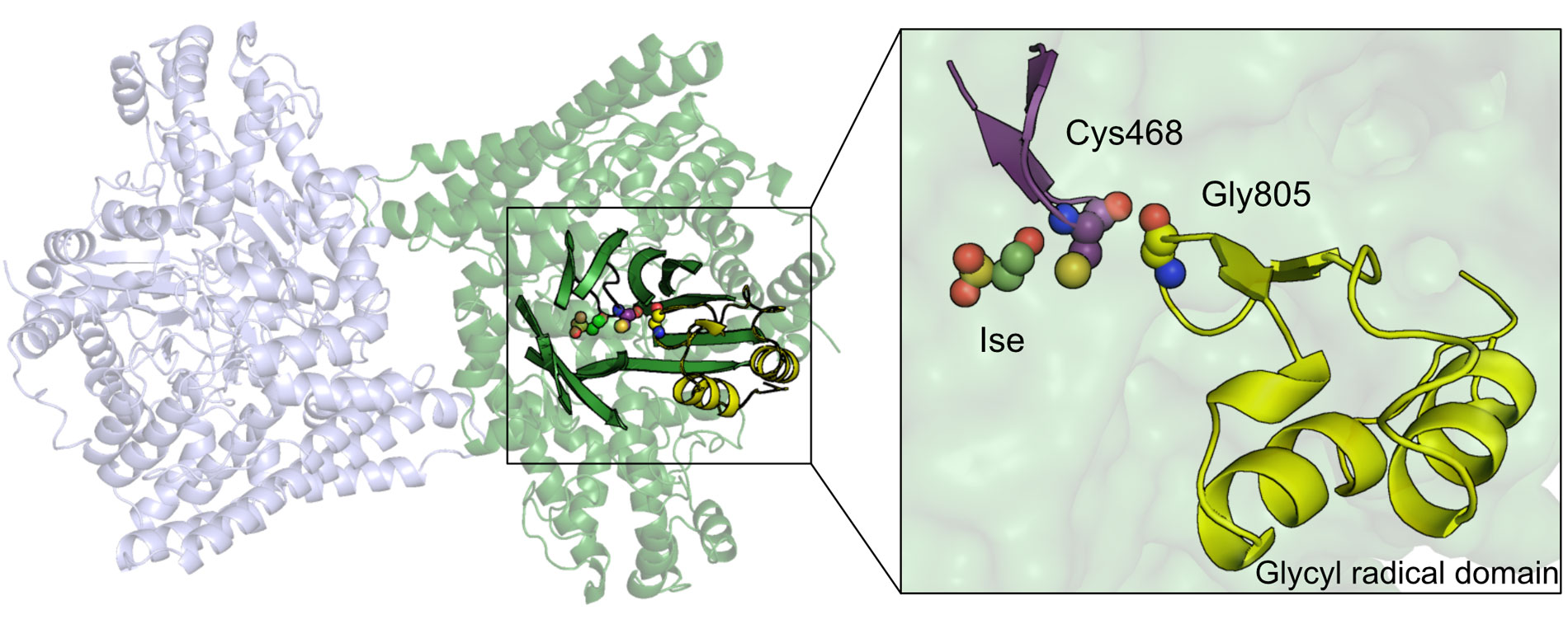
IslA is a member of a large family of enzymes, known as glycyl radical enzyme (GREs). Scientists can learn a lot from examining the way GREs bind to other molecules, according to Christopher Dawson PhD ’20, the study’s co-first author.
GREs contain a binding site (or “active site”) where they latch onto their respective substrates to perform chemical reactions. “Understanding GREs better will aid in drug design efforts to combat the deleterious effects of some of these enzymes,” Dawson says. “It will also help to engineer enzymes that perform diverse, challenging reactions to expand the toolkit for chemical synthesis.”
To this end, Dawson wanted to compare IslA’s active site — where it binds to isethionate to break the C-S bond — to other enzymes in the GRE family. He used X-ray crystallography to visualize this interaction at the level of individual atoms. The GREs he examined shared similar “barrel-like” structures in their active sites, but used these core features in different ways, depending on the substrates they bound. For instance, isethionate bound higher in IslA’s active site compared to the way other GREs bind their respective substrates. While this aberrant binding behavior is quite unique — even among GREs — another group had found something similar when they elucidated IslA’s structure in a different bacterium. And, the Drennan lab suspects this pattern could be prevalent in other classes of enzymes as well.
Next, Dawson and his colleagues wanted to investigate how IslA goes about cleaving the C-S bond once the enzyme has bound to isethionate. Others had predicted this process would occur via a “migration” reaction. In that scenario, the sulfite leaving group first migrates to another carbon atom and then that C-S bond is cleaved to release it. However, after co-first author Stephania Irwin generated multiple IslA variants, the Kulik lab performed computational analyses, and the researchers completed structural comparisons, the team concluded that migration was not occurring. Instead, IslA appeared to be performing an “elimination” reaction that severed the C-S bond without forming another one via migration.
Now that they know more about IslA — and GREs in general — the researchers hope their insights will aid drug design.
“Understanding how pathogens use enzymes like these to extract sulfite from their hosts and fuel hydrogen sulfide production has very clear therapeutic implications,” Dawson says. “And that’s one of the things I like best about this story.”
Citation
“Molecular Basis of C-S Bond Cleavage in the Glycyl Radical Enzyme Isethionate Sulfite-Lyase” Cell Chemical Biology, online March 26, 2021,
DOI: 10.1016/j.chembiol.2021.03.001
Christopher D. Dawson, Stephania M. Irwin, Lindsey R. F. Backman, Chip Le, Jennifer X. Wang, Vyshnavi Vennelakanti, Zhongyue Yang, Heather J. Kulik, Catherine L. Drennan, and Emily P. Balskus
Members of MIT Biology came together with alumni, industry representatives, and supporters to review the department’s challenges and accomplishments.
March 9, 2021
Seychelle Vos investigates how the genome is organized so it can fit inside the cell — and how that careful organization affects gene expression.
February 24, 2021
Eva Frederick | Whitehead Institute
July 28, 2020
In order to grow and thrive, cells need sugar. A repertoire of cellular mechanisms turn unwieldy molecules of glucose and fructose into versatile building blocks for making useful molecules such as lipids, and energy to fuel necessary processes in the cell. But for any of these things to happen, the cells need to sense when sugars are present in the first place — and scientists are still unraveling how they do it.
Now, in a new paper online July 27 in Nature Metabolism, researchers in the lab of Whitehead Institute Member David Sabatini, identify a key molecule that signals to the cell’s growth-triggering complex mTORC1 when there is sugar to be had, leading to a metabolic response. “This discovery puts us another step closer to understanding the biology of mTORC1 and its effects on cellular growth and metabolism,” said Sabatini, who is also a professor of biology at Massachusetts Institute of Technology and investigator with the Howard Hughes Medical Institute.
mTORC1 — short for “mechanistic target of rapamycin complex 1” — is a complex of proteins involved in regulating cell growth and metabolism. Jose Orozco, a fifth-year M.D./Ph.D student in Sabatini’s lab, describes mTORC1 as a sort of cellular licensing board. In order for other parts of the cell to grow and create new products, they must first be “approved” by mTORC1. If there are enough building blocks in the cell to create a certain product, mTORC1 will add a phosphate group to the appropriate “builders” — a signal that allows the building to begin.
“The builders in this case are metabolic pathways responsible for the creation of proteins, the regulation of nucleotides, regulation of glycolysis, regulation of fatty acid synthesis,” he says. “None of these builders can sense everything. But mTORC1 can, and it makes this sort of unified decision for the cell, ‘Yes, we have everything we need to grow.’”
One essential component for cellular building is glucose. That means mTORC1 has a sweet tooth by necessity: the complex is only active when there is enough glucose in the cell. When there’s glucose to go around, mTORC1 is “on” and binds to a lysosome, a structure that serves as the cell’s “digestive system”, where it perches to perform its phosphorylation duties. When a cell is starved for glucose, the complex falls off the lysosome, inactive.
Since the early 2010s, scientists have known one way that mTOR proteins sense glucose: when there is no glucose available, the cell inhibits the action of mTORC1 through a pathway involving the protein AMPK. But another study suggested that even without AMPK, mTORC1 can still sense an absence of glucose. “I think a lot of people had written it off as ‘Oh, [the signal] must just be AMPK,’” Orozco says. “But when we tested that hypothesis, we showed that even cells that didn’t have any AMPK were still able to sense glucose availability. That was the observation that started this project.”
To find the mysterious second sugar-sensing process, Orozco and colleagues created cells in which the known signalling protein AMPK was out of the picture. Using these modified cells, they began looking for specific traits of the glucose molecule that might be triggering the response. The team found that sugars that could be broken down by the cell, such as mannose, glucosamine and fructose, were able to activate mTORC1. Non-metabolizable sugars had no effect.
This suggested that the signaling molecule was not glucose itself, but something produced when glucose is taken apart during glycolysis — the biochemical process that breaks down the sugar into usable building blocks. With this in mind, the researchers next combed step by step through glycolysis products to see which ones could be the signal molecule.
The team identified a step of glycolysis that seemed to be key, zeroing in on a glycolysis product called dihydroxyacetone phosphate, or DHAP. Even in the complete absence of glucose, the researchers could turn on mTORC1 by adding DHAP.
It is difficult to prove exactly why the cell relies on DHAP as a signal, but Orozco has some ideas. For one thing, DHAP later goes on to serve as the backbone of lipids, which are built by a process controlled by mTORC1 — so it would make sense that mTORC1 would respond to its presence or absence. Also, DHAP levels are extremely sensitive to changes in the amount of cellular glucose, more so than any other glycolysis intermediary. Also, DHAP is a product of both glucose and fructose, which are both important sugars in the human diet.
In the future, the team hopes to understand more. “We don’t know the biochemical details of how DHAP [conveys its message],” Orozco says. “We don’t know the sensor, we don’t know what proteins bind it, and we don’t know if that causes conformational changes in [associated proteins]. That we sort of leave as the enticing next question that we want to tackle.”
At the moment, studying the glucose sensing pathway is purely foundational research. But while there are no clear applications yet, surprises could lurk just around the corner. “Targeting nutrient sensing in mTOR has shown some promise in, of all things, regulating depression and mood,” Orozco says. “That’s interesting, and we don’t really understand why that is the case. How is glucose targeting going to be important? We don’t know yet. But we think it has a lot of potential.”
***
Written by Eva Frederick
Citation:
Orozco, J.M., Krawczyk, P.A., Scaria, S.M. et al. Dihydroxyacetone phosphate signals glucose availability to mTORC1. Nat Metab (2020). https://doi.org/10.1038/s42255-020-0250-5
A biophysicist employs super-resolution microscopy to peer inside living cells and witness never-before-seen phenomena.
Raleigh McElvery | Department of Biology
July 23, 2020
How do cells use physics to carry out biological processes? Biophysicist Ibrahim Cissé explores this fundamental question in his interdisciplinary laboratory, leveraging super-resolution microscopy to probe the properties of living matter. As a postdoc in 2013, he discovered that RNA polymerase II, a critical protein in gene expression, forms fleeting (“transient”) clusters with similar molecules in order to transcribe DNA into RNA. He joined the Department of Physics in 2014, and was recently granted tenure and a joint appointment in biology. He sat down to discuss how his physics training led him to rewrite the textbook on biology.
Q: How does your work revise conventional models describing how RNA polymerases carry out their cellular duties?
A: My interest in biology has always been curiosity-driven. As a physicist reading biology textbooks, I thought that transcription — the process by which DNA is made into RNA — was fully understood. It’s so basic, and the textbooks write about it with such confidence. Come to find out, most of what we know about the cell nucleus, where gene expression starts, comes from people studying these processes outside the cell, inside a test tube. I started to wonder: Do we actually know how they work in a living cell?
The textbook models say that when a specific gene is being activated, RNA polymerase and dozens of other molecules are recruited to the DNA to begin transcription. If you don’t look closely enough, the polymerases appear to be uniformly distributed and acting randomly throughout the nucleus. However, my single-molecule and “super-resolution” microscopy methods allowed me to see something different when I looked inside live cells: polymerase clusters, which are very dynamic. In the mid-’90s, people had observed similar clusters in so-called “fixed” cells that were chemically frozen. But these findings were dismissed as possible artifacts of the fixation procedure. However, when we saw these same protein clusters in living cells that were not treated with harsh chemicals, it suggested that the textbook explanation may be incomplete.
Q: How has your background in physics given you a unique perspective on the mechanics of living cells?
A: When I arrived at the University of Illinois at Urbana-Champaign to begin my PhD in physics, I hadn’t enrolled in a biology class since high school. I was really taken with the interdisciplinary work of one physics professor, Taekjip Ha, who became my PhD mentor. He had developed single-molecule fluorescence resonance energy transfer techniques, to study with unprecedented sensitivity when two biomolecules are close to each other and monitor the distance between them in real time.
Taekjip graciously accepted me into his lab despite my limited biology background, and I never looked back. His work mirrored my interest in condensed matter physics, but the material we were looking at wasn’t from the inanimate world, it was living matter.
Between 2006 and 2008, as I was working on my PhD, super-resolution microscopy really took off from the single-molecule microscopes I used in grad school. It was a natural progression, in my mind, to learn cell biology during my postdoc fellowship at École Normale Supérieure in Paris, and to try to visualize weak and transient interactions directly in living cells using single-molecule and super-resolution imaging. You could now pinpoint molecules with nanometer accuracy; you could “turn on” and “off” molecules to observe them individually and ensure there was no overlap between those that were side-by-side.
Thanks to these new techniques, we saw clusters of RNA polymerases in living cells for the first time during my postdoc, and I pushed the technique further to reveal the cluster dynamics. But the fact that you had to turn individual molecules on and off made it really hard to see these clusters assembling or disassembling. I didn’t want to trade temporal resolution for spatial resolution. So I came up with an approach called Time-Correlated Photoactivated Localization Microscopy (tcPALM). It allowed us to measure the lifetimes of these ephemeral polymerase clusters, and we found that they last just a few seconds.
Once I arrived at MIT, we wanted to test whether the clusters could be fleeting but still biologically relevant. We pushed a dual-color super-resolution technique where we correlated the clusters with gene activity. With RNA live-imaging experts at Howard Hughes Medical Institute’s Janelia Research Campus, Brian English and Tim Lionnet, and my postdoc, Wonki Cho, we found that roughly 80 to 100 polymerases form a cluster on a gene where transcription is about to start. Although the cluster is only there for a few seconds, that’s enough time to load a handful of polymerases and generate “bursts” of RNA transcription. In fact, there was a linear correlation between the clusters’ transient dynamics and the number of messenger RNAs made in each burst.
Q: What is it like to be a physicist working with biologists?
A: Even though I joined MIT as a physics hire, I was lucky enough to get lab space in Building 68 alongside amazing biologists. They were the perfect people to talk to about my crazy ideas. And it turned out that renowned researchers like Rick Young and Phil Sharp actually had similar theories. They had genomic evidence for clusters of gene regulators, which they call “super enhancers,” that we all thought could relate to what my lab was seeing. That’s led to hours of exciting discussions between our labs, and has evolved into one of my most rewarding collaborations — and revealed that clusters associate as tiny transcriptional condensates with properties of liquid droplets.
Now, students and postdocs in my lab are wondering about the clusters’ functions and mechanisms of action, and whether protein clustering extends beyond transcription. For instance, clustering could explain some aspects of neurodegeneration. One perplexing idea that came out of this work is that perhaps it gets harder for our cells to clear protein condensates as we age, leading to Parkinson’s, Alzheimer’s, and other diseases. It’s becoming clearer that physics may be just as important as biology for understanding how cells work. The physics of how condensates and droplets form in the inanimate world is increasingly helpful in determining how living cells can evolve to regulate the same process for specific biological functions like transcription. Nature uses physics in much more elaborate ways than we initially anticipated.
Education
PhD, 2014, University of North Carolina at Chapel Hill
BA, 2008, Biology, Franklin and Marshall College
Research Summary
We study how cells regulate the spatial organization of signaling molecules at the plasma membrane to control downstream signaling. For example, receptor clustering and higher-order assembly with cytoplasmic proteins can create compartments with unique biochemical and biophysical properties. We use quantitative experimental approaches from biochemistry, molecular biophysics, and cell biology to study transmembrane signaling pathways and how they are misregulated in diseases like cancer.
Awards
NSF Career Award, 2025
Searle Scholar, 2022
NIH Director’s New Innovator Award, 2022
AFOSR Young Investigator Award, 2021
Brown-Goldstein Award, 2020
Damon Runyon-Dale F. Frey Breakthrough Scientist, 2020
July 2, 2020
In the lab, Biology Professor Amy Keating researches the interactions of proteins with a mix of modeling and synthetic lab work and diverse minds
School of Science
June 11, 2020
Almost everything in biology is a multistep process, from the metabolization of carbohydrates and fats as fuel to information transcription from DNA and RNA. Without proteins and their interactions, cells couldn’t perform any of these biological tasks. But how do proteins establish their individual roles? And how do they interact with each other? These questions drive Professor Amy Keating’s research, and both lab experiments and computational modeling are helping her reveal the mysteries behind the basic functions of life.
In Keating’s field of research, as with most areas of science, the use of artificial intelligence is a relatively new – and growing – trend. “It’s pretty scary how fast new methods in machine learning are changing the landscape,” says Keating, who holds appointments in both the Department of Biology and the Department of Biological Engineering. “I think that we will see a disruptive change in protein modeling over the coming years.” She has found that incorporating basic machine learning methods in her own work has generated some success in uncovering how protein sequences determine their interactions.
However, there are limits to using only computational modeling due to the complexities of protein-protein interaction and a general need for empirical data to calibrate the models. Her lab group integrates computation with biological engineering in a laboratory setting. Keating’s team often starts by using computational modeling to narrow down their search from a massive collection of protein structure models. This step limits their output from an effectively infinite space (~1030) to something on the order of 106 potential promising molecules that can be experimentally tested. They can feed the results of experiments into other algorithms that help designate the specific features of the protein that prove important. This process is cyclical, and Keating emphasizes that experimental efforts are crucial for improving the success rate of this kind of work. That is where the lab comes in. There, they do what the computer cannot: they build proteins.
With the disruption of the COVID-19 crisis the Keating lab has focused their attention on computational projects, as well as on reviewing the literature and writing up papers and theses. The members are also using their time at home to brainstorm and plan their research. “We are having multiple group meetings per week by Zoom, including a ‘Keating Group Idea Lab,’ at which everyone throws out ideas, ranging from practical suggestions about current projects to out-there new concepts, for group discussion,” says Keating. “We are confident that we can use this time productively, to advance our science, even as we make long lists of things that we are eager to do as soon as we can get back into the laboratory.”
A topic of current interest to Keating and her group members is interactions among proteins with “short linear motifs” or SLiMs, which are abundant –more than one hundred thousand such motifs are thought to exist in one human. One family of these SLiM-binding proteins regulates movement of cells within the body and is implicated in the spread of cancer cells to a secondary location (metastasis). The lab’s novel mini-protein and peptide designs aim to disrupt these protein interactions and could be useful for eventually disrupting and treating cancer and other diseases.
FOSTERING MULTIPLE INTERACTIONS
Currently, Keating’s research team consists of six students who have backgrounds in almost as many different cultures. Her students’ diversity, which stems not just from different focuses in formal training but also from life experiences, is integral to their success, according to Keating. She wishes that more women like herself and members of underrepresented minority groups who love STEM would consider pursuing academic careers. “It’s hard work, but it’s very rewarding,” she entices. The best thing about being a faculty member, she believes, is having a team of bright minds who contribute unique ideas and insights to a problem and provide information beyond her own areas of expertise.
“I learn facts that they know and I do not. I learn interesting ways of thinking about science and also ways of doing science,” she says, noting that novel ideas in methodology lead to advances in research. “I’ve learned a lot of things about computer science from my students. I’m happy that one of my former biology students is [now] a professor of computer science,” she admits, appreciating his expertise as a benefit in frequent collaborations. “I love that students at MIT question everything.” Keating’s ever-expanding knowledge builds on top of a diverse background gleaned during her time as a student.
Keating’s bachelor’s degree from Harvard University is in physics. During her PhD at University of California, Los Angeles, she shifted to chemistry — specifically computational physical organic chemistry. When browsing for a postdoctoral position, she discovered the work of former MIT Department of Biology faculty and Whitehead Institute member Peter Kim and joined him. She maintained her interest in computation as a tool for biological research, concurrently co-advised by MIT Professor of Electrical Engineering and Computer Science Bruce Tidor. It was somewhat down to chance that her academic job search led her to MIT. “I certainly never thought I would be a biology professor, especially at MIT,” she remarks of her convoluted career path through the wide world of science.
But it is an unexpected result for which Keating is grateful. “My undergrad self would have been surprised by the MIT School of Science,” she muses, which makes MIT “so much more than ‘just’ the world’s best engineering school.” That is something of a common misconception about the Institute, she feels. “I think a lot of people outside of MIT don’t know how outstanding our basic science programs are.” Keating is a part of the strong science education at MIT, which is constantly adapting to keep up with the digital age, which led to her receiving the most recent Fund for the Future of Science Award.
“I was thrilled, and pretty surprised, to receive the award; my fantastic colleagues in the School of Science are not people that you want to be competing with.” This support is invaluable to her research on the foundations of biological interactions, and to ensure a robust team that has what it needs to develop important advances. The curious minds with which she collaborates are equally as invaluable.
“The people at MIT are amazingly smart, curious, and focused on things that I value,” Keating adds, “like good ideas, intellectual rigor, discovering new things, and education.”