Education
- PhD, 1987, Case Western Reserve University; MD, 1989, Case Western Reserve University
- BS, 1981, Chemistry with Concentration in Solid-State and Polymer Physics, Cornell University
Research Summary
Our goal is to understand how signaling pathways are integrated at the molecular and systems levels to control cellular responses. We have two main focuses: First, we study signaling pathways and networks that control cell cycle progression and DNA damage responses in cancer and cancer therapy. Second, we examine the cross-talk between inflammation, cytokine signaling and cancer. Much of our work focuses on how modular protein domains and kinases work together to build molecular signaling circuits, and how this information can be used to design synergistic drug combinations for the personalized treatment of human disease.
Awards
- MacVicar Faculty Fellow, 2021
- Fellow, Association of American Physicians, 2021
- Teaching with Digital Technology Award, 2018
Education
- PhD, 1998, University of California, Los Angeles
- SB, 1992, Physics, Harvard University
Research Summary
Our goal is to understand, at a high level of detail, how the interaction properties of proteins are encoded in their sequences and structures. We investigate protein-protein interactions by integrating data from high throughput assays, structural modeling, and bioinformatics with biochemical and biophysical experiments. Much of our work focuses on α-helical coiled-coil proteins, Bcl-2 apoptosis-regulating proteins, and protein domains that bind to short linear motifs.
Education
- PhD, 1976, California Institute of Technology
- BS, 1971, Chemistry, Tulane University
Research Summary
We use genetic, biochemical, physiologic, chemical, cellular and molecular biological methods to study cell surface receptor structure and function. We focus on lipoprotein receptors — in particular, the High Density Lipoprotein (HDL) receptor called Scavenger Receptor, Class B, Type I (SR-BI). Our analyses have provided insight into basic biological processes, contributed to our understanding of atherosclerosis and coronary heart disease (CHD) and have uncovered an unexpected connection between cholesterol and mammalian female infertility.
No longer accepting new students.
Awards
- Tulane University School of Science and Engineering Outstanding Alumnus Award, 2010
- National Academy of Sciences, Member, 2009
- Outstanding Achievement Award for Contributions to Atherosclerosis Research, International Atherosclerosis Society, 2009
- Margaret MacVicar Faculty Fellow, 1993-2003
Education
- PhD, 2002, Stanford University
- BS, 1997, Molecular Biology, University of California, San Diego
Research Summary
We study the biological mechanisms and evolution of how cells process information to regulate their own growth and proliferation. Using bacteria as a model organism, we aim to elucidate the detailed molecular basis for this remarkable regulatory capability, and understand the selective pressures and mechanisms that drive the evolution of signaling pathways. Our work is rooted in a desire to develop a deeper, fundamental understanding of how cells function and evolve, but it also has important medical implications since many signaling pathways in pathogenic bacteria are needed for virulence.
Awards
- Howard Hughes Medical Institute, HHMI Investigator, 2015
- National Science Foundation, Presidential Early Career Award for Scientists and Engineers, 2010
- Howard Hughes Medical Institute, Early Career Scientist, 2009
Education
- PhD, 2010, Harvard University
- SB, 2004, Physics, National Tsinghua University
Research Summary
We seek to understand the optimization of bacterial proteomes at both mechanistic and systems levels. Our work combines high-precision assays, genome-wide measurements, and quantitative/biophysical modeling. Ongoing projects focus on the design principles of transcription, translation, and RNA maturation machineries in the face of competing cellular processes.
Awards
-
Smith Odyssey Award, 2020
-
MIT Committed to Caring Award, 2020
- NSF Career Award, 2019
- Pew Biomedical Scholar, 2017
- Smith Family Award for Excellence in Biomedical Research, 2017
- NIGMS R35 Maximizing Investigator Research Award, 2017
- Sloan Research Fellowship, 2016
- Searle Scholar, 2016
- NIH Pathway to Independence Award, 2013
Education
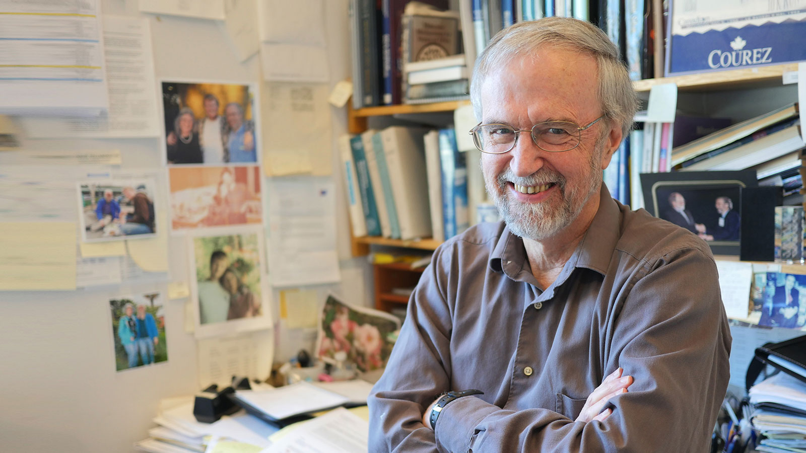
- PhD, 1974, University of Illinois
- BS, 1970, Chemistry, Carleton University
Research Summary
Our research is concentrated in two major areas. First, we aim to understand how the proteins involved in DNA repair, mutagenesis and other cellular responses to DNA damage are regulated. Some of our discoveries have the potential to improve chemotherapy. Second, we probe how nitrogen-fixing nodules develop on legumes, and the relationship between rhizobial functions required for nodule invasion/infection and mammalian pathogenesis.
Awards
- Revolutionizing Innovative, Visionary Environmental health Research (RIVER), R35 Outstanding Investigator Award, 2017
- National Academy of Sciences, Member, 2013
- Howard Hughes Medical Institute, HHMI Professor, 2010
- University of Guelph, Doctor of Science, honoris causa, 2010
- American Association for the Advancement of Science, Fellow, 2008
- Environmental Mutagen Society, EMS Award, 2006
- American Academy of Arts and Sciences, Fellow, 2004
- American Cancer Society, Research Professor, 2002
- Howard Hughes Medical Institute, HHMI Professor, 2002
- Charles Ross Scholar, 2000-2003
- American Academy of Microbiology, Fellow, 1994
- Margaret MacVicar Faculty Fellow, 1992-2002
- John Simon Guggenheim Memorial Foundation, Guggenheim Fellowship, 1984
- Massachusetts Institute of Technology, MacVicar Faulty Fellow, 1984
- Rita Allen Foundation, Career Development Award, 1978
Education
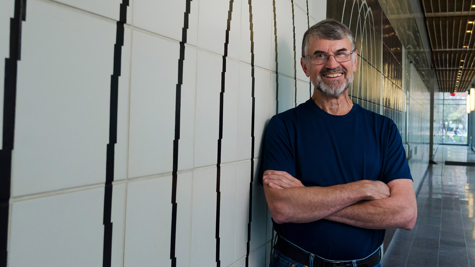
- PhD, 1979, Harvard University
- BA, 1972, Biophysics, Amherst College
Research Summary
Before closing his lab, Bob Sauer studied the relationship between protein structure, function, sequence and folding. He focused on the molecular machines that degrade or remodel proteins, targeting proteins for these ATP-dependent reactions. His experimental tools included biochemistry and single-molecule biophysics, structural biology, protein design and engineering, and molecular genetics.
Awards
- Protein Society, Stein and Moore Award, 2013
- Protein Society, Hans Neurath Award, 2007
- Protein Society, Amgen Award, 2001
- National Academy of Sciences, Member, 1996
- American Academy of Arts and Sciences, Fellow, 1993
Education
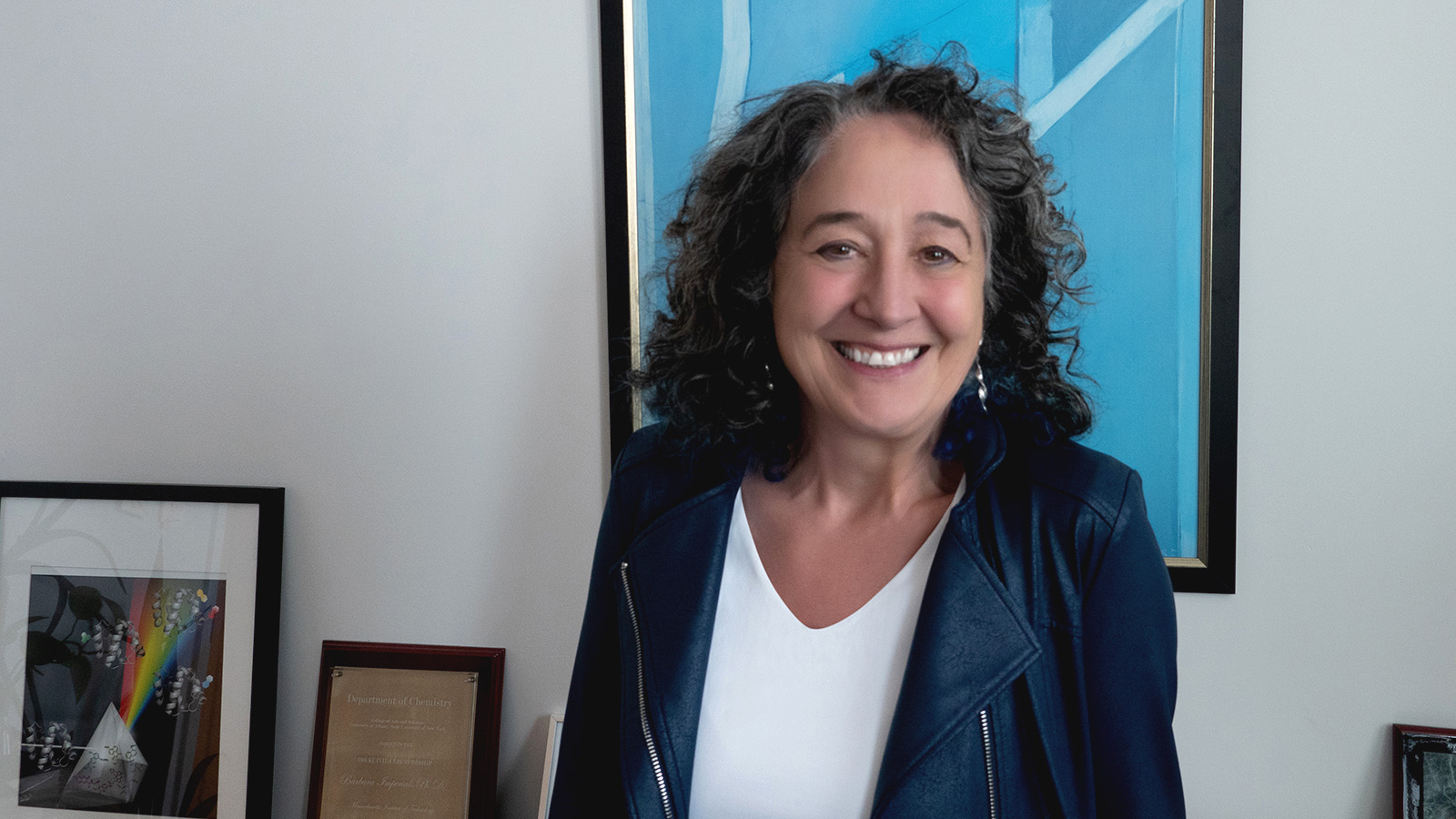
- PhD, 1983, MIT
- BSc, 1979, Medicinal Chemistry, University College London
Research Summary
We study diverse aspects of protein structure and function and employ multidisciplinary approaches to address fundamental problems at the interface of chemistry and biology. We are fascinated by the amazing complexity and myriad functions of glycoconjugates in human health and disease. Still more enthralling are the intricate membrane-associated pathways that lead to the cellular biogenesis of these important macromolecules. Our group applies approaches and technologies from a wide range of synergistic fields including chemical biology (for inhibitor and probe development), biochemistry and biophysics (for analyses within and beyond native and model membranes), and cellular, molecular and microbiology to unravel these pathways. Ultimately we seek to decipher the molecular logic of glycoconjugate biosynthesis and to identify processes to target in the study of infectious disease.
Awards
- National Academy of Sciences, Member, 2010
- Fellow of the Royal Society of Chemistry (FRSC) 2006
- American Chemical Society – Breslow Award for Achievement in Biomimetic Chemistry 2006
- Protein Society – Kaiser Award, 2006
- Margaret MacVicar Faculty Fellow, 2003-2013
- American Academy of Arts and Sciences, Fellow, 2001
Education
- PhD, 2000, Free University of Berlin
- MS, 1996, Biochemistry, Free University of Berlin
- BS, 1993, Biochemistry, Free University of Berlin
Research Summary
Our primary goal is to understand how signals and molecules are transmitted between the nucleus and cytoplasm across the nuclear envelope. We work to decipher the mechanism and structure of the machinery that executes these cellular processes.
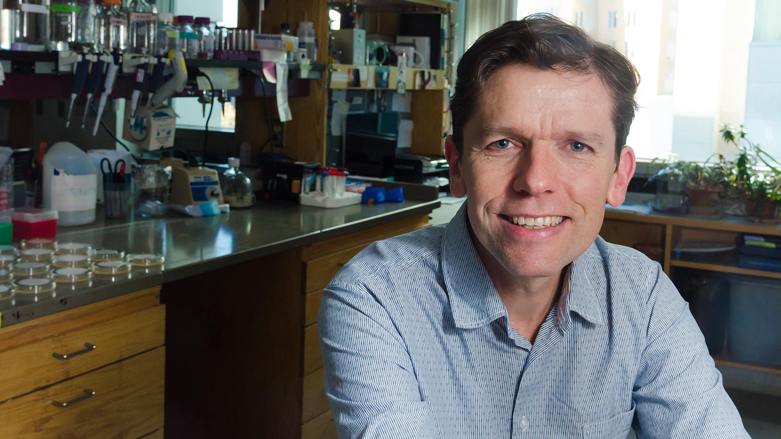
Education
- PhD, 2011, The Johns Hopkins University School of Medicine
- BS, 2002, Molecular Biology and Biotechnology, Millersville University
Research Summary
In the Lamason lab, we investigate how intracellular bacterial pathogens hijack host cell processes to promote infection. In particular, we study how Rickettsia parkeri and Listeria monocytogenes move through our tissues via a process called cell-to-cell spread. We utilize cellular, molecular, genetic, biochemical and biophysical approaches to elucidate the mechanisms of spread in order to reveal key aspects of pathogenesis and host cell biology.
Awards
- NIH Pathway to Independence Award, 2015