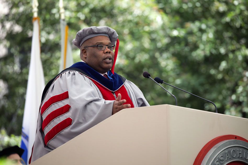
Biochemist Squire Booker PhD ’94 says MIT’s new doctoral graduates will “grow as future leaders” by giving back.
Peter Dizikes | MIT News Office
Distinguished biochemist Squire Booker PhD ’94 emphasized the importance of opportunity for all, in his keynote speech at today’s 2019 Investiture of Doctoral Hoods and Degree Conferral, a ceremony for MIT’s new doctoral degree holders.
While congratulating MIT’s doctoral graduates, Booker also urged them to give back to society and to take responsibility for helping others accomplish their own goals — however daunting those goals, such as a PhD, may seem.
“Almost anyone can excel if given the chance,” Booker said. “Take advantage of opportunities, and make the most of them. But also, work to provide opportunities for others. That’s how you will grow as a future leader.”
Reflecting on his own trajectory, from a childhood when he knew no one working in the sciences to a career on the front lines of discovery, Booker called himself “just an average guy from southeast Texas, no different than anyone else.” But he said new opportunities had “made all the difference” in his career. One key moment of opportunity, Booker said, was his graduate training at the Institute.
“MIT gave me my first real opportunity to explore scientific research and realize my passion for discovery and working with people from all over the world to solve problems,” Booker said. He credited his mentors with “helping me to achieve goals that I didn’t even know existed when I undertook this journey, or that I didn’t even have for myself. I can honestly say my cup runneth over today.”
Booker is the Evan Pugh Professor of chemistry and of biochemistry and molecular biology and Eberly Family Distinguished Chair in Science at Penn State University. He is also an investigator with the Howard Hughes Medical Institute, and in April of this year was elected to the National Academy of Sciences.
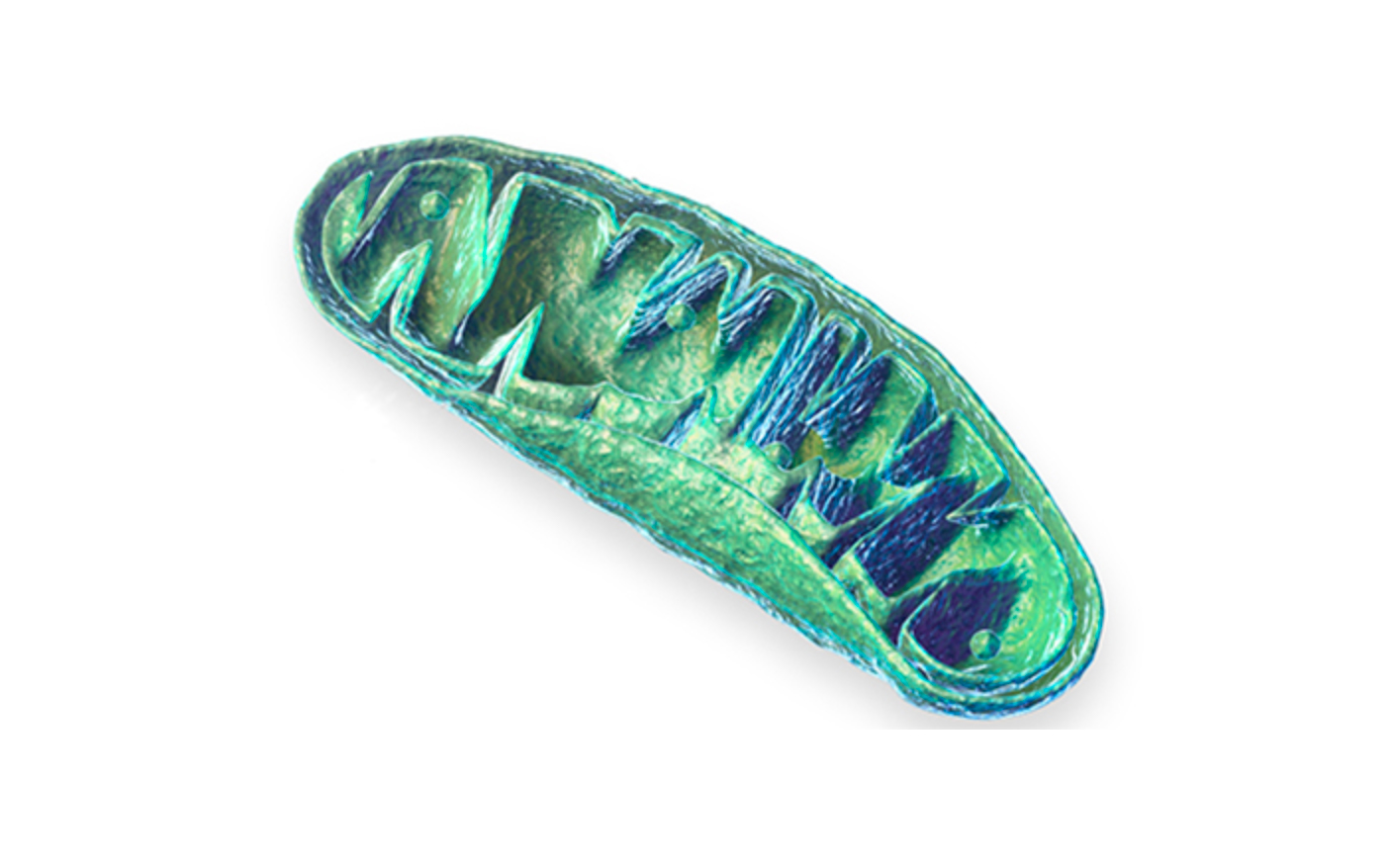
During his career, Booker has conducted significant research uncovering the ways enzymes catalyze reactions within cells, a line of work with applications ranging from medicine to biofuels.
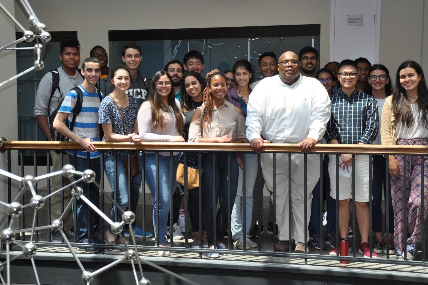
The ceremony honors graduate students who have earned their doctoral degrees within this academic year. It was held this year in MIT’s Killian Court, where a large audience of family members and friends filled the seats. Killian Court is also the site of Friday’s 2019 Commencement exercises.
Graduates from 26 departments, programs, and centers at the Institute, as well as MIT’s joint program with the Woods Hole Oceanographic Institution, received degrees on Thursday. MIT faculty — who wear the brightly colored formal garb of the universities where they received their own doctorates — placed doctoral hoods, a part of the formal academic clothing, over the shoulders of the new graduates.
In his remarks, Booker said he shared the experience this year’s doctoral graduates have gone through, and understood how hard they have worked at the Institute.
“I don’t just imagine the blood, the sweat, the tears, and the immense amount of time that you put into arriving at this point in your careers and your lives,” Booker said. “I actually experienced it firsthand as a graduate student here at MIT between 1987 and 1994.” He cited his graduate advisor, JoAnne Stubbe, as an important influence on his career.
Booker infused his remarks with self-deprecating humor, joking that he first thought MIT had ask him to speak by mistake. But he also spoke earnestly about the serious hurdles he had faced in his life.
Booker grew up in what he described as a segregated environment in Beaumont, Texas. He noted that it was not uncommon for him to hear teachers make disparaging remarks about the abilities of African-Americans, adding, “A career in science was about as likely as winning the lottery … largely because there were no role models.”
Raised by a grandmother with the help of three uncles, Booker earned his undergraduate degree in chemistry at Austin College in Sherman, Texas, and first came to MIT in 1986, as part of the Institute’s MIT Summer Research Program, which now supports 40 interns every year from underrepresented backgrounds.
That stint at MIT helped lead Booker to enter the graduate program, where he studied biochemistry. It also gave him a greater awareness of the travails of black scientists who had gone before him — partly through the work of MIT’s Kenneth Manning, the Thomas Maloy Professor in Rhetoric, whose 1983 book, “Black Apollo of Science: The Life of Ernest Everett Just,” chronicled the life of a pioneering black researcher excluded from American academia.
In his speech, Booker outlined the lives of both Just and Percy Lavon Julian, an innovative 20th-century African-American research chemist who also spent decades excluded from a conventional professorship in academia.
“We’re still trying to recover from the bigotry and misogyny of the past, some of which still exist,” Booker said. In that vein, he noted, in 2008, he became the first Afrcian-American professor in Penn State’s chemistry department.
‘That it took so long is completely tragic,” said Booker, observing that countless talented people had been excluded from promising careers and fulfilling lives as a result of prejudice.
“America’s strength is its people,” Booker said. “And there is so much untapped potential in people who have been traditionally disenfranchised, including people of color, women, the LGBTQ community, and the differently abled.”
At the same time, Booker added, “In fact, first-generation white students, or students from modest socioeconomic backgrounds, are the ones that I have impacted the greatest, directly, at Penn State. And you can’t imagine how appreciative they have been to have been given the chance, and some direction.”
Booker was introduced by MIT Chancellor Cynthia Barnhart SM ’86, PhD ’88, the Ford Foundation Professor of Engineering, who briefly delivered her own remarks to the graduates.
“Today is about honoring the accomplishment and success of all of you, our doctoral candidates,” Barnhart said. “Congratulations. Each and every one of you have succeeded. … You were curious and creative, determined to problem-solve, to collaborate, and to innovate.”
Barnhart also called the doctoral hooding ceremony a “delightfully hopeful moment where infinite possibilities stretch out in front of you,” and asked the graduates to rise in appreciation of their friends and families who have supported their efforts.
This is the fifth year in a row that MIT’s doctoral hooding ceremony has had a keynote speaker — who is annually drawn from the ranks of past MIT doctoral graduates. Booker was chosen with input from the MIT community.
The festive, bright regalia of the doctoral ceremony represents a mix of old traditions and recent changes. Formal academic wear, at least of the kind seen at commencement ceremonies, dates to the 1400s, if not earlier. However, American universities did not agree to standards for such gowns and hoods until 1893.
At MIT, the doctoral degree robes were redesigned as recently as 1995. MIT gowns feature a silver-gray robe with a cardinal red velvet front panel, and are embellished by cardinal red velvet bars on the sleeves. Additional color markings signify whether graduates have received the Doctor of Philosophy degree (PhD) or the Doctor of Science degree (ScD). Silver-gray academic caps complement the gowns. The doctoral hoods are an accessory to the main robe ensemble.
After Barnhart’s introductory remarks and Booker’s speech, all doctoral graduates had their names announced as they walked across the stage one by one. The newly minted degree holders then had the hoods draped over their shoulders by their department or program heads.
The names of all the new doctoral degree holders were read aloud, one after another, by two MIT staff members: Monica Lee, a senior communications officer in the Department of Facilities; and Steven M. Lanou, a project manager in the Office of Sustainability.