To be long-lived or short-lived?
Nicole Davis | Whitehead
February 20, 2020

Genes are often imagined as binary actors: on or off. Yet such a simple view ignores the fact that genes’ activities, exerted by their corresponding proteins, can run the gamut from barely perceptible to off the charts. This rheostat-like range is due in part to molecular controls that determine how long the protein-making instructions for any given gene — known as messenger RNA (mRNA) — can persist before being destroyed.

Now, in a pair of papers published online in Molecular Cell, Whitehead Institute member David Bartel and his colleagues take a deep and systematic look at the dynamics of mRNA decay across thousands of genes. Their analysis — the most extensive to date — reveals surprising variability in the rate at which the ends (or “tails”) of mRNAs are shortened. In addition, the researchers uncover a link between this rate of shortening and how quickly the short-tailed mRNAs decay.

“Ultimately, these dynamics are responsible for determining how much mRNA is present for each gene, and that, of course, is really important for determining cell identity — for example, whether a cell is cancerous or a normal, healthy cell,” says Bartel, who is also a professor of biology at the Massachusetts Institute of Technology and an investigator at the Howard Hughes Medical Institute. “There is a thousand-fold difference in how long mRNAs stick around. That has a very profound effect on the amount of protein that gets made.”

TOWARDS A GLOBAL VIEW OF MRNA DEGRADATION

The anatomy of a typical mRNA consists of three key parts: a body, which contains the protein-making instructions; at one end, a string of repeating A’s known as the poly(A) tail; and at the other end, a protective biochemical cap.

Prior to the Molecular Cell studies, the future of a mRNA was known be linked to the length of its poly(A) tail — the longer the string of A’s, the longer the mRNA tends to persist. However, the speed that tails shorten as they age, and the rate at which mRNAs decay when their tails become short was known for just a handful of mRNAs.

To gain a more global picture, Bartel and his team, most recently led by graduate student Timothy Eisen, combined a set of techniques for high-throughput analyses of mRNA. These include a method for chemically modifying mRNAs as they are being made in order to distinguish newly synthesized mRNAs from those that are older, as well as sequencing-based approaches for measuring both the length of poly(A) tails and the amount of mRNA that was recently made. In addition, Eisen used computational methods to model the data they gathered and make predictions about them.

“All of the work in these papers involves time as an axis,” says Eisen. “The power of our approach is that it allowed us to plot and visualize how things change over time — and to infer for mRNAs from thousands of genes the rate at which the tail shortens and the subsequent rate at which the mRNA is destroyed.”

THE TAIL WAGS THE MRNA

By leveraging these techniques, Bartel, Eisen and their colleagues explored the mRNA dynamics for thousands of genes. One key observation is that mRNAs enter the cytoplasm with diverse poly(A) tail lengths. That variability encompasses not only the mRNAs from different genes but even those that correspond to the same gene.

“Previously, there wasn’t any reason to think there would be any differences, so people just assumed that the initial tail lengths would be the same,” says Bartel. “But it turns out there’s quite a bit of variability there.”

The Whitehead team also uncovered a striking amount of variation in the rate at which poly(A) tails are shortened. For some mRNAs, the tail shortens at a rate of about 30 nucleotides per minute. With an average tail length of around 200 nucleotides, that translates to the tail lasting just a few minutes. Other mRNAs have much more durable tails, with shortening rates of just a nucleotide or two an hour.

“That’s a thousand-fold difference,” says Eisen. Previously, researchers had shown that tail-shortening rates could vary, but they had observed only a 60-fold difference.

Bartel and his colleagues also found some striking differences among mRNAs once their poly(A) tails became short. “If we consider just those mRNA molecules that have tails of only 20 nucleotides, the ones that come from certain genes disappear much more rapidly than those coming from other genes — again spanning a thousand-fold range,” says Bartel.

That finding challenges long-held views about mRNA stability, as it had been generally assumed that short tails equaled short lives, and that all mRNAs whose tails had been shortened decay at the same rate. But it turns out that both processes are important: the rate at which mRNA tails are shortened (a process known as deadenylation), and the rate at which mRNAs decay after this shortening. Moreover, Bartel and his colleagues find that these two processes are coupled —  the more rapidly deadenylated mRNAs also degrade more rapidly once they have short tails.

“This coupling between rate of decay of short-tailed mRNAs and the rate of deadenylation is important because it prevents a large build-up of short-tailed versions of mRNAs that had undergone rapid deadenylation,” says Bartel. “Because these short-tailed versions do not build up, the thousand-fold difference that we observe in deadenylation rates can impart a thousand-fold difference in mRNA stabilities.”

SHINING A LIGHT ON MICRORNAS

MicroRNAs are small, regulatory RNA molecules that play critical roles in human biology. Their primary job is to recruit molecular machinery that shortens the poly(A) tails of mRNAs, thereby accelerating mRNA degradation, which reduces gene activity.

But strikingly, when Eisen and his colleagues harnessed their elegant system to examine microRNA activity, it appeared that these regulatory RNAs were leaving the tails of their targets completely unaltered — despite the fact that those mRNAs were being more rapidly degraded.

“That really left us scratching our heads wondering, ‘How could this be?’” adds Eisen. “It’s been known for quite some time that microRNAs operate by influencing poly(A) tail length.”

The team decided to look at the dynamics of this process, focusing on newly generated mRNAs. In this context, they observed that microRNAs accelerate both tail-shortening of target mRNAs and the subsequent decay of those mRNAs once their tails become short. “This second aspect of microRNA activity really hadn’t been appreciated before,” says Bartel. “But it’s a critical part of the story because it helps explain why we don’t see a build-up of short-tailed mRNAs.”

These findings, as well as the other results described here, significantly enhance what is known about mRNA decay and the factors that can influence it. With this expanded knowledge, Bartel and his colleagues, together with other research teams can work to uncover the molecular components and cellular contexts that cause mRNAs to have such drastically different lifetimes.

***

Written by Nicole Davis

***

Citations:

Eisen T, et al. The Dynamics of Cytoplasmic mRNA MetabolismMolecular Cell. Published online January 2, 2020.

Eisen T, et al. MicroRNAs Cause Accelerated Decay of Short-Tailed Target mRNAsMolecular Cell. Published online January 2, 2020.

Why C. difficile infection spreads despite increased sanitation practices

Research underscores infection is not a common hospital transmission.

Maria Iacobo | Department of Civil and Environmental Engineering
February 20, 2020

New research from MIT suggests the risk of becoming colonized by Clostridium difficile (C. difficile) increases immediately following gastrointestinal (GI) disturbances that result in diarrhea.

Once widely considered an antibiotic- and hospital-associated pathogen, recent research into C. difficile has shown the infection is more frequently acquired outside of hospitals. Now, a team of researchers has shown that GI disturbances, such as those caused by food poisoning and laxative abuse, trigger susceptibility to colonization by C. difficile, and carriers remain C. difficile-positive for a year or longer.

“Our work helps show why the hospital and antibiotic association of C. difficile infections is an oversimplification of the risks and transmission patterns, and helps reconcile a lot of the observations that have followed the more recent revelation that transmission within hospitals is uncommon,” says David VanInsberghe PhD ’19, a recent graduate of the MIT Department of Biology and lead author of the study. “Diarrheal events can trigger long-term Clostridium difficile colonization with recurrent blooms” in Nature Microbiology, published on Feb. 10.

The researchers analyzed human gut microbiome time series studies conducted on individuals who had diarrhea illnesses and were not treated with antibiotics. Observing the colonization of C. difficile soon after the illnesses were acquired, they tested this association directly by feeding mice increasing quantities of laxatives while exposing them to non-pathogenic C. difficile spores. Their results suggest that GI disturbances create a window of susceptibility to C. difficile colonization during recovery.

Further, the researchers found that carriers shed C. difficile in highly variable amounts day-to-day; the number of C. difficile cells shed in a carrier’s stool can increase by over 1,000 times in one day. These recurrent blooms likely influence the transmissibility of C. difficile outside of hospitals, and their unpredictability questions the reliability of single time-point diagnostics for detecting carriers.

“In our study, two of the people we followed with high temporal resolution became carriers outside of the hospital,” says VanInsberghe, who is now a postdoc in the Department of Pathology at Emory University. “The observations we made from their data helped us understand how people become susceptible to colonization and what the short- and long-term patterns in C. difficile abundance in carriers look like. Those patterns told us a lot about how C. difficile can spread between people outside of hospitals.”

“I believe that there is a lot of rethinking of C. diff infections at the moment and I hope our study will help contribute to ultimately better manage the risks associated with it,” says Martin Polz, senior author of the study and a visiting professor in MIT’s Parsons Laboratory for Environmental Science and Engineering within the MIT Department of Civil and Environmental Engineering.

The research team also included Joseph A. Elsherbini, a graduate student in the MIT Department of Biology; Bernard Varian, a researcher in MIT’s Division of Comparative Medicine; Theofilos Poutahidis, a professor in the Department of Pathology within the College of Veterinary Medicine at Aristotle University in Greece; and Susan Erdman, a principal research scientist in MIT’s Division of Comparative Medicine.

Pediatric Radiologist Looks to AI to Learn More from Scans
Alison F. Takemura | Slice of MIT
February 12, 2020

There’s a moment during a routine pregnancy ultrasound when everything can change. When the sonographer pauses—and frowns in concentration. Inside the fetus’s brain, did she see an abnormality? Were the cavities, called ventricles, larger than normal? In such moments, the skills of doctors like Sarah Sarvis Milla ’96 are needed. Milla is a pediatric radiologist who specializes in neuroradiology, using imaging to help diagnose and direct treatment for young patients with health problems, particularly those related to the brain and spinal cord.

Milla most often uses magnetic resonance imaging (MRI) to clarify what’s happening inside her patients, including those still in utero. She says the power of MRI can amaze even seasoned professionals. “Sometimes, consulting doctors will just walk by and say, ‘Oh my goodness, you can really see the fetus!’” she says. The scans are breathtakingly detailed; you can make out bone, tissue, brain, and eyes.

An MRI of the fetus helps not only the doctor but also the family to understand what’s happening. Families may not realize how severe an abnormality is just from the ultrasound, Milla says. “So we really try to show them the more detailed MRI images, talk to them about what they’re seeing, and answer their questions.”

These conversations are a privilege—and, sometimes, a sorrow, says Milla, who has two sons of her own, ages 9 and 11. “I have definitely cried with families when there’s been bad news.” Milla’s position has taught her gratitude for “the amazing things that happen for us to develop normally. I’ve basically seen every way that development can go wrong.”

AT MIT, Milla majored in biology, but she was also drawn to art and worked closely with two professors, Ritsuko Taho and Dennis Adams, then on the faculty of the MIT Program in Art, Culture, and Technology. Her interests in science and art fused when, as a medical student at Duke University, she was assigned to a group advised by a radiology professor. Holding up radiographs, CT scans, and MRIs of patients’ chests, he started describing what he saw and could infer about their medical conditions.

“From a contemporary artist’s standpoint, it kind of blew my mind,” says Milla. “You could put someone through a tube [for a CT scan] in one minute, take pictures, and be able to look at all their organs, and essentially be able to tell what was wrong with them—without surgery. For me, that was incredible. It was such a visual field. Radiology was like a slam dunk for me.”

Today, Milla is a clinician, teacher, and researcher at Emory University in Atlanta, Georgia. She teaches pediatric radiology and neuroradiology not just to medical students but to people anywhere along the medical career path: interns, fellows, and other physicians. “All physicians are lifelong learners,” she says.

Milla has travelled to Africa and South America to share her knowledge. In November, she visited Peru through a visiting professorship from the American Society of Neuroradiology to deliver lectures on her field—including research she and her colleagues published in 2013 on applying a new MRI protocol in children, who can have trouble staying still during a scan, that renders much clearer pictures than the conventional MRI protocol.

One of the persistent challenges of radiology, Milla explains, is that findings on imaging studies can be so subtle—a radiologist might have more of a feeling about an image than something she can pinpoint. “It’s like the game where there are two pictures, and you have to find five things different in one of the pictures,” says Milla. “We have to know what normal looks like, and then our eyes are trained to look for things out of the norm. When we find them, the brain has to use knowledge, deduction, and experience to figure out what the abnormality is.”

Now, Milla has teamed up with Matthew Gombolay SM ’13, PhD ’17, an assistant professor of interactive computing at Georgia Tech, to investigate what images are most helpful for training students—and machines—to identify these subtleties. In one project, they’re harnessing machine learning to find abnormalities responsible for epilepsy. Epilepsy is a common reason why MRIs are done in children, says Milla. About half a million children in the US have the condition. But it’s estimated that about 70 percent of those patients don’t show a discernible cause for their epilepsy on MRI scans.

“I’ve been wanting to do this project for years,” she says. Being able to spot what’s abnormal can enable surgery to cure the epilepsy or improve the patient’s condition.

Milla’s outlook on radiology’s future, bolstered by machine learning, is bright. “As a scientist, I’m always excited for new technologies that may help patients, and that is how I view the field of artificial intelligence,” she says. While she doesn’t believe algorithms will perform perfectly on their own, “AI will help doctors do their jobs more accurately and efficiently. And that’ll allow us to give results to families, in person, quicker, and with more certainty.”

Gerald Fink awarded the Genetic Society of America’s Thomas Hunt Morgan Medal

Award recognizes scientists for lifetime achievement in genetics research who has a strong history as a mentor.

Merrill Meadow | Whitehead Institute
February 10, 2020

Gerald R. Fink, Whitehead Institute founding member and former director and professor of molecular genetics in the MIT Department of Biology, has been awarded the 2020 Thomas Hunt Morgan Medal, bestowed by the Genetics Society of America (GSA). The award recognizes a distinguished scientist who has a lifetime achievement in the field of genetics and a strong history as a mentor to fellow geneticists. The GSA is an international community of more than 5,000 scientists who advance the field of genetics.

Fink, who is also the Herman and Margaret Sokol Professor at Whitehead Institute, is a former GSA president and the 1982 recipient of the GSA Medal. In honoring him with the Thomas Hunt Morgan Medal, GSA is recognizing Fink’s discovery of principles central to genome organization and regulation in eukaryotic cells.

This year, the Morgan Medal will also be awarded to David Botstein, chief scientific officer for Calico Labs and professor emeritus of molecular biology at the Lewis-Sigler Institute for Integrative Genomics at Princeton University, in recognition of his multiple contributions to genetics, including the collaborative development of methods for defining genetic pathways, mapping genomes, and analyzing gene expression.

“These awards to Gerry and David are richly deserved and I am so pleased they are being honored together,” says Whitehead Institute Director David Page. “Gerry Fink has fundamentally changed the way researchers approach biological problems, and his many discoveries have significantly shaped modern science. David Botstein has helped drive modern genetics, establishing the ground rules for human genetic mapping.” Page has worked closely with both men: beginning his research career as an investigator in Botstein’s lab, and collaborating with Fink for more than three decades at Whitehead Institute.

The medals will be formally presented to Fink and Botstein at the Allied Genetics Conference in April.

Whitehead Institute Member David Sabatini Receives the 2020 Sjöberg Prize from the Royal Swedish Academy Of Sciences
Whitehead Institute
February 4, 2020

The Royal Swedish Academy of Sciences has announced that Whitehead Institute Member David M. Sabatini is co-recipient of the 2020 Sjöberg Prize, which promotes scientific research on cancer, health, and the environment. Sabatini, who is also a professor of biology at Massachusetts Institute of Technology (MIT) and an Investigator of the Howard Hughes Medical Institute, is being recognized for discovering the mTOR protein and its role in controlling cell metabolism and growth.

Throughout his career, Sabatini has made insightful and important discoveries, beginning when, as a graduate student, he identified the mTOR protein. In mammalian cells, mTOR—which stands for “mechanistic target of Rapamycin,” an immunosuppressant drug that inhibits cell growth—is the keystone molecule in a pathway that regulates cellular metabolic processes in response to nutrients.

Sabatini’s lab has since identified most of the components of the mTOR pathway and shown how they contribute to the function of cells and organisms. In the last 10 years, his lab has deciphered the mechanisms through which the pathway senses nutrients. These discoveries have opened avenues for identifying disease vulnerabilities and treatment targets for diverse conditions—notably including key metabolic vulnerabilities in pancreatic and ovarian cancer cells and neurodevelopmental defects. He is currently working to exploit those vulnerabilities as targets for new therapies.

“Research has suggested that 60 percent of cancers have some mechanism for turning on the mTOR pathway,” Sabatini says. “I could never have imagined the implications of that first discovery. And I am grateful that, with the Sjöberg Prize, the Academy has provided us with substantial new resources for our continuing and expanding research.”

In recent weeks, Sabatini—who is also a Member of the David H. Koch Institute for Integrative Cancer Research at MIT—has been the co-recipient of the BBVA Foundation’s Frontiers of Knowledge Award in Biology and Biomedicine for discovery of the mTOR protein, and of Columbia University’s Louisa Gross Horwitz Prize for his contributions to understanding mTOR’s role in physiology and oncogenesis.

“David Sabatini’s discoveries have helped transform cellular physiology,” says Whitehead Institute director David Page, “and they have profound implications for our understanding of cancer’s development—as well as for uncovering the processes underlying neurodegenerative disease, diabetes, and aging.”

This is the fourth time the Sjöberg Prize has been awarded. One of the first winners—James P. Allison of the MD Anderson Cancer Center—was subsequently awarded the Nobel Prize in Physiology or Medicine in 2018.

The official Sjöberg Prize Lecture will be delivered by Sabatini and fellow Laureate Michael Hall at the Karolinska Institute on March 30, 2020.

Singing for joy and service

After surgery to correct childhood hearing loss, Swarna Jeewajee discovered a a desire to be a physician-scientist, and a love of a cappella music.

Shafaq Patel | MIT News correspondent
February 3, 2020

Swarna Jeewajee grew up loving music — she sings in the shower and blasts music that transports her to a happy state. But until this past year, she never felt confident singing outside her bedroom.

Now, the senior chemistry and biology major spends her Saturdays singing around the greater Boston area, at hospitals, homes for the elderly, and rehabilitation centers, with the a cappella group she co-founded, Singing For Service.

Jeewajee says she would not have been able to sing in front of people without the newfound confidence that came after she had transformative ear surgery in the spring of 2018.

Jeewajee grew up in Mauritius, a small island off the east coast of Madagascar, where she loved the water and going swimming. When she was around 8 years old, she developed chronic ear infections as a result of a cholesteatoma, which caused abnormal skin growth in her middle ear.

It took five years and three surgeries for the doctors in Mauritius to diagnose what had happened to Jeewajee’s ear. She spent some of her formative years at the hospital instead of leading a normal childhood and swimming at the beach.

By the time Jeewajee was properly diagnosed and treated, she was told her hearing could not be salvaged, and she had to wear a hearing aid.

“I sort of just accepted that this was my reality,” she says. “People used to ask me what the hearing aid was like — it was like hearing from headphones. It felt unnatural. But it wasn’t super hard to get used to it. I had to adapt to it.”

Eventually, the hearing aid became a part of Jeewajee, and she thought everything was fine. During her first year at MIT, she joined Concourse, a first-year learning community which offers smaller classes to fulfill MIT’s General Institute Requirements, but during her sophomore year, she enrolled in larger lecture classes. She found that she wasn’t able to hear as well, and it was a problem.

“When I was in high school, I didn’t look at my hearing disability as a disadvantage. But coming here and being in bigger lectures, I had to acknowledge that I was missing out on information,” Jeewajee says.

Over the winter break of her sophomore year, her mother, who had been living in the U.S. while Jeewajee was raised by her grandmother in Mauritius, convinced Jeewajee to see a specialist at Massachusetts Eye and Ear Hospital. That’s when Jeewajee encountered her role model, Felipe Santos, a surgeon who specializes in her hearing disorder.

Jeewajee had sought Santos’ help to find a higher-performing hearing aid, but instead he recommended a titanium implant to restore her hearing via a minimally invasive surgery. Now, Jeewajee does not require a hearing aid at all, and she can hear equally well from both ears.

“The surgery helped me with everything. I used to not be able to balance, and now I am better at that. I had no idea that my hearing affected that,” she says.

These changes, she says, are little things. But it’s the little things that made a large impact.

“I gained a lot more confidence after the surgery. In class, I was more comfortable raising my hand. Overall, I felt like I was living better,” she says.

This feeling is what brought Jeewajee to audition for the a cappella group. She never had any formal training in singing, but in January, during MIT’s Independent Activities Period, her friend mentioned that she wanted to start an a cappella group and convinced Jeewajee to help her launch Singing For Service.

Jeewajee describes Singing For Service as her “fun activity” at MIT, where she can just let loose. She is a soprano singer, and the group of nine to 12 students practices for about three hours a week before their weekly performances. They prepare three songs for each show; a typical lineup is a Disney melody, Josh Groban’s “You Raise Me Up,” and a mashup from the movie “The Greatest Showman.”

Her favorite part is when they take song requests from the audience. For example, Singing For Service recently went to a home for patients with multiple sclerosis, who requested songs from the Beatles and “Bohemian Rhapsody.” After the performance, the group mingles with the audience, which is one of Jeewajee’s favorite parts of the day.

She loves talking with patients and the elderly. Because Jeewajee was a patient for so many years growing up, she now wants to help people who are going through that type of experience. That is why she is going into the medical field and strives to earn an MD-PhD.

“When I was younger, I kind of always was at the doctor’s office. Doctors want to help you and give you a treatment and make you feel better. This aspect of medicine has always fascinated me, how someone is literally dedicating their time to helping you. They don’t know you, they’re not family, but they’re here for you. And I want to be there for someone as well,” she says.

Jeewajee says that because she grew up with a medical condition that was poorly understood, she wants to devote her career to search for answers to tough medical problems. Perhaps not surprisingly, she has gravitated toward cancer research.

She discovered her passion for this field after her first year at MIT, when she spent the summer conducting research in a cancer hospital in Lyon, through MISTI-France. There, she experienced an “epiphany” as she watched scientists and physicians come together to fight cancer, and was inspired to do the same.

She cites the hospital’s motto, “Chercher et soigner jusqu’à la guérison,” which means “Research and treat until the cure,” as an expression of what she will aspire to as a physician-scientist.

Last summer, while working at The Rockefeller University investigating mechanisms of resistance to cancer therapy, she developed a deeper appreciation for how individual patients can respond differently to a particular treatment, which is part of what makes cancer so hard to treat. Upon her return at MIT, she joined the Hemann lab at the Koch Institute for Integrative Cancer Research, where she conducts research on near-haploid leukemia, a subtype of blood cancer. Her ultimate goal is to find a vulnerability that may be exploited to develop new treatments for these patients.

The Koch Institute has become her second home on MIT’s campus. She enjoys the company of her labmates, who she says are good mentors and equally passionate about science. The walls of the lab are adorned with science-related memes and cartoons, and amusing photos of the team’s scientific adventures.

Jeewajee says her work at the Koch Institute has reaffirmed her motivation to pursue a career combining science and medicine.

“I want to be working on something that is challenging so that I can truly make a difference. Even if I am working with patients for whom we may or may not have the right treatment, I want to have the capacity to be there for them and help them understand and navigate the situation, like doctors did for me growing up,” Jeewajee says.

Testing the waters

MIT sophomore Rachel Shen looks for microscopic solutions to big environmental challenges.

Lucy Jakub | Department of Biology
January 28, 2020

In 2010, the U.S. Army Corps of Engineers began restoring the Broad Meadows salt marsh in Quincy, Massachusetts. The marsh, which had grown over with invasive reeds and needed to be dredged, abutted the Broad Meadows Middle School, and its three-year transformation fascinated one inquisitive student. “I was always super curious about what sorts of things were going on there,” says Rachel Shen, who was in eighth grade when they finally finished the project. She’d spend hours watching birds in the marsh, and catching minnows by the beach.

In her bedroom at home, she kept an eye on four aquariums furnished with anubias, hornwort, guppy grass, amazon swords, and “too many snails.” Now, living in a dorm as a sophomore at MIT, she’s had to scale back to a single one-gallon tank. But as a Course 7 (Biology) major minoring in environmental and sustainability studies, she gets an even closer look at the natural world, seeing what most of us can’t: the impurities in our water, the matrices of plant cells, and the invisible processes that cycle nutrients in the oceans.

Shen’s love for nature has always been coupled with scientific inquiry. Growing up, she took part in Splash and Spark workshops for grade schoolers, taught by MIT students. “From a young age, I was always that kid catching bugs,” she says. In her junior year of high school, she landed the perfect summer internship through Boston University’s GROW program: studying ant brains at BU’s Traniello lab. Within a colony, ants with different morphological traits perform different jobs as workers, guards, and drones. To see how the brains of these castes might be wired differently, Shen dosed the ants with serotonin and dopamine and looked for differences in the ways the neurotransmitters altered the ants’ social behavior.

This experience in the Traniello lab later connected Shen to her first campus job working for MITx Biology, which develops online courses and educational resources for students with Department of Biology faculty. Darcy Gordon, one of the administrators for GROW and a postdoc at the Traniello Lab, joined MITx Biology as a digital learning fellow just as Shen was beginning her first year. MITx was looking for students to beta-test their biochemistry course, and Gordon encouraged Shen to apply. “I’d never taken a biochem course before, but I had enough background to pick it up,” says Shen, who is always willing to try something new. She went through the entire course, giving feedback on lesson clarity and writing practice problems.

Using what she learned on the job, she’s now the biochem leader on a student project with the It’s On Us Data Sciences club (formerly Project ORCA) to develop a live map of water contamination by rigging autonomous boats with pollution sensors. Environmental restoration has always been important to her, but it was on her trip to the Navajo Nation with her first-year advisory group, Terrascope, that Shen saw the effects of water scarcity and contamination firsthand. She and her peers devised filtration and collection methods to bring to the community, but she found the most valuable part of the project to be “working with the people, and coming up with solutions that incorporated their local culture and local politics.”

Through the Undergraduate Research Opportunities Program (UROP), Shen has put her problem-solving skills to work in the lab. Last summer, she interned at Draper and the Velásquez-García Group in MIT’s Microsystems Technologies Laboratories. Through experiments, she observed how plant cells can be coaxed with hormones to reinforce their cell walls with lignin and cellulose, becoming “woody” — insights that can be used in the development of biomaterials.

For her next UROP, she sought out a lab where she could work alongside a larger team, and was drawn to the people in the lab of Sallie “Penny” Chisholm in MIT’s departments of Biology and Civil and Environmental Engineering, who study the marine cyanobacterium Prochlorococcus. “I really feel like I could learn a lot from them,” Shen says. “They’re great at explaining things.”

Prochlorococcus is one of the most abundant photosynthesizers in the ocean. Cyanobacteria are mixotrophs, which means they get their energy from the sun through photosynthesis, but can also take up nutrients like carbon and nitrogen from their environment. One source of carbon and nitrogen is found in chitin, the insoluble biopolymer that crustaceans and other marine organisms use to build their shells and exoskeletons. Billions of tons of chitin are produced in the oceans every year, and nearly all of it is recycled back into carbon, nitrogen, and minerals by marine bacteria, allowing it to be used again.

Shen is investigating whether Prochlorococcus also recycles chitin, like its close relative Synechococcus that secretes enzymes which can break down the polymer. In the lab’s grow room, she tends to test tubes that glow green with cyanobacteria. She’ll introduce chitin to half of the cultures to see if specific genes in Prochlorococcus are expressed that might be implicated in chitin degradation, and identify those genes with RNA sequencing.

Shen says working with Prochlorococcus is exciting because it’s a case study in which the smallest cellular processes of a species can have huge effects in its ecosystem. Cracking the chitin cycle would have implications for humans, too. Biochemists have been trying to turn chitin into a biodegradable alternative to plastic. “One thing I want to get out of my science education is learning the basic science,” she says, “but it’s really important to me that it has direct applications.”

Something else Shen has realized at MIT is that, whatever she ends up doing with her degree, she wants her research to involve fieldwork that takes her out into nature — maybe even back to the marsh, to restore shorelines and waterways. As she puts it, “something that’s directly relevant to people.” But she’s keeping her options open. “Currently I’m just trying to explore pretty much everything.”

The new front against antibiotic resistance

Deborah Hung shares research strategies to combat tuberculosis as part of the Department of Biology's IAP seminar series on microbes in health and disease.

Lucy Jakub | Department of Biology
January 21, 2020

After Alexander Fleming discovered the antibiotic penicillin in 1928, spurring a “golden age” of drug development, many scientists thought infectious disease would become a horror of the past. But as antibiotics have been overprescribed and used without adhering to strict regimens, bacterial strains have evolved new defenses that render previously effective drugs useless. Tuberculosis, once held at bay, has surpassed HIV/AIDS as the leading cause of death from infectious disease worldwide. And research in the lab hasn’t caught up to the needs of the clinic. In recent years, the U.S. Food and Drug Administration has approved only one or two new antibiotics annually.

While these frustrations have led many scientists and drug developers to abandon the field, researchers are finally making breakthroughs in the discovery of new antibiotics. On Jan. 9, the Department of Biology hosted a talk by one of the chemical biologists who won’t quit: Deborah Hung, core member and co-director of the Infectious Disease and Microbiome Program at the Broad Institute of MIT and Harvard, and associate professor in the Department of Genetics at Harvard Medical School.

Each January during Independent Activities Period, the Department of Biology organizes a seminar series that highlights cutting-edge research in biology. Past series have included talks on synthetic and quantitative biology. This year’s theme is Microbes in Health and Disease. The team of student organizers, led by assistant professor of biology Omer Yilmaz, chose to explore our growing understanding of microbes as both pathogens and symbionts in the body. Hung’s presentation provided an invigorating introduction to the series.

“Deborah is an international pioneer in developing tools and discovering new biology on the interaction between hosts and pathogens,” Yilmaz says. “She’s done a lot of work on tuberculosis as well as other bacterial infections. So it’s a privilege for us to host her talk.”

A clinician as well as a chemical biologist, Hung understands firsthand the urgent need for new drugs. In her talk, she addressed the conventional approaches to finding new antibiotics, and why they’ve been failing scientists for decades.

“The rate of resistance is actually far outpacing our ability to discover new antibiotics,” she said. “I’m beginning to see patients [and] I have to tell them, I’m sorry, we have no antibiotics left.”

The way Hung sees it, there are two long-term goals in the fight against infectious disease. The first is to find a method that will greatly speed up the discovery of new antibiotics. The other is to think beyond antibiotics altogether, and find other ways to strengthen our bodies against intruders and increase patient survival.

Last year, in pursuit of the first goal, Hung spearheaded a multi-institutional collaboration to develop a new high-throughput screening method called PROSPECT (PRimary screening Of Strains to Prioritize Expanded Chemistry and Targets). By weakening the expression of genes essential to survival in the tuberculosis bacterium, researchers genetically engineered over 400 unique “hypomorphs,” vulnerable in different ways, that could be screened in large batches against tens of thousands of chemical compounds using PROSPECT.

With this approach, it’s possible to identify effective drug candidates 10 times faster than ever before. Some of the compounds Hung’s team has discovered, in addition to those that hit well-known targets like DNA gyrase and the cell wall, are able to kill tuberculosis in novel ways, such as disabling the bacterium’s molecular efflux pump.

But one of the challenges to antibiotic discovery is that the drugs that will kill a disease in a test tube won’t necessarily kill the disease in a patient. In order to address her second goal of strengthening our bodies against disease-causing microbes, Hung and her lab are now using zebrafish embryos to screen small molecules not just for their extermination of a pathogen, but for the survival of the host. This way, they can investigate drugs that have no effect on bacteria in a test tube but, in Hung’s words, “throw a wrench in the system” and interact with the host’s cells to provide immunity.

For much of the 20th century, microbes were primarily studied as agents of harm. But, more recent research into the microbiome — the trillions of organisms that inhabit our skin, gut, and cavities — has illuminated their complex and often symbiotic relationship with our immune system and bodily functions, which antibiotics can disrupt. The other three talks in the series, featuring researchers from Harvard Medical School, delve into the connections between the microbiome and colorectal cancer, inflammatory bowel disease, and stem cells.

“We’re just starting to scratch the surface of the dance between these different microbes, both good and bad, and their role in different aspects of organismal health, in terms of regeneration and other diseases such as cancer and infection,” Yilmaz says.

For those in the audience, these seminars are more than just a way to pass an afternoon during IAP. Hung addressed the audience as potential future collaborators, and she stressed that antibiotic research needs all hands on deck.

“It’s always a work in progress for us,” she said. “If any of you are very computationally-minded or really interested in looking at these large datasets of chemical-genetic interactions, come see me. We are always looking for new ideas and great minds who want to try to take this on.”

Putting a finger on the switch of chronic parasite infection
Greta Friar | Whitehead Institute
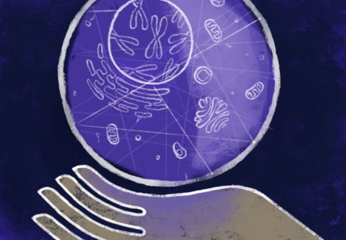
January 16, 2020

Toxoplasma gondii (T. gondii) is a parasite that chronically infects up to a quarter of the world’s population, causing toxoplasmosis, a disease that can be dangerous, or even deadly, for the immunocompromised and for developing fetuses. One reason that T. gondii is so pervasive is that the parasites are tenacious occupants once they have infected a host. They can transition from an acute infection stage into a quiescent life cycle stage and effectively barricade themselves inside of their host’s cells. In this protected state, they become impossible to eliminate, leading to long term infection. Researchers used to think that a combination of genes were involved in triggering the parasite’s transition into its chronic stage, due to the complexity of the process and because a gene essential for differentiation had not been identified. However, new research from Whitehead Institute Member Sebastian Lourido, who is also an assistant professor of biology at the Massachusetts Institute of Technology (MIT), and graduate student Benjamin Waldman has identified a sole gene whose protein product is the master regulator, which is both necessary and sufficient for the parasites to make the switch. Their findings, which appear online in the journal Cell on January 16, illuminate an important aspect of the parasite’s biology and provide researchers with the tools to control whether and when T. gondii transitions, or undergoes differentiation. These tools may prove valuable for treating toxoplasmosis, since preventing the parasites from assuming their chronic form keeps them susceptible to both treatment and elimination by the immune system.

T. gondii spreads when a potential host, which can be any warm-blooded animal, ingests infected tissue from another animal—in the case of humans, by eating undercooked meat or unwashed vegetables—or when the parasite’s progeny are shed by an infected cat, T. gondii’s target host for sexual reproduction. When T. gondii parasites first invade the body, they are in a quickly replicating part of their life cycle, called the tachyzoite stage. Tachyzoites invade a cell, isolate themselves by forming a sealed compartment from the cell’s membrane, and then replicate inside of it until the cell explodes, at which point they move on to another cell to repeat the process. Although the tachyzoite stage is when the parasites do the most damage, it’s also when they are easily targetable by the immune system and medical therapies. In order for the parasites to make their stay more permanent, they must differentiate into bradyzoites, a slow-growing stage, during which they are less susceptible to drugs and have too little effect on the body to trigger the immune system. Bradyzoites construct an extra thick wall to isolate their compartment in the host cell and encyst themselves inside of it. This reservoir of parasites remains dormant and undetectable, until, under favorable conditions, they can spring back into action, attacking their host or spreading to new ones.

Although the common theory was that multiple genes collectively orchestrate the transition from tachyzoite to bradyzoite, Lourido and Waldman suspected that there was instead a single master regulator.

“Differentiation is not something a parasite wants to do halfway, which could leave them vulnerable,” Waldman says. “Multiple genes means more chances for things to go wrong, so you would want a master regulator to ensure that differentiation happens cleanly.”

To investigate this hypothesis, Waldman used CRISPR-based screens to knock out T. gondii genes, and then tested to see if the parasite could still differentiate from tachyzoite to bradyzoite. Waldman monitored whether the parasites were differentiating by developing a strain of T. gondii that fluoresces in its bradyzoite stage. The researchers also performed a first of its kind single-cell RNA sequencing of T. gondii in collaboration with members of Alex Shalek’s lab in the department of chemistry at MIT. This sequencing allowed the researchers to profile the genes’ activity at each stage in unprecedented detail, shedding light on changes in gene expression during the parasite’s cell-cycle progression and differentiation.

The experiments identified one gene, which the researchers named Bradyzoite-Formation Deficient 1 (BFD1), as the only gene both sufficient and necessary to prevent the transition from tachyzoite to bradyzoite: the master regulator. Not only was T. gondii unable to make the transition without the BFD1 protein, but Waldman found that artificially increasing its production induced the parasites to become bradyzoites, even without the usual stress triggers required to cue the switch. This means that the researchers can now control Toxoplasma differentiation in the lab.

These findings may inform research into potential therapies for toxoplasmosis, or even a vaccine.

Toxoplasma that can’t differentiate is a good candidate for a live vaccine, because the immune system can eliminate an acute infection very effectively,” Lourido says.

The researchers’ findings also have implications for food production. T. gondii and other cyst-forming parasites that use BFD1 can infect livestock. Further research into the gene could inform the development of vaccines for farm animals as well as humans.

“Chronic infection is a huge hurdle to curing many parasitic diseases,” Lourido says. “We need to study and figure out how to manipulate the transition from the acute to chronic stages in order to eradicate these diseases.”

This study was supported by an NIH Director’s Early Independence Award (1DP5OD017892), a grant from the Mathers Foundation, the Searle Scholars Program, the Beckman Young Investigator Program, a Sloan Fellowship in Chemistry, the NIH (1DP2GM119419, 2U19AI089992, 5U24AI118672), and the Bill and Melinda Gates Foundation.

Written by Greta Friar

***

Sebastian Lourido’s primary affiliation is with Whitehead Institute for Biomedical Research, where his laboratory is located and all his research is conducted. He is also an assistant professor of biology at the Massachusetts Institute of Technology.

***

Citation:

“Identification of a master regulator of differentiation in Toxoplasma”

Cell, online January 16, DOI: 10.1016/j.cell.2019.12.013

Benjamin S. Waldman (1,2), Dominic Schwarz (1,3), Marc H. Wadsworth II (4,5,6), Jeroen P. Saeij (7), Alex K. Shalek (4,5,6), Sebastian Lourido (1,2)

1. Whitehead Institute for Biomedical Research, Cambridge, MA 02142, USA

2. Department of Biology, Massachusetts Institute of Technology, Cambridge, MA 02142, USA

3. Institute of Pharmacy and Molecular Biotechnology, University of Heidelberg, Im Neuenheimer Feld 364, 69120 Heidelberg, Germany

4. Institute for Medical Engineering & Science (IMES), Department of Chemistry, and Koch Institute for Integrative Cancer Research, Massachusetts Institute of Technology, Cambridge, MA 02139, USA.

5. Broad Institute of MIT and Harvard, Cambridge, MA 02142, USA

6. Ragon Institute of MGH, MIT and Harvard, Cambridge, MA 02319, USA

7. Department of Pathology, Microbiology and Immunology, School of Veterinary Medicine, University of California, Davis, Davis, CA 95616, USA

With these neurons, extinguishing fear is its own reward
Picower Institute
January 16, 2020

When you expect a really bad experience to happen and then it doesn’t, it’s a distinctly positive feeling. A new study of fear extinction training in mice may suggest why: The findings not only identify the exact population of brain cells that are key for learning not to feel afraid anymore, but also show these neurons are the same ones that help encode feelings of reward.

The study, published Jan. 14 in Neuron by scientists at MIT’s Picower Institute for Learning and Memory, specifically shows that fear extinction memories and feelings of reward alike are stored by neurons that express the gene Ppp1r1b in the posterior of the basolateral amygdala (pBLA), a region known to assign associations of aversive or rewarding feelings, or “valence,” with memories. The study was conducted by Xiangyu Zhang, a graduate student, Joshua Kim, a former graduate student, and Susumu Tonegawa, Professor of Biology and Neuroscience at RIKEN-MIT Laboratory of Neural Circuit Genetics at the Picower Institute for Learning and Memory at MIT and Howard Hughes Medical Institute.

“We constantly live at the balance of positive and negative emotion,” Tonegawa said. “We need to have very strong memories of dangerous circumstances in order to avoid similar circumstances to recur. But if we are constantly feeling threatened we can become depressed. You need a way to bring your emotional state back to something more positive.”

Overriding fear with reward

In a prior study, Kim showed that Ppp1r1b-expressing neurons encode rewarding valence and compete with distinct Rspo2-expressing neurons in the BLA that encode negative valence. In the new study, Zhang, Kim and Tonegawa set out to determine whether this competitive balance also underlies fear and its extinction.

In fear extinction, an original fearful memory is thought to be essentially overwritten by a new memory that is not fearful. In the study, for instance, mice were exposed to little shocks in a chamber, making them freeze due to the formation of fearful memory. But the next day, when the mice were returned to the same chamber for a longer period of time without any further little shocks, freezing gradually dissipated and hence this treatment is called fear extinction training. The fundamental question then is whether the fearful memory is lost or just suppressed by the formation of a new memory during the fear extinction training.

While the mice underwent fear extinction training the scientists watched the activity of the different neural populations in the BLA. They saw that Ppp1r1b cells were more active and Rspo2 cells were less active in mice that experienced fear extinction. They also saw that while Rspo2 cells were mostly activated by the shocks and were inhibited during fear extinction, Ppp1r1b cells were mostly active during extinction memory training and retrieval, but were inhibited during the shocks.

These and other experiments suggested to the authors that the hypothetical fear extinction memory may be formed in the Ppp1r1b neuronal population and the team went on to demonstrate this vigorously. For this, they employed the technique previously pioneered in their lab for the identification and manipulation of the neuronal population that holds specific memory information, memory “engram” cells.  Zhang labeled Ppp1r1b neurons that were activated during retrieval of fear extinction memory with the light-sensitive protein channelrhodopsin. When these neurons were activated by blue laser light during a second round of fear extinction training it enhanced and accelerated the extinction. Moreover, when the engram cells were inhibited by another optogenetic technique, fear extinction was impaired because the Ppp1r1b engram neurons could no longer suppress the Rspo2 fear neurons. That allowed the fear memory to regain primacy.

These data met the fundamental criteria for the existence of engram cells for fear extinction memory within the pBLA Ppp1r1b cell population: activation and reactivation by recall and enduring and off-line maintenance of the acquired extinction memory.

Because Kim had previously shown Ppp1r1b neurons are activated by rewards and drive appetitive behavior and memory, the team sequentially tracked Ppp1r1b cell activity in mice that eagerly received water reward followed by food reward followed by fear extinction training and fear extinction memory retrieval. The overlap of Ppp1r1b neurons activated by fear extinction vs. water reward was as high as the overlap of neurons activated by water vs. food reward. And finally, artificial optogenetic activation of Ppp1r1b extinction memory engram cells was as effective as optogenetic activation of Ppp1r1b water reward-activated neurons in driving appetitive behaviors. Reciprocally, artificial optogenetic activation of water-responding Ppp1r1b neurons enhanced fear extinction training as efficiently as optogenetic activation of fear extinction memory engram cells. These results demonstrate that fear extinction is equivalent to bona fide rewards and therefore provide the neuroscientific basis for the widely held experience in daily life: omission of expected punishment is a reward.

What next?

By establishing this intimate connection between fear extinction and reward and by identifying a genetically defined neuronal population (Ppp1r1b) that plays a crucial role in fear extinction this study provides potential therapeutic targets for treating fear disorders like PTSD and anxiety, Zhang said.

From the basic scientific point of view, Tonegawa said, how fear extinction training specifically activates Ppp1r1b neurons would be an important question to address. More imaginatively, results showing how Ppp1r1b neurons override Rspo2 neurons in fear extinction raises an intriguing question about whether a reciprocal dynamic might also occur in the brain and behavior. Investigating “joy extinction” via these mechanisms might be an interesting research topic.

The research was supported by the RIKEN Brain Science Institute, the Howard Hughes Medical Institute and the JPB Foundation funded the research.