Placement: Promote to Homepage
Picower Institute
June 29, 2020
Scientific discoveries can sometimes seem like products on a store shelf. Packaged neatly in the wrapping of a journal article or maybe a news story, there remain few hints of what they really took to produce – the struggle and surprises, the ingenuity and serendipity, the toil and triumph. Perhaps it’s no wonder that many members of the public (7 in 10, by one National Science Foundation measure) feel at least somewhat unclear about how scientists know what they know or do what they do.
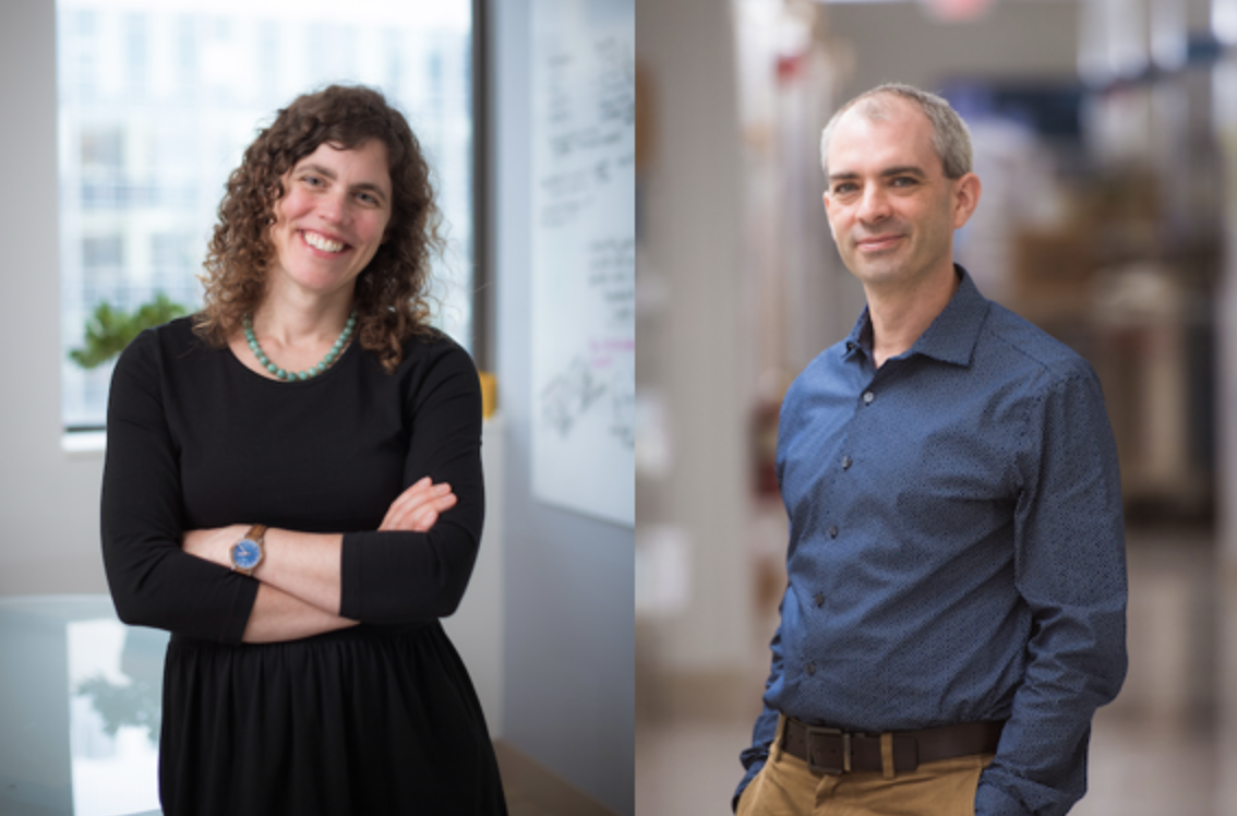
To deconstruct and perhaps demystify discovery, let’s unwrap the inside story of a paper published by the lab of Troy Littleton, Menicon Professor of Neuroscience in the Departments of Biology and Brain and Cognitive Sciences. The study reported important findings both about a possible mechanism of seizures in epilepsy, which affects 60 million people worldwide, and also the underappreciated relationship between neurons and the brain cells called glia that help them function. Through four years of work led by former postdoc Shirley Weiss, Littleton’s team thoroughly unraveled the complex breakdown that makes fruit flies with a genetic mutation prone to seizures and showed multiple ways to intervene, including with human medicines. Published in April 2019 in eLife, a far-reaching “open access” journal that is free for all to read, the paper has since been viewed thousands of times.
Figuring out the puzzle of exactly how the mutation made glial cells fail to prevent seizures was a source of particular excitement for Weiss. Littleton adds that the discovery could open up new strategies for developing drugs to address epilepsy in humans, which the flies model well.
“One of the long-term motivations for the field in general, not just our lab, is there might be pharmacological access to glial cells that might have less side effects than would happen if you target neurons directly,” he said.
Too much excitement
First, a little biological background. Neurons are electrical. Their participation in the brain circuits that guide behavior, emotion, reasoning and memory depends on how they build up or dissipate electrical charge by taking in or ejecting ions of calcium, potassium and sodium. If they remain too electrically charged up because of an imbalance of these ions, they can become hyperactive and, in groups, produce seizures. In this study, it turned out that a certain kind of glial cells were responsible for regulating the balance of potassium ions around their neuronal neighbors to help govern their electrical charge and activity. The mutation, Weiss discovered, caused the glia to leave too much potassium outside the neurons, making it harder for the neurons to get rid of the potassium they had built up inside when they were electrically active. Without the ability to get their potassium out, the neurons stayed too excited, producing seizures.
The lab first discovered the mutation, which the Louisianan Littleton named “zydeco,” in 2005 when a team led by Zhuo Guan used genetic screening techniques Littleton learned when he was a postdoc in the 1990s at the University of Wisconsin. The team’s broader goal was to learn about how neurons communicate with each other, so they looked for flies with mutations that either shut the process down, leading to a readily observable symptom of paralysis, or amped it way up, leading to seizures. Zydeco fell into that second category, making fruit flies seize dramatically when stressed by heat or by getting jostled around.
“It was so striking, it was hard to ignore,” Littleton said. “Whatever this gene was, it was doing something very important in the brain.”
When Weiss joined the Littleton lab after earning her PhD at Hebrew University in Jerusalem in 2013, the lab had just published a new paper about zydeco. It was a long-awaited follow-up. Zydeco disrupted a gene on the fly’s X chromosome which at the time was poorly understood. Led by former graduate student Jan Melom, the lab finally was able to clone the gene that was mutated in Zydeco and showed that it specifically affected “cortex glial” cells and that it caused them to retain too much calcium. But what remained completely unclear was how this made the neurons that those glia contact so susceptible to seizures.
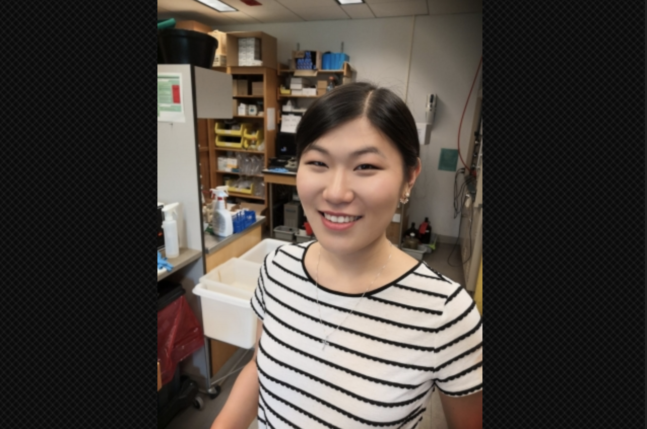
Though Weiss had a research specialty in studying calcium in brain cells, at first she worked on a few other ideas. But because Melom had left the lab, Weiss soon picked up the zydeco baton. In so doing, she was taking on what would become an especially extensive effort involving scores of experiments and a vast array of techniques, some of which she would have to learn along the way.
No hypothesis needed or heeded
Based on the 2013 findings, Littleton had formulated a working hypothesis about what might be going on to cause the seizures. He figured the excess of calcium in the cortex glia probably caused them to emit too much of some kind of signal to the neurons, in turn causing them to remain too active.
That turned out to be wrong, Littleton acknowledges with a smile.
“What Shirley did was to disprove my very strong impression of what was actually happening,” Littleton said. “Sometimes that’s very difficult to do. Once you have an idea of how you think the biology is working, that can reinforce the sorts of experiments you do and affects how you think about the project. It was very exciting in the end that Shirley was able to get past my pre-conceived notions and figure out what was really happening.”
The team didn’t fall into that trap because the experimental approach they chose didn’t depend on what they thought. Weiss’s key initial inquiries were based on a wide-open, free-ranging manipulation of the zydeco flies’ genes. Her strategy was to “knock down” or interfere with the cell’s ability to make use of 847 different genes covering a wide variety of potentially relevant glial cell functions. If knocking down any particular gene stopped the seizures, that would give them a huge clue about how the seizures happen. And whatever worked, if anything, would work regardless of anyone’s guesses up front.
“The great thing about using forward genetics is you don’t have to have a very strong hypothesis,” Weiss said. “You can let the genetics lead the way. I tried to be hypothesis free and to be as unbiased as I could be.”
The knockdown screening yielded about 50 genes where interference totally or partially alleviated the seizures. One in particular squared well with what Melom had observed about a specific cellular process (scientists call it a “pathway”)that related to handling calcium.
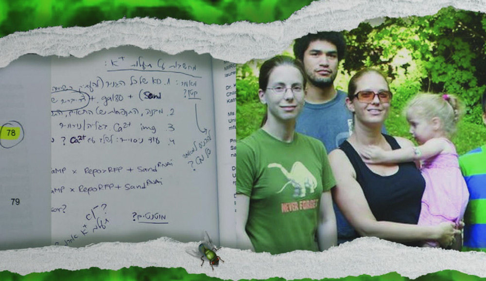
Around that time, though, life outside the lab intervened. In February 2015 Weiss and her husband Kfir Sharabi, also a postdoc, and their then four-and-a-half year old daughter, Amit, welcomed their second daughter, Ma’ayan, to the world. With two young kids and the rest of her family in Israel, Weiss came back from maternity leave and got back to investigating the most promising hits of the knockdown screen.
A calcium conundrum
The particular hit related to calcium that caught Weiss’s eye was a gene was called CanB2. Zydeco flies with that gene knocked down experienced no more seizure troubles at all. Moreover, she found that it was specifically helpful to knock it down in cortex glia and that knocking it down in healthy flies didn’t do any discernable harm.
So what does CanB2 do? In general the gene, along with two others, make a protein called calcineurin. No one had ever characterized what calcineurin does in glia. If Weiss could become the first to figure that out, she could whatever problems the zydeco mutation causes.
By manipulating all three calcineurin genes, Weiss was able to confirm that calcineurin activity was indeed crucial for zydeco seizures. She engineered cortex glia so that a glowing green protein would start to be expressed when calcineurin was active. She could see the protein light up under the microscope. This told her there was a lot more calcineurin activity in zydeco mutant brains than in normal fly brains. Apparently, the excess calcium in the cortex glia correlated with increased calcineurin activity.
There are human medicines that ratchet back calcineurin activity. They are typically used to suppress the immune system after a transplant. Weiss wanted to see whether they could reduce seizures in the zydeco flies. When she fed them the drugs the seizures did subside, providing a clear demonstration that intervening in this glial pathway could hold promise for drug development.
Any such effort, to be truly well targeted, would require more than just an association between calcineurin and seizures. Weiss was determined to discover the mechanism that linked the two.
Figuring out what that mechanism was and how it led to seizures, would turn out to be the heart of the discovery and the most challenging phase of her four year endeavor.
A potassium epiphany
Weiss needed to find out what process this excess calcineurin activity might be putting into overdrive. She went back to genetics. She performed a screen to knock down direct targets of calcineurin. It didn’t appear to yield anything helpful. She did another screen of pathways where calcineurin was implicated. In that case, a process called “endocytosis” came up and sure enough, Weiss found that by inhibiting the process in the cortex glia she could again stop seizures in the zydeco flies. Endocytosis is how cells ingest material from their surroundings, including regulating the content of their cell surface membrane proteins. The process can therefore affect the proteins they employ on the membrane to interact with the environment outside the cell. Excess endocytosis could mean that the way by which cortex glia interact with their environment is altered in zydeco, perhaps affecting the neurons they support. But how might that matter in this case?
Weiss struggled with this question for months. She received feedback and advice in meetings with Littleton and in lab meetings where the members discuss, challenge and refine ideas. The discussions were helpful, but it turns out that the key breakthrough came from Weiss attending a conference in Cold Spring Harbor, N.Y. in July, 2018.
Among Weiss’s genetic screen results of calcineurin targets was a gene called “SAND” that makes a protein in flies called “sandman” (the human version is called TRESK). Sandman, when deployed to the cell membrane, forms a channel (picture a portal though the cell’s surface) that allows a cell to bring in potassium ions from outside. At first this result didn’t strike Weiss as all that notable, but at the conference, potassium channels kept coming up as a topic in talks. An idea started to percolate as she took notes. Then at the conference posters she started talking with a scientist who said that problems with potassium channels in glial cells have been linked to epilepsy. Potassium channels apparently merited another look.

“I already had the result,” Weiss said. “I just didn’t connect the two dots.”
One of her screens indeed showed that knocking down SAND in healthy flies caused seizures just like the ones seen in zydeco mutants. Further genetic manipulations confirmed that SAND knockdown and zydeco affected the same pathway in cortex glia cells.
By September 2018, a new hypothesis was emerging: Elevated calcium in cortex glia triggered excess calcineurin activity, which spurred increased endocytosis that hindered sandman’s intake of potassium. This came at a good time as Weiss was nearing the point where she had to start thinking of wrapping up her postdoctoral appointment at MIT. The hypothesis, and the evidence she’d built up, seemed enough to submit a paper to a journal.
Always mindful that a paper was the goal, Weiss had been writing as she went and developing the key figures. When she had a draft done, Littleton then set to polishing it and giving her feedback. eLife wasn’t the first journal they submitted the paper to, but the editors received it enthusiastically. All three of the scientists who reviewed the manuscript for eLife, however, said the same thing: If endocytosis was pulling sandman back from the membrane of the cortex glia, thereby disrupting its ability to take in potassium ions, they wanted to see it happening. Weiss and Littleton not only agreed with that critique, they had even anticipated it.
“You sort of know your own holes in the story,” Littleton said. “This is what we were planning to do next anyway.”
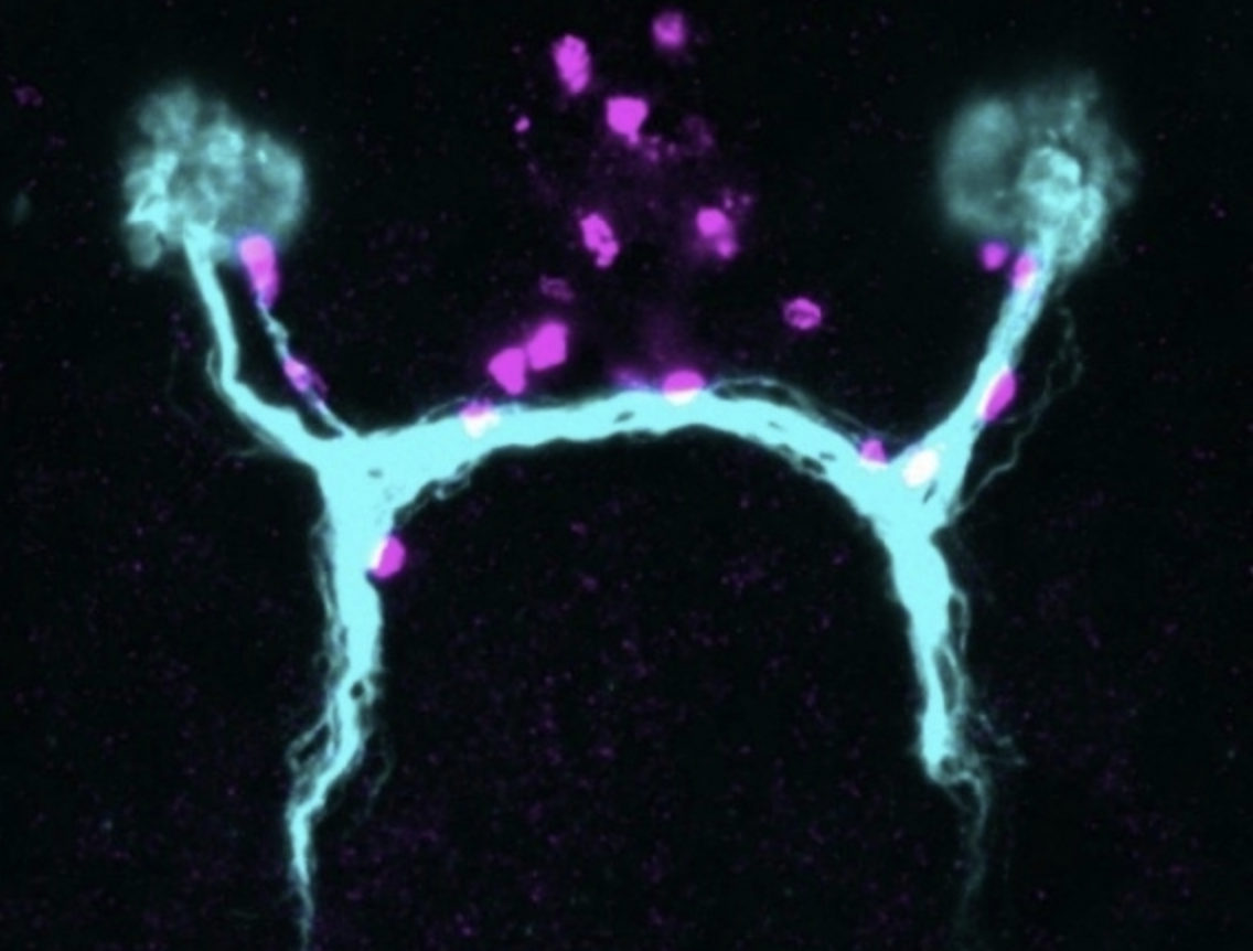
Since sending in the paper, Weiss had already produced those smoking gun images, showing that in zydeco cortex glia, sandman was much less abundant on the membranes than in the non-mutant flies. This cemented the argument, neatly wrapped up in the paper, that neurons become more susceptible to seizing when zydeco cortex glia, saddled with too much calcium and resulting calcineurin activity, overdo the endocytosis of sandman potassium channels, leaving too much potassium outside of neurons, causing increased excitability and the onset of seizures.
Since publication, the paper has garnered some mentions in the scientific press. It has also earned a new National Institutes of Health grant for Littleton’s lab, where they are following in Melom’s and Weiss’ footsteps to study how calcium levels in glia affect the flux of membrane proteins, not just in disease, but as a matter of course in healthy cells. And for Weiss, the paper impressed funders in Israel, providing her with the money to support her new position where she continues studying glia, calcium and seizures.
It was a hard-earned success. Though their end product is knowledge, scientists spend the vast majority of their time with the unknown. Between the lines of most every paper are years of effort in which scientists persistently asked open questions with open minds so that the evidence could lead them to a discovery they could share with the world.
Whitehead Institute
June 26, 2020
At the heart of Whitehead Institute are its Members, a group of world-class investigators who propel science forward through their research discoveries and technical advances. Recently, Whitehead Institute singled out two Members for special recognition: appointment to endowed professorships, which provide financial support for their research programs and serve as an endorsement of their scientific visions.
Whitehead Institute Member Mary Gehring, who is also an associate professor of Biology at Massachusetts Institute of Technology (MIT), has been appointed to the Landon T. Clay Career Development Chair. “Mary is an emerging superstar in plant biology and a respected leader within the Whitehead Institute community,” says Institute director David C. Page. “I know that Landon held her in high regard, and I think he would be very pleased that Mary is this chair’s first incumbent.”
Gehring recalls, “I met Landon on my very first day at Whitehead Institute. His curiosity, insight, and wide-ranging knowledge made that initial conversation absorbing — as was every subsequent discussion we had. His impact as an Institute board member was formidable, and I will hold the Clay Career Development Chair with pride.”
Gehring’s research focuses on plant epigenetics — the heritable information that influences cellular function but is not encoded in the DNA sequence itself. Primarily using the model plant Arabidopsis thaliana, Gehring has determined that altering the methylation state of a single gene is sufficient to cause changes in seed weight and in the timing of certain aspects of seed development. Methylation patterns can be passed from one cell generation to the next and from one plant to its offspring. By studying the epigenetic difference between multiple generations of plants, Gehring seeks to learn if epigenetic responses to environmental factors can ultimately lead to evolutionary changes. Her work has tremendous implications for addressing food security in a period of significant climate change.
Institute Member Iain Cheeseman has been appointed to the Margaret and Herman Sokol Chair in Biomedical Research. He succeeds Member and former director Gerald Fink, who held the chair since it was established in 2006. “Iain is an extraordinary scientist, a dedicated teacher, and a wonderful colleague,” says Page. “He is, therefore, the perfect person to succeed Gerry in the Sokol Chair.”
“This is a great honor,” says Cheeseman, who is also a professor of Biology at MIT. “I am humbled to hold a chair named for Margaret and Herman Sokol, who were among Whitehead Institute’s earliest and most ardent supporters; and a chair that has so long been associated with Gerry Fink.”
Cheeseman’s research focuses on the kinetochore — a central player in directing chromosome segregation — which comprises more than 100 different proteins. Although the kinetochore’s importance has long been appreciated, the molecular basis for its many activities remains poorly understood. His lab has helped identify dozens of the kinetochore’s molecular components and their specific roles, and is defining how the attachments between kinetochores and spindle microtubules are regulated throughout cell division. Because many cancers may be driven by errors in chromosome segregation, Cheeseman’s studies may inform cancer research — and may contribute to development of more effective treatments for leukemia and other diseases.
Earlier this spring, Whitehead Institute announced appointments to two other endowed chairs.
Cell biologist Jonathan Weissman, who recently became a Member, is the inaugural incumbent of the Landon T. Clay Chair of Biology at Whitehead Institute. And in September 2020, developmental biologist Yukiko Yamashita will join the Institute and become the inaugural incumbent of the Susan Lindquist Chair for Women in Science at Whitehead Institute.
Eva Frederick | Whitehead Institute
June 25, 2020
If anything happens to the eyes of the tiny, freshwater-dwelling planarian Schmidtea mediterranea, they can grow them back within just a few days. How they do this is a scientific conundrum — one that Peter Reddien’s lab at Whitehead Institute has been studying for years.
The lab’s latest project offers some insight: in a paper published in Science June 25, researchers in Reddien’s lab have identified a new type of cell that likely serves as a guidepost to help route axons from the eyes to the brain as the worms complete the difficult task of regrowing their neural circuitry.
Schmidtea mediterranea’s eyes are composed of light-capturing photoreceptor neurons connected to the brain with long, spindly processes called axons. They use their eyes to respond to light to help navigate their environment.
The worms, which are popular models for research into regeneration, can regrow pretty much any part of their body; eyes are an interesting part to study because regenerating the visual system requires the worms rewire their neurons to connect them to the brain.
When neural systems develop in embryos, the first nerve fibers, called pioneer axons, snake their way through tissue to form the circuitry needed to perceive and interpret external stimuli. The axons are helped along their way by specialized cells called guidepost cells. These special cells are positioned at choice points — places where the axon’s path could fork in different directions.
In many organisms, these guidepost cells aren’t a priority anymore once development is finished, and typically are not renewed through adulthood. That’s one reason why, when humans experience brain or nerve damage, the injury is usually permanent.
“This is a fundamental mystery of regeneration that we hadn’t even been thinking about,” says Reddien, the senior author of the paper who is also a professor of biology at Massachusetts Institute of Technology and an investigator with the Howard Hughes Medical Institute. “How can an adult animal regenerate a functional nervous system when the original development of the nervous system typically involves a number of cues that are thought to be transient?”
Then, in 2018, Reddien Lab scientist Lucila Scimone found something surprising in adult planarians: groups of mysterious cells that looked like they might play a role in guiding growing axons. She’d noticed this group of cells because they co-expressed two genes not often seen together and some were conspicuously close to the eyes.
“I was captivated by these cells,” she says. They appeared in very small numbers (a normal worm might have around 5; a large one might have up to 10) in every planarian she examined. They were divided into two distinct groups: some around the flatworms’ eyes, and others spaced out along the path to the brain center. When she traced the path of existing axons leading from the planarians’ eyes to their brain, they coincided with the positions of these cells without exception.
When the researchers characterized the cells, they found that they did not express any of the genes that are hallmarks of photoreceptor neurons; instead, they had markers often found in muscle tissue. “That was very striking, because muscle cells — that’s not what they do in most animals,” Scimone says.
In other organisms, guidepost cells are often neurons or glia. It would be unusual for muscle cells to serve as guideposts; but past work in the Reddien Lab had shown that planarian muscle cells played other special roles, such as secreting the extracellular matrix. The researchers now wondered whether they could add the role of guidepost to the long list of planarian muscle cell functions.
To test their hypothesis, the researchers designed a series of experiments. “We developed an eye transplantation method where you can take an eye from an animal and transplant it into another animal,” says Reddien Lab postdoc Kutay Deniz Atabay. “When you do this, the axonal projections from that eye will basically, if positioned appropriately, correctly wire themselves into the brain, producing a functional state.”
The researchers also created genetically engineered planarians that had the muscle cells, but no eyes, and then transplanted eyes onto their eyeless heads. Sure enough, the neurons grew as normal, snaking towards the cells and then adjusting their trajectories after encountering them.
Without the cells, it was a different story. When the researchers transplanted eyes to distant parts of planarians’ bodies without a population of these muscle cells, the photoreceptor neurons did not connect to the brain center. Likewise, when they transplanted eyes into planarians that had been modified to not have these muscle cells, their photoreceptor neurons still grew — but they did not wire properly to reach the brain.
These findings combined suggested that the cells were fully independent of the visual system — they did not form because of eyes or photoreceptor neurons, but likely established themselves before the neurons grew — which provided more evidence for the guidepost role.
The guidepost-like activity of these cells then begged the question: how do the cells themselves know where to be? “We found that there’s a pattern of signaling molecules in muscle that is setting where these cells should be,” Reddien says. “If we perturb the global positional information of the system, these cells get placed in the wrong positions, and then axons go to the wrong positions — so we think there’s a positional information framework that places the cells during regeneration, and that allows them to work as guideposts in the correct locations.”
At this point, the researchers don’t know exactly how the cells are able to communicate with growing axons to serve as guideposts. They could be releasing some sort of signaling molecule that attracts the axons, or they could be communicating by using trans-membrane proteins.
“That will be an exciting direction for the future,” Reddien says. “We have now identified the transcriptome for the cells, which means we know all the genes that these cells express. That provides us with an intriguing list of genes that can be probed functionally, to try to see which ones are mediating the functions of these cells.”
This study is a step forward in a body of work that aims to expand the capabilities of regenerative medicine. “Imagine a scenario where someone experiences a spinal cord injury or an eye injury or stroke that leads to the loss of a neural circuit,” says Atabay. “The reason we can’t fully cure these cases today is that we lack fundamental information regarding how these systems can regenerate. Looking at regenerative organisms provides a lot of insights. From this case, we see that regenerating the lost system may not be enough; you may also need to regenerate systems that are properly patterning that system.”
***
Written by Eva Frederick
***
Scimone, M. L. et al. “Muscle and neuronal guidepost-like cells facilitate planarian visual system regeneration.” Science, June 25, 2020.
Whitehead Institute
June 17, 2020
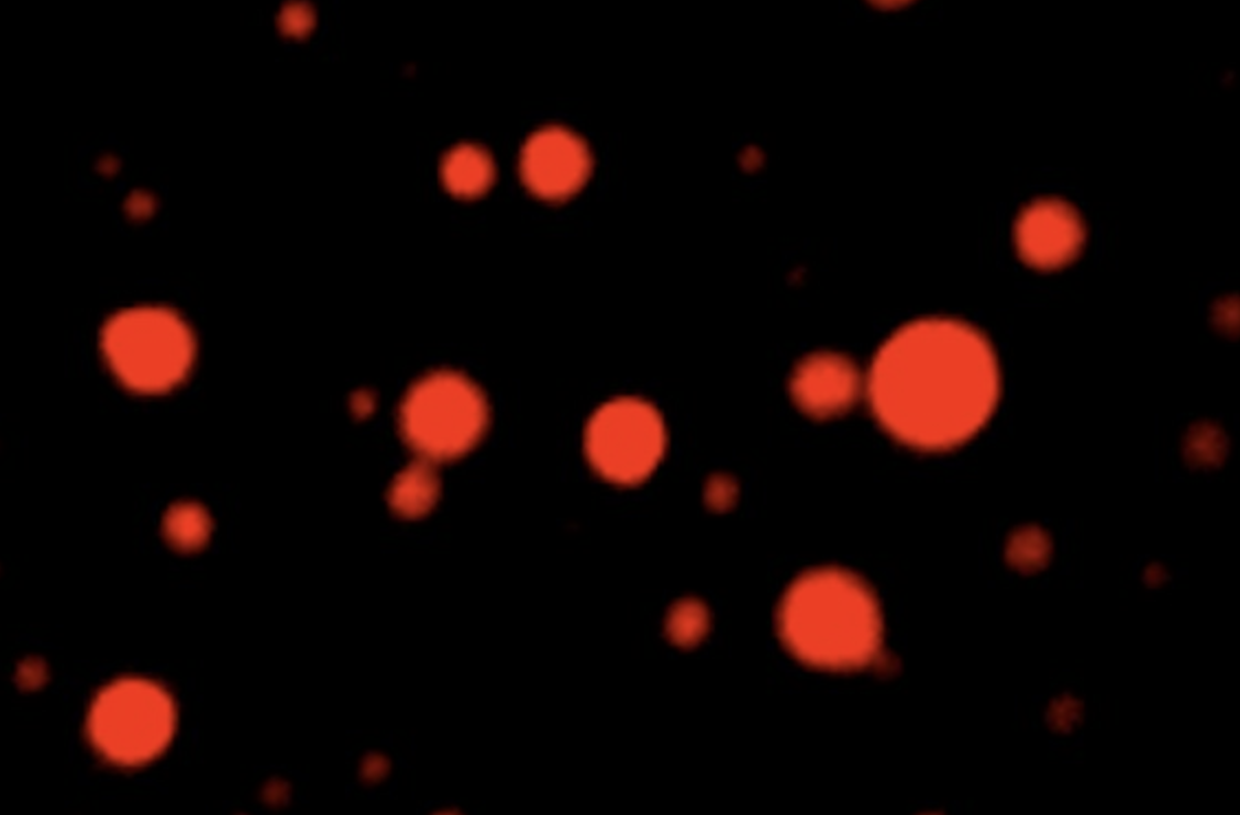
In the watery inside of a cell, complex processes take place in tiny functional compartments called organelles. Energy-producing mitochondria are organelles, as is the frilly golgi apparatus, which helps to transport cellular materials. Both of these compartments are bound by thin membranes.
But in the past few years, research at Whitehead Institute and elsewhere has shown that there are other cellular organelles held together without a membrane. These organelles, called condensates, are tiny droplets which keep certain proteins close together amidst the chaos of the cell, allowing complex functions to take place within. “We know of about 20 types of condensate in the cell so far,” says Isaac Klein, a postdoc in Richard Young’s lab at Whitehead Institute and oncologist at the Dana-Farber Cancer Institute.
Now, in a paper published in Science on June 19, Klein and Ann Boija, another postdoc in Young’s lab, show the mechanism by which small molecules, including cancer drugs, are concentrated in these cellular droplets — a finding that could have implications for the development of new cancer therapeutics. If researchers could tailor a chemical to seek out and concentrate in one kind of droplet in particular, it might have a positive effect on the delivery efficiency of the drug. “We thought, maybe that’s an avenue by which we can improve cancer treatments and discover new ones,” says Klein.
“This [research] is part of a revolutionary new way of looking at the organization within cells,” says Phillip Sharp, a professor at the Massachusetts Institute of Technology’s Koch Institute for Integrative Cancer Research and a co-author on the study. “Cells are not little pools of soup, all mixed together. They are actually highly organized, compartmentalized units, and that organization is important in their function and in their diseases. We’ve just started to understand that, and this new paper is a really important step, using that insight, to understand how to potentially treat diseases differently.”
CONDENSATES AND DRUG DELIVERY
To explore how different properties of condensates inside the cell’s nucleus affected the delivery of cancer drugs, Boija and Klein selected a few example condensates to study. These included splicing speckles, which store cellular materials needed for RNA splicing, nucleoli, where ribosomes are formed, and a new kind of droplet Young’s lab discovered in 2018 called a transcriptional condensate. These new condensates bring together all the different proteins needed to successfully transcribe a gene.
The researchers created their own suite of four different fluorescently-labeled condensates by adding glowing tags to marker proteins specific to each kind of droplet. For example, transcriptional condensates are marked by the droplet-forming protein MED1, splicing speckles by a protein called SRSF2, and nucleoli by FIB1 and NPM1.
Now that they could tell individual droplets apart by their cellular purpose, the team, along with the help of Nathanael Gray, a chemical biologist at Harvard University and the Dana-Farber Cancer Institute, created fluorescent versions of clinically important drugs. The tested drugs included cisplatin and mitoxantrone, two anti-tumor medicines commonly used in chemotherapy. These therapeutics were the perfect test subjects, because they both target proteins that lie within nuclear condensates.
The researchers added the cancer drugs to a mixture containing various droplets (and only droplets, none of the actual drug targets), and found that the drugs sorted themselves into specific condensates. Mitoxantrone concentrated in condensates marked by MED1, FIB1 and NPM1, selectively avoiding the others. Cisplatin, too, showed a particular affinity for droplets held together by MED1.
“The big discovery with these in vitro studies is that a drug can concentrate within transcriptional condensate independent of its target,” Boija says. “We used to think that drugs come to the right place because their targets are there, but in our in vitro system, the target is not there. That’s really informative — it shows the drug is actually being concentrated in a different way than we thought.”
To understand why some drugs were drawn into transcriptional condensates, they screened a panel of chemically-modified dyes and found that the important part of many drugs — the part that led them to concentrate in transcriptional condensates — is the molecules’ aromatic ring structure. Aromatic rings are stable, ring-shaped groupings of carbon atoms. The aromatic ring in some drugs are thought to stack with rings in MED1’s amino acids, leading the drug to concentrate in transcriptional condensates.
Being able to tailor a drug to enter a certain condensate is a powerful tool for drug developers. “We found that if we add an aromatic group to a molecule, it becomes concentrated within the transcriptional condensate,” Boija says. “It’s that type of interaction that is important when we design new drugs to enter transcriptional condensates — and maybe we can improve existing drugs by modifying their structure. This will be very exciting to look into.”
WHERE DRUGS CONCENTRATE AFFECTS HOW WELL THEY FIGHT CANCER
In order for this tool to be practically useful in drug development, the researchers had to make sure that concentration in specific droplets would actually impact the drugs’ performance. Boija and Klein decided to test this using cisplatin, which is drawn to transcriptional condensates by MED1 and works to fight cancer by adding clunky platinum molecules to DNA strands. This damages tumor cells’ genetic material. When the researchers administered cisplatin to a mixture of different condensates, both in the test tube and in cells, the drug preferentially altered DNA that lay within transcriptional condensates.
This could explain why cisplatin and other platinum drugs are effective against so many diverse cancers, says Young, who is also a professor of biology at MIT; cancer-causing genes often carry regions of DNA called super enhancers, which are extremely active in transcription, leading to very large transcriptional condensates. “We now think the reason that drugs like cisplatin can work well in patients with diverse cancers is because they’re becoming selectively concentrated at the cancer-causing genes, where these large transcriptional condensates occur,” he said. “The effect is to have the drug home in on the gene that’s causing each cancer to be so deadly.”
A DRUG RESISTANCE MYSTERY, SOLVED
The new insights in condensate behavior also provided some answers to another question in cancer research: why people become immune to the breast cancer drug tamoxifen.Tamoxifen works by attaching itself to estrogen receptors in the cancer cells, preventing them from getting the hormones they need to grow and eventually slowing or stopping the formation of new cancer cells altogether. The drug is one of the most effective treatments for the disease, reducing recurrence rates for ER+ breast cancers by around 50%.
Unfortunately, many patients quickly develop a resistance to tamoxifen — sometimes as soon as a few months after they start taking it. This happens in a variety of ways — for example, sometimes the cancer cells will mutate to be able to kick the tamoxifen out of the cells, or simply produce fewer estrogen receptors for the drug to bind. One form of resistance was associated with an overproduction of the protein MED1, but scientists didn’t know why.
With their newfound knowledge of how a drug’s activity is affected by where it concentrates, Boija and Klein had a hypothesis: the extra MED1 might increase the size of the droplets, effectively diluting the concentration of tamoxifen and making it more difficult for the drug to bind its targets. When they tested this in the laboratory, the team found that more MED1 did indeed cause larger droplets, leading to lower concentrations of tamoxifen.
A NEW TOOLSET FOR DRUG DESIGNERS
The ability to better understand the behavior of drugs in cancer cells — how they concentrate, and why the cancer could become resistant to them — may provide drug developers with a new arsenal of tools to craft efficient therapeutics. “This study suggests that we should be exploring whether we can design or isolate drugs that are concentrated in a given condensate, and to understand how existing drugs are concentrated in the cell,” says Phil Sharp. “I think this is really important for drug development — and I think [figuring it out] is going to be fun.”

In honor of #ShutDownSTEM, students, faculty, and staff facilitated virtual discussions to understand and combat anti-Black racism in academia.
June 18, 2020
On June 10, as part of the #ShutDownSTEM, #ShutDownAcademia, and #Strike4BlackLives national initiative, members of the Department of Biology took the day to engage in open conversations about racial bias, diversity, and inclusion.
The #ShutDownSTEM.MITbio program, organized by trainees, postdocs, and staff, included 13 virtual sessions on topics ranging from allyship and white privilege to antiblackness in Boston and the history of racism in science. The goal was to provide a space for white and non-Black People of Color (POC) to educate themselves and offer support to Black colleagues, as well as determine ways to make the Biology community more equitable.
In a letter to the department publicizing the June 10 event, the organizers wrote: “We have a responsibility as scientists to educate ourselves and initiate and continue difficult but necessary conversations on race and how systemic racism impacts ourselves and our field, particularly through the lens of recent events and how we can better support, amplify, and listen to our Black community members within the department and within our larger communities.”
Although the event came together in just a few days, more than 45 community members volunteered to help facilitate — and over 200 participated in concurrent sessions at any given time throughout the day.
Graduate student Talya Levitz heard about the #ShutDownSTEM initiative through various student activism channels a week prior. She brought the idea to department affinity groups, including the Biology Diversity Community (BDC), and ultimately aggregated over nine co-organizers. Other departments, labs, and centers across MIT developed their own initiatives, and Levitz’s team worked closely with their counterparts in the Department of Chemistry to share resources.
When they built the day’s agenda, Levitz says they had two main goals. “First, we wanted people to think about how their own identities intersect with anti-Blackness and anti-racism efforts,” she says. “The other big goal was to meet people where they are, and recognize that everyone is at a different place on their personal growth trajectories.”
Meghann Kasal, graduate student and co-founder of the BDC, responded to Levitz’s call to action immediately. “The #ShutDownSTEM program seemed like a great way to continue conversations that the BDC was already having, and transform dialogue into action,” she says. “It was a chance to empower people to make changes on an individual level and have those personal commitments ripple out to the larger community.”
SaRa Kim, administrative assistant and research technician, joined Levitz, Kasal, and others to help encourage other staff members to get involved. “The onus to make changes shouldn’t fall solely on those experiencing injustices,” she says, “and many of the co-organizers already had an active network of peers ready to provide support.”
Before the event, the team sent out a list of relevant resources, and afterwards they collated a docket of action items to ensure that the conversation would continue — especially regarding recruiting and retaining Black and non-Black POC graduate students, staff, and faculty. Plans are also coalescing to apply for a Quality of Life grant to sponsor similar programs in the future, and students have spearheaded a faculty-matched donation drive within the department.
Graduate student and co-organizer, Gerardo Perez Goncalves, aims to take the day’s discussions and turn them into tangible plans with concrete timelines. “We need to hold each other accountable, and make sure those goals don’t get lost in committees,” he says. “Even though I’m just one person, I can be involved in a number of different ways, such as helping to craft actionable plans to spread awareness of current initiatives to those near me. The whole department needs to be made aware of these initiatives and plans so that we can establish community accountability.”
Sora Kim, a fellow graduate student and co-organizer, adds that scientists are often expected to separate their personal lives from their work. “You’re not supposed to bring what you personally think into the workplace,” Kim says, “but we know from history and current events that these things bleed into one another, and not talking about them creates a culture of silence and isolation.”
In the past, students have voiced concerns via anonymous polls and surveys, but there have been few opportunities for the entire community to come together, acknowledge current issues, and brainstorm solutions collaboratively.
The #ShutDownSTEM.MITbio event marked the beginning of what the organizing committee hopes will become substantive action to combat anti-racism and build a more diverse, inclusive, and equitable community within both the department and the Institute. Already, open letters and petitions are circulating asking for concrete actions from leadership.
“#ShutDownSTEM was not the start of these conversations for many people, but a continuation of ongoing discussions,” Kasal says. “We’ve wanted to hold these kinds of events before, but didn’t have the bandwidth in the BDC. This has given me hope that people will come together and help, and that it’s possible to organize something like this with just a few days of planning.”
Image credit: shutdownstem.com
Posted: 6.18.20
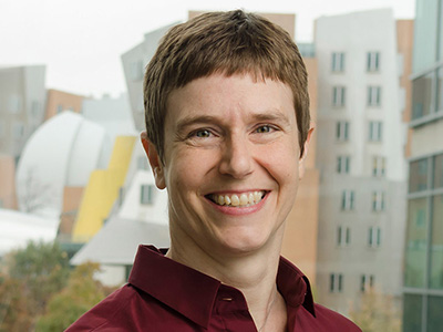
In the lab, Biology Professor Amy Keating researches the interactions of proteins with a mix of modeling and synthetic lab work and diverse minds
School of Science
June 11, 2020
Almost everything in biology is a multistep process, from the metabolization of carbohydrates and fats as fuel to information transcription from DNA and RNA. Without proteins and their interactions, cells couldn’t perform any of these biological tasks. But how do proteins establish their individual roles? And how do they interact with each other? These questions drive Professor Amy Keating’s research, and both lab experiments and computational modeling are helping her reveal the mysteries behind the basic functions of life.
In Keating’s field of research, as with most areas of science, the use of artificial intelligence is a relatively new – and growing – trend. “It’s pretty scary how fast new methods in machine learning are changing the landscape,” says Keating, who holds appointments in both the Department of Biology and the Department of Biological Engineering. “I think that we will see a disruptive change in protein modeling over the coming years.” She has found that incorporating basic machine learning methods in her own work has generated some success in uncovering how protein sequences determine their interactions.
However, there are limits to using only computational modeling due to the complexities of protein-protein interaction and a general need for empirical data to calibrate the models. Her lab group integrates computation with biological engineering in a laboratory setting. Keating’s team often starts by using computational modeling to narrow down their search from a massive collection of protein structure models. This step limits their output from an effectively infinite space (~1030) to something on the order of 106 potential promising molecules that can be experimentally tested. They can feed the results of experiments into other algorithms that help designate the specific features of the protein that prove important. This process is cyclical, and Keating emphasizes that experimental efforts are crucial for improving the success rate of this kind of work. That is where the lab comes in. There, they do what the computer cannot: they build proteins.
With the disruption of the COVID-19 crisis the Keating lab has focused their attention on computational projects, as well as on reviewing the literature and writing up papers and theses. The members are also using their time at home to brainstorm and plan their research. “We are having multiple group meetings per week by Zoom, including a ‘Keating Group Idea Lab,’ at which everyone throws out ideas, ranging from practical suggestions about current projects to out-there new concepts, for group discussion,” says Keating. “We are confident that we can use this time productively, to advance our science, even as we make long lists of things that we are eager to do as soon as we can get back into the laboratory.”
A topic of current interest to Keating and her group members is interactions among proteins with “short linear motifs” or SLiMs, which are abundant –more than one hundred thousand such motifs are thought to exist in one human. One family of these SLiM-binding proteins regulates movement of cells within the body and is implicated in the spread of cancer cells to a secondary location (metastasis). The lab’s novel mini-protein and peptide designs aim to disrupt these protein interactions and could be useful for eventually disrupting and treating cancer and other diseases.
FOSTERING MULTIPLE INTERACTIONS
Currently, Keating’s research team consists of six students who have backgrounds in almost as many different cultures. Her students’ diversity, which stems not just from different focuses in formal training but also from life experiences, is integral to their success, according to Keating. She wishes that more women like herself and members of underrepresented minority groups who love STEM would consider pursuing academic careers. “It’s hard work, but it’s very rewarding,” she entices. The best thing about being a faculty member, she believes, is having a team of bright minds who contribute unique ideas and insights to a problem and provide information beyond her own areas of expertise.
“I learn facts that they know and I do not. I learn interesting ways of thinking about science and also ways of doing science,” she says, noting that novel ideas in methodology lead to advances in research. “I’ve learned a lot of things about computer science from my students. I’m happy that one of my former biology students is [now] a professor of computer science,” she admits, appreciating his expertise as a benefit in frequent collaborations. “I love that students at MIT question everything.” Keating’s ever-expanding knowledge builds on top of a diverse background gleaned during her time as a student.
Keating’s bachelor’s degree from Harvard University is in physics. During her PhD at University of California, Los Angeles, she shifted to chemistry — specifically computational physical organic chemistry. When browsing for a postdoctoral position, she discovered the work of former MIT Department of Biology faculty and Whitehead Institute member Peter Kim and joined him. She maintained her interest in computation as a tool for biological research, concurrently co-advised by MIT Professor of Electrical Engineering and Computer Science Bruce Tidor. It was somewhat down to chance that her academic job search led her to MIT. “I certainly never thought I would be a biology professor, especially at MIT,” she remarks of her convoluted career path through the wide world of science.
But it is an unexpected result for which Keating is grateful. “My undergrad self would have been surprised by the MIT School of Science,” she muses, which makes MIT “so much more than ‘just’ the world’s best engineering school.” That is something of a common misconception about the Institute, she feels. “I think a lot of people outside of MIT don’t know how outstanding our basic science programs are.” Keating is a part of the strong science education at MIT, which is constantly adapting to keep up with the digital age, which led to her receiving the most recent Fund for the Future of Science Award.
“I was thrilled, and pretty surprised, to receive the award; my fantastic colleagues in the School of Science are not people that you want to be competing with.” This support is invaluable to her research on the foundations of biological interactions, and to ensure a robust team that has what it needs to develop important advances. The curious minds with which she collaborates are equally as invaluable.
“The people at MIT are amazingly smart, curious, and focused on things that I value,” Keating adds, “like good ideas, intellectual rigor, discovering new things, and education.”
This article appeared in the Summer 2020 issue of Science@MIT

Institute ends negotiations for a new journals contract in the absence of a proposal aligning with the MIT Framework for Publisher Contracts.
MIT Libraries
June 10, 2020
Standing by its commitment to provide equitable and open access to scholarship, MIT has ended negotiations with Elsevier for a new journals contract. Elsevier was not able to present a proposal that aligned with the principles of the MIT Framework for Publisher Contracts.
Developed by the MIT Libraries in collaboration with the Ad Hoc Task Force on Open Access to MIT’s Research and the Committee on the Library System in October 2019, the MIT Framework is grounded in the conviction that openly sharing research and educational materials is key to the Institute’s mission of advancing knowledge and bringing that knowledge to bear on the world’s greatest challenges. It affirms the overarching principle that control of scholarship and its dissemination should reside with scholars and their institutions, and aims to ensure that scholarly research outputs are openly and equitably available to the broadest possible audience, while also providing valued services to the MIT community.
“I am disappointed that we were not able to reach a contract with Elsevier that honors the principles of the MIT Framework, but I am proud knowing that the MIT community — as well as hundreds of colleagues across the country — stand by the importance of these principles for advancing the public good and the progress of science,” said Chris Bourg, director of the MIT Libraries. “In the face of these unprecedented global challenges, equitable and open access to knowledge is more critical than ever.”
More than 100 institutions, ranging from multi-institution consortia to large research universities to liberal arts colleges, decided to endorse the MIT Framework in recognition of its potential to advance open scholarship and the public good.
“We’ve seen widespread support in all quarters of the MIT community — faculty, students, postdocs, and staff alike — for the core grounding of the framework: that the value in published scholarship originates in our work and in the institutions that support us,” says Roger Levy, associate professor in the Department of Brain and Cognitive Sciences and chair of the Committee on the Library System (CLS). “CLS was unanimous in its recommendation to end negotiations. We are publicly committed to supporting the rights of MIT community members to freely share the scholarship we create, and stand by the principles articulated in the MIT Framework in our recommendation.”
“We hope to be able to resume productive negotiations if and when Elsevier is able to provide a contract that reflects our community’s needs and values and advances MIT’s mission,” said Bourg. “In the meantime, we will continue to use the framework to pursue new paths to achieving open access to knowledge. The groundbreaking agreement we reached with the Association for Computing Machinery in collaboration with the University of California, Carnegie Mellon University, and Iowa State University is one such example of building the business models of the future.”
MIT has long been a leader in open access. Adopted in 2009, the MIT Faculty Open Access Policy was one of the first and most far-reaching initiatives of its kind in the United States. Forty-seven percent of faculty journal articles published since the adoption of the policy are freely available to the world. In 2017, the Institute announced a new policy under which all MIT authors — including students, postdocs, and staff — can opt in to an open access license. The Ad Hoc Task Force on Open Access to MIT’s Research, first convened by Provost Martin Schmidt in 2017, released its final recommendations in October 2019. An implementation team, led by Bourg, is working to prioritize and enact the task force’s recommendations, which range from policy to incentives to national and global advocacy.
Information for the MIT community about access to Elsevier articles can be found on the MIT Libraries’ website.
June 11, 2020