Fellowship funds graduate studies for outstanding immigrants and children of immigrants.
Julia Mongo | Office of Distinguished Fellowships | MIT Career Advising and Professional Development
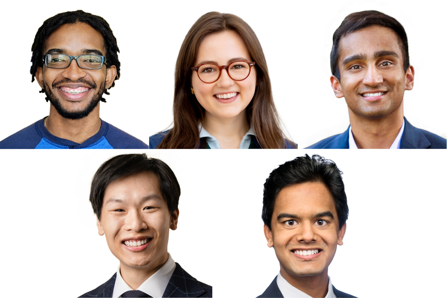
MIT graduate students Kat Kajderowicz and Shomik Verma, alumni Desmond Edwards ’22 and Steven Truong ’20, and Vaibhav Mohanty, an MD-PhD student in the Harvard-MIT Program in Health Sciences and Technology, are among the 30 recipients of this year’s Paul and Daisy Soros Fellowships for New Americans.
The P.D. Soros Fellowships for New Americans program honors the contributions of immigrants and children of immigrants to the United States. The program recognizes the potential of immigrants to make significant contributions to U.S. society, culture, and academia by providing $90,000 in graduate school financial support over two years.
Students interested in applying to the P.D. Soros Fellowship may contact Kim Benard, associate dean of distinguished fellowships in Career Advising and Professional Development.
Desmond Edwards ’22
Desmond Edwards graduated from MIT in 2022 with a double major in biological engineering and biology and a minor in French. As a National Science Foundation GRFP Fellow and Jamaica’s first Knight-Hennessy Scholar, Edwards is currently a PhD student in microbiology and immunology at Stanford University’s School of Medicine, where he researches immunity to infectious diseases. He intends to lead a scientific career not only contributing to groundbreaking academic research, but also ensuring that the fruits of this research have their maximal benefit to society through public policy, outreach, and education.
Born and raised in Jamaica, Edwards lived in rural St. Mary and attended school in urban Kingston. His constant childhood illnesses prompted his interest in better understanding human disease and his desire to develop novel therapeutic options for their treatment and prevention. At MIT, Edwards conducted host-pathogen research in Professor Rebecca Lamason’s lab, focusing on characterizing mutants of interest and developing novel genetic tools for use in the tick-borne pathogen Rickettsia parkeri. As an Amgen Scholar, he also worked with Professor Viviana Gradinaru at Caltech to engineer solutions for a novel gene therapy for Rett syndrome, a neurodevelopmental disorder primarily seen in girls.
Interested not only in the technical details of scientific research but also in its societal impact, Edwards has dedicated himself to serving the community through roles in the MIT Biotech Group, student representation and advocacy, and teaching and mentorship. For his academic achievements and commitments to community and nation-building, he was awarded a 2022 Prime Minister’s National Youth Award for Excellence, the highest national award bestowed on Jamaicans between the ages of 15 and 29 by the Prime Minister of Jamaica.
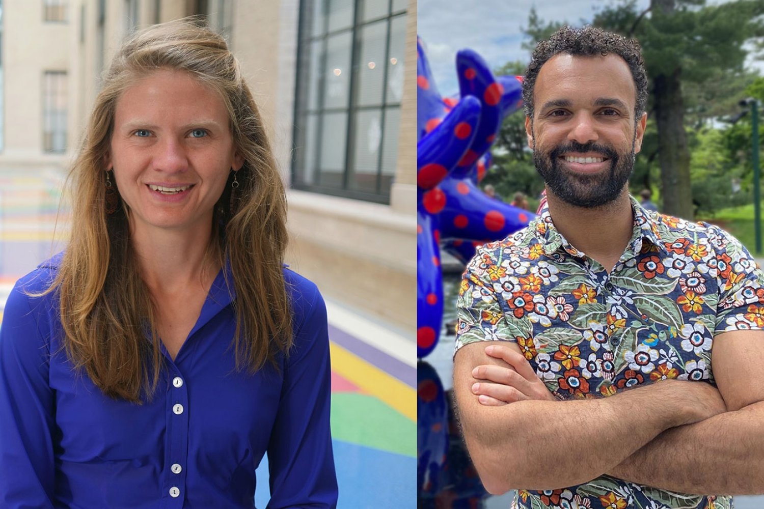
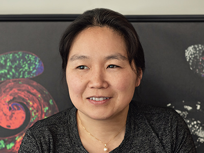
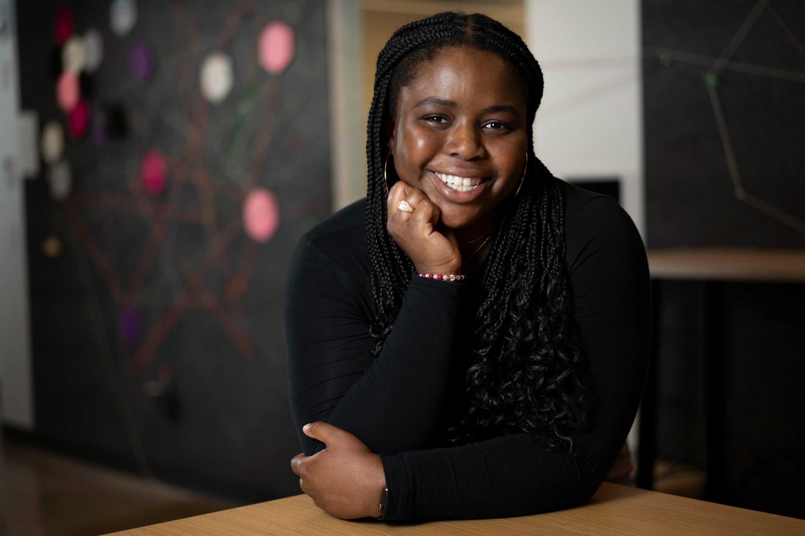
Kathrin (Kat) Kajderowicz
Kat Kajderowicz is a neuroscience PhD student in the Department of Brain and Cognitive Sciences at MIT. She is co-advised by professors Sinisa Hrvatin and Jonathan Weissman at the Whitehead Institute for Biomedical Research and is researching how cells from hibernating organisms can survive cold temperatures to engineer human cells to do the same. Kajderowicz envisions her work improving organ transplantation and therapeutic hypothermia technologies. She hopes to someday lead a research group developing technologies that allow humans to safely enter and exit “hibernation-like” stasis for medical treatment purposes.
Kajderowicz grew up in Chicago’s large Polish-immigrant community. Her parents fled communist Poland in the early 1980s, arriving in the United States with no savings, college degrees, or knowledge of English. Kajderowicz’s mother worked as a housekeeper, while her father worked in construction. As undocumented immigrants fighting to obtain green cards, they spent most of their savings on the American naturalization process and rarely sought medical care because they couldn’t afford insurance and feared deportation. To help financially, Kajderowicz began working as a golf caddie at age 14. Her golf clients connected her with shadowing and interning opportunities at technology and biotechnology companies and hospitals, which inspired her career interests.
As an undergraduate at Cornell University, Kajderowicz worked at the Lab of Ornithology, where she built computational pipelines to better understand songbird communication. During undergraduate summers at Harvard University, she worked on comparative genomics and population genetics projects using plants, fruit flies, and butterflies. As a post-baccalaureate researcher at Harvard Medical School, she built imaging tools to visualize the development of different types of mouse retinal neurons.
In 2020, Kajderowicz’s father passed away from metastatic lung cancer. Kajderowicz served as his caregiver and medical proxy. Her greatest source of comfort was her hospital waiting room community. Inspired by the power of communities, Kajderowicz founded DNA Deviants, a 2,000-plus member biotechnology group that hosts podcasts on Twitch to discuss breakthrough research and organizes career mentorship programs.
Vaibhav Mohanty
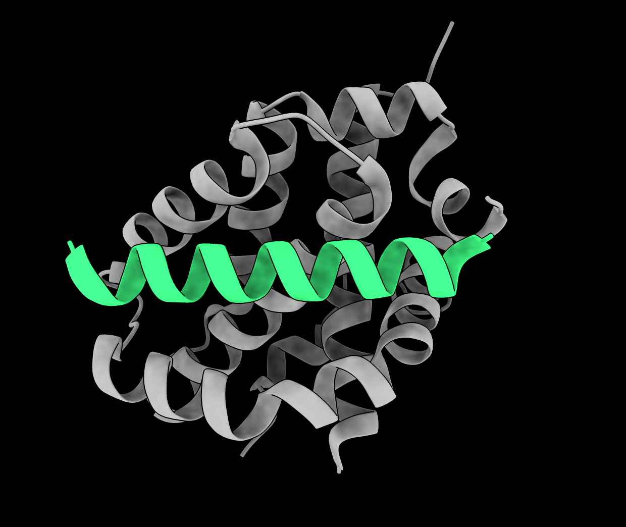
Vaibhav Mohanty is pursuing an MD-PhD in the Harvard-MIT Program in Health Sciences and Technology, where he is earning a second PhD (in chemistry). His goal is to extend his physics-based theories of evolution to understand how molecular-level structural changes in proteins can induce changes in evolutionary fitness of viruses and cancers. Mohanty aspires to develop novel therapeutic approaches to combat diseases subject to evolution on fast timescales, and to treat patients with such diseases.
Mohanty was born in Durham, North Carolina, and grew up in Charleston, South Carolina. His parents emigrated from Odisha, India, to the United States to pursue academic research careers in biology. Accepted to Harvard College at age 15, Mohanty graduated in 2019 with a master’s degree in chemistry (theory) and a bachelor’s degree summa cum laude in chemistry and physics and a minor in music. He was inducted into the Phi Beta Kappa academic honor society and received a 2018 Barry Goldwater Scholarship for his physics research. As an undergraduate and master’s student, Mohanty’s published research papers spanned a number of interdisciplinary topics across the sciences and even music, including diffusion MRI physics, time-dependent quantum mechanics of graphene, and mathematical and geometrical models of voice leading in music theory. In 2022, Mohanty earned a PhD in theoretical physics as a Marshall Scholar at the University of Oxford’s Rudolf Peierls Centre for Theoretical Physics, where he worked in the Condensed Matter Theory Group to use statistical physics and spin glass theory to investigate fundamental properties of biological evolution.
In addition to being a scientific researcher, Mohanty is an award-winning classical and jazz music composer, arranger, pianist, and saxophonist. His large wind ensemble and chamber works are distributed and performed regularly around the United States and in many parts of the world. He actively performs as a jazz pianist.
Steven Truong ’20
Steven Truong graduated from MIT in 2020 with a double major in biological engineering and writing. He was inducted into Phi Beta Kappa and Tau Beta Pi and was also named a Barry Goldwater Scholar. As a Marshall Scholar in the United Kingdom, Truong completed an MPhil in computational biology at Cambridge University and an MA in creative writing at Royal Holloway, University of London. Currently, he is an MD-PhD student at Stanford University. In his future career, Truong aspires to help solve and treat metabolic disorders such as diabetes. He hopes to make these discoveries accessible — especially for communities traditionally underrepresented and underserved in medicine — as a physician-scientist, science communicator, and storyteller.
Truong was born in St. Paul, Minnesota, to Vietnamese refugees. His parents and relatives pooled their resources to start a family-owned nail salon. Truong spent his evenings after school at the salon, where he assisted in the business’s operations and finished homework between helping customers. In his free time, he avidly read science fiction and fantasy, which evolved into a passion for science. Truong eventually realized he could use science to address diabetes, a disease that affects much of his family and community.
During his undergraduate years at MIT, Truong worked in the Langer-Anderson Lab to develop smart insulins, and in the Lauffenburger Lab to study the link between the immune system and diabetes. With funding from MIT’s Undergraduate Research Opportunities Program, he started a study investigating the genetic basis of diabetes with colleagues at the University of Medicine and Pharmacy at Ho Chi Minh City. The data from this study were published, associating single nucleotide polymorphisms to Type 2 diabetes in Vietnamese individuals. Truong and his colleagues eventually secured a grant to expand their studies through the National Foundation for Science Technology and Development. The grant currently funds Vietnam’s largest genome-wide association study, which he co-leads.
Shomik Verma
Shomik Verma is pursuing a PhD in mechanical engineering at MIT with Professor Asegun Henry, where he is working on energy storage to make variable renewable energy sources such as solar more reliable, and on a next-generation power plant based on thermophotovoltaic power conversion. After his PhD, Verma hopes to use his skill set to decarbonize industry and make cheap, clean, and reliable energy available to all.
Growing up in Sugar Land, Texas, Verma maintained a deep connection to Indian culture. There was a strong emphasis on education, and he spent many weekends at math competitions with fellow Asian Americans. Verma started noticing some interesting patterns at the math competitions he attended — oil and gas companies would often sponsor them, and the conversations his petroleum engineer father had with his friends often turned to the geopolitics of energy. Verma was struck by the realization that he lived in the oil and gas capital of the world, with parents who were from the coal capital of India. He was caught between two worlds — the fossil fuel industry that enabled his way of life, and the growing threat of global warming he learned about in school.
When Verma lost his uncle to black lung, he decided it was time to devote his life to clean energy. While studying mechanical engineering at Duke University, he helped lead the Duke Electric Vehicles team to two Guinness world records for fuel efficiency, for both battery electric and fuel cell vehicles. In the U.K., as a Marshall Scholar, he completed an MPhil in materials science and conducted research at Imperial College London and the University of Cambridge, working on improving the efficiency of solar cells.