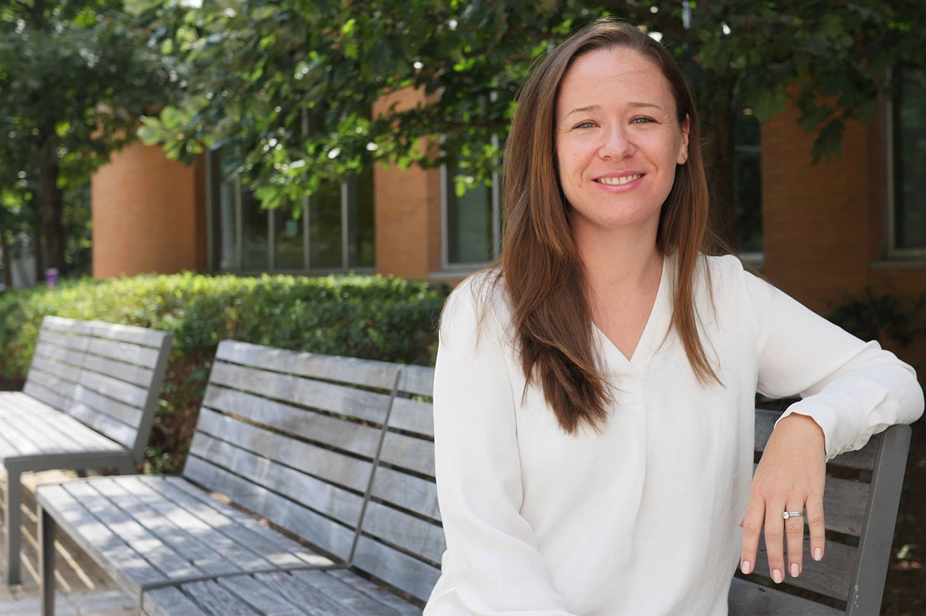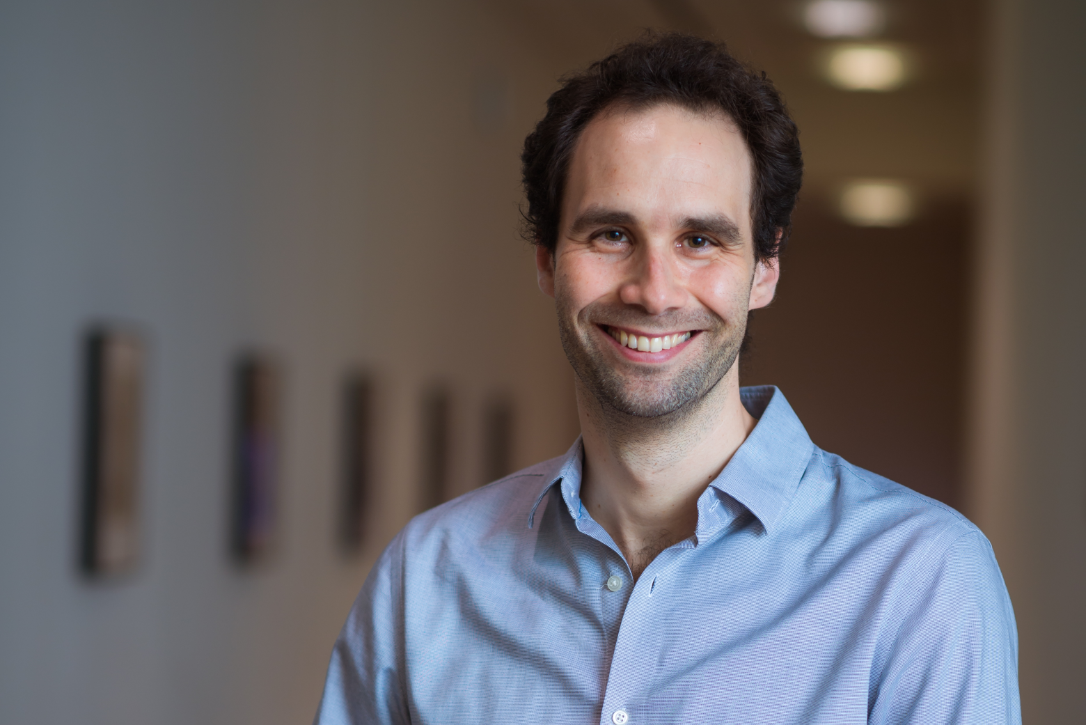
Now an assistant professor at UT Dallas, Nicole De Nisco draws on love of problem solving and interdisciplinary skills honed as an undergraduate and graduate student at MIT
Lillian Eden | Department of Biology
June 12, 2023
There were early signs that Nicole De Nisco, SB ‘07, PhD ‘13, might become a scientist. She ran out of science classes to take in high school and fondly remembers the teacher that encouraged her to pursue science instead of the humanities. But she ended up at MIT, in part, out of spite.
“I applied because my guidance counselor told me I wouldn’t get in,” she said. The rest, as they say, is history for the first-generation college student from Los Angeles.
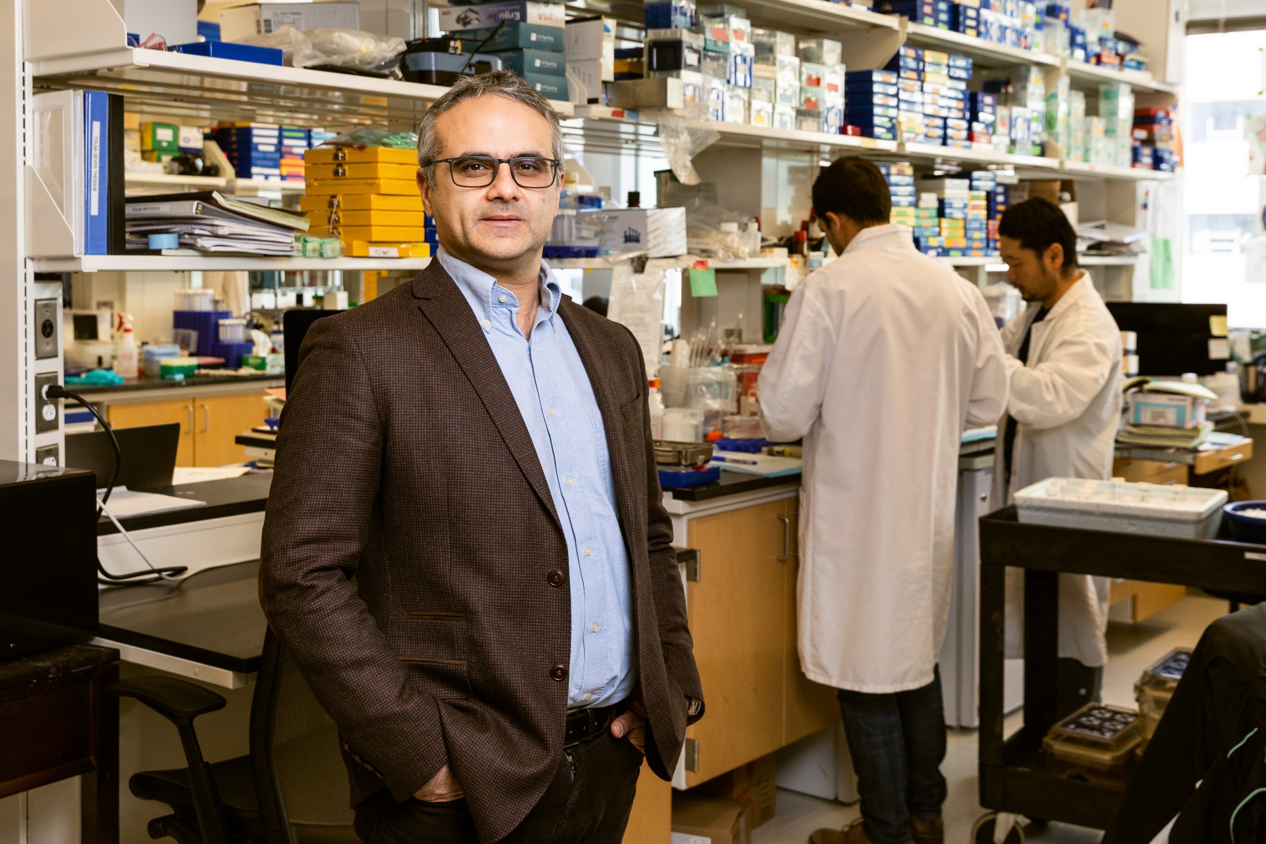
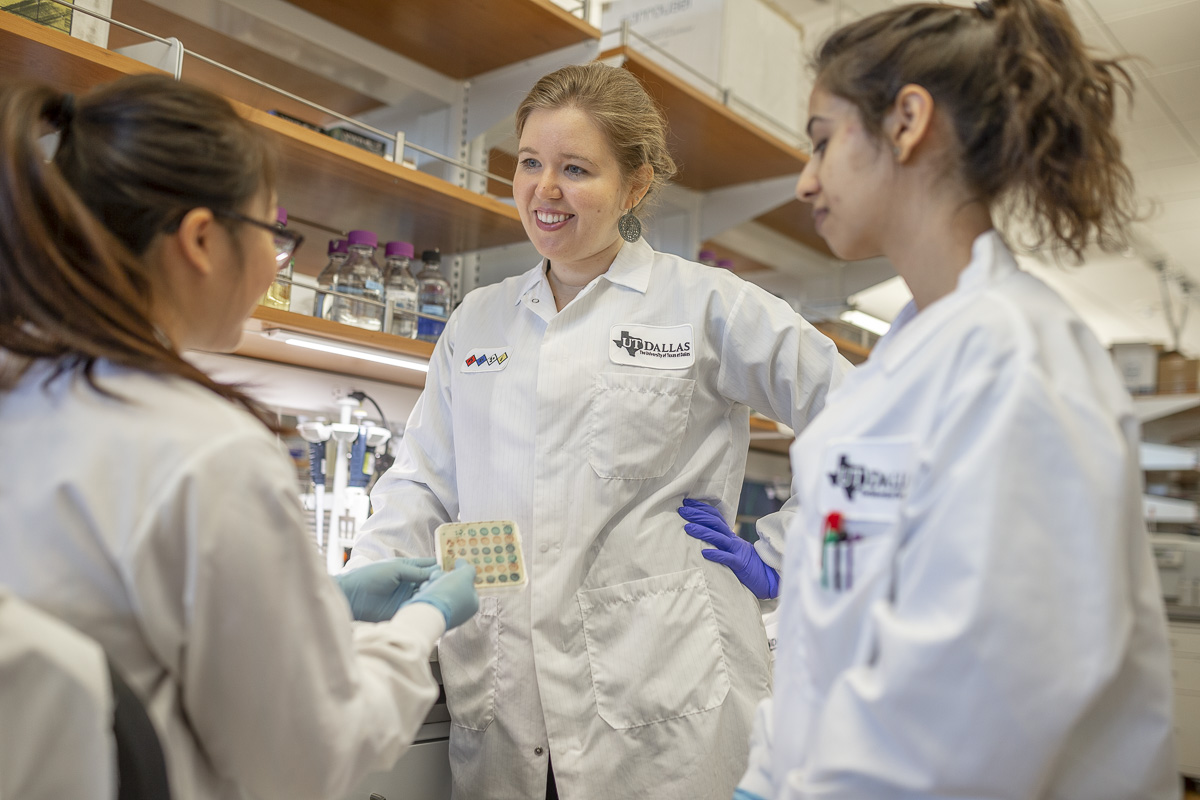
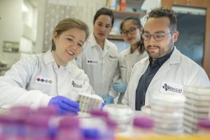
Now, she’s an assistant professor of biological sciences at UT Dallas studying urinary tract infections (UTIs) and the urinary microbiome in postmenopausal women.
De Nisco has already made some important advancements in the field: she developed a new technique for visualizing bacteria in the bladder and used it to demonstrate that bacteria form reservoirs in human bladder tissue, leading to chronic or recurrent UTIs.
It was known that in mice, bacteria are able to create communities within the bladder tissue, forming reservoirs and staying there long term—but no one had shown that occurring in human tissue before.
De Nisco found that reservoirs of tissue-resident bacteria exist in human patients with recurring UTIs, a condition which may ultimately lead to women needing to have their bladder removed. De Nisco now mostly works with postmenopausal women who have been suffering from decades of recurring UTIs.
There was a big gap in the field, De Nisco explained, so entering the field of urology was also an opportunity to make new discoveries and find new ways to treat those recurring infections.
De Nisco said she’s in the minority, both as a woman studying urology and as someone studying diseases that affect female patients. Most researchers in the urology field are men, and most focus on the prostate.
But things are changing.
“I think there are a lot of women in the field who are now pushing back, and I actually collaborate with a lot of other female investigators in the field. We’re trying to support each other so that we can survive and, hopefully, actually advance the science—instead of it being in the same place it was 15 years ago,” De Nisco says.
De Nisco first fell in love with biomedical research as an undergrad doing a UROP in Catherine Drennan’s lab, back when Drennan was still located in the chemistry building.
“Cathy herself was incredibly encouraging, and is probably the main reason I decided to pursue a career in science—or felt that I could,” De Nisco said.
De Nisco became fascinated with the dialogue between a microbe and a host organism during an undergraduate course in microbial physiology with Graham Walker, which led to De Nisco’s decision to remain at MIT for her PhD work and to perform her doctoral research in rhizobia legume symbiosis in Walker’s lab.
De Nisco said during her time at MIT, Drennan and Walker gave her a lot of encouragement and “room to do my own thing,” fostering a love of discovery and problem solving. It’s a mentoring style she’s using now with her own graduate students; she currently has eight working in her lab.
“Every student is different: some just want a project and they want to know what they’re doing, and some want to explore,” she said. “I was the type that wanted to do my own thing and so they gave me the room and the patience to be able to explore and find something new that I was interested in and excited about.”
As a low-income student sending financial help home, she also pursued teaching opportunities outside of her usual duties; Walker was very supportive of pursuing other teaching opportunities. De Nisco was a graduate student tutor for Next House watching over 40 undergrads, served as a teaching fellow with the Harvard Extension School, and worked with Eric Lander to help launch the course 7.00x Introduction to Biology – The Secret of Life for EdX, one of the most highly rated MOOCs of all time.
She said MIT definitely prepared her for a life as a professor, teacher, and mentor; the most important thing about graduate school isn’t choosing “the most cutting-edge research project,” but making sure you have a good training experience and an advisor who can provide that.
“You don’t need to start building your name in the field when you’re a grad student. The lab environment is much more important than the topic. It’s easy to get burned out or to be turned off to a career in academia altogether if you have the wrong advisor,” she said. “You need to learn how to be a scientist, and you have plenty of time later in your career to follow whatever path you want to follow.”
She knows this from experience: her current research is somewhat parallel but unrelated to her previous research experience.
“I think my motivation for being a scientist is rooted in my desire to help people doing something I enjoy,” she said. “I was not doing this kind of research as a graduate student, and that doesn’t mean that I wasn’t able to end up at this point in my career where I’m doing research that is focused on improving the lives of women, specifically.”
She did her postdoctoral work at UT Southwestern Medical Center studying Vibrio parahaemolyticus, a human pathogen that causes gastroenteritis. The work was a marriage of her interests in biochemistry and host-microbiome interactions.
She said MIT prepared her well for the type of interdisciplinary work that she does every day: At UT Dallas, all the research buildings are fully integrated, with engineers, chemists, physicists, and biologists sharing lab spaces in the same building. Her closest collaborators are mathematicians, chemists, and engineers.
Although she may not be fully literate, she has a common language with the people she works with thanks to MIT’s undergraduate course requirements in many different topics and MIT’s focus on interdisciplinary research, which is “how real advancement is made.”
Ultimately, De Nisco said she is glad to this day that she attended MIT.
“Getting that acceptance letter to attend MIT probably changed the trajectory of my life,” she said. “You never know, on paper, what someone is going to achieve eventually, and what kind of force they’re going to be. I’m always grateful to whoever was on the admissions committees that made the decision to accept me—twice.”