The first Live μ in the country will reveal fleeting sub-cellular events in high resolution
Saima Sidik
February 1, 2023
Inside cells, events can unfold quickly. Sub-cellular compartments constantly re-arrange while proteins move along structural fibers and membranes fuse and divide. By attaching fluorescent tags to sub-cellular structures, researchers can watch events unfold in real time using light microscopes. But to see the finest details of these processes, scientists need to shift from using light microscopy to using beams of electrons to generate even higher resolution images using a technique called electron microscopy. Using these techniques together is a powerful and rapidly growing strategy called correlative light electron microscopy (CLEM). In CLEM, light microscopy images are used to target regions of interest, and then the same sample is interrogated with electron microscopy to see the same areas at higher resolution.
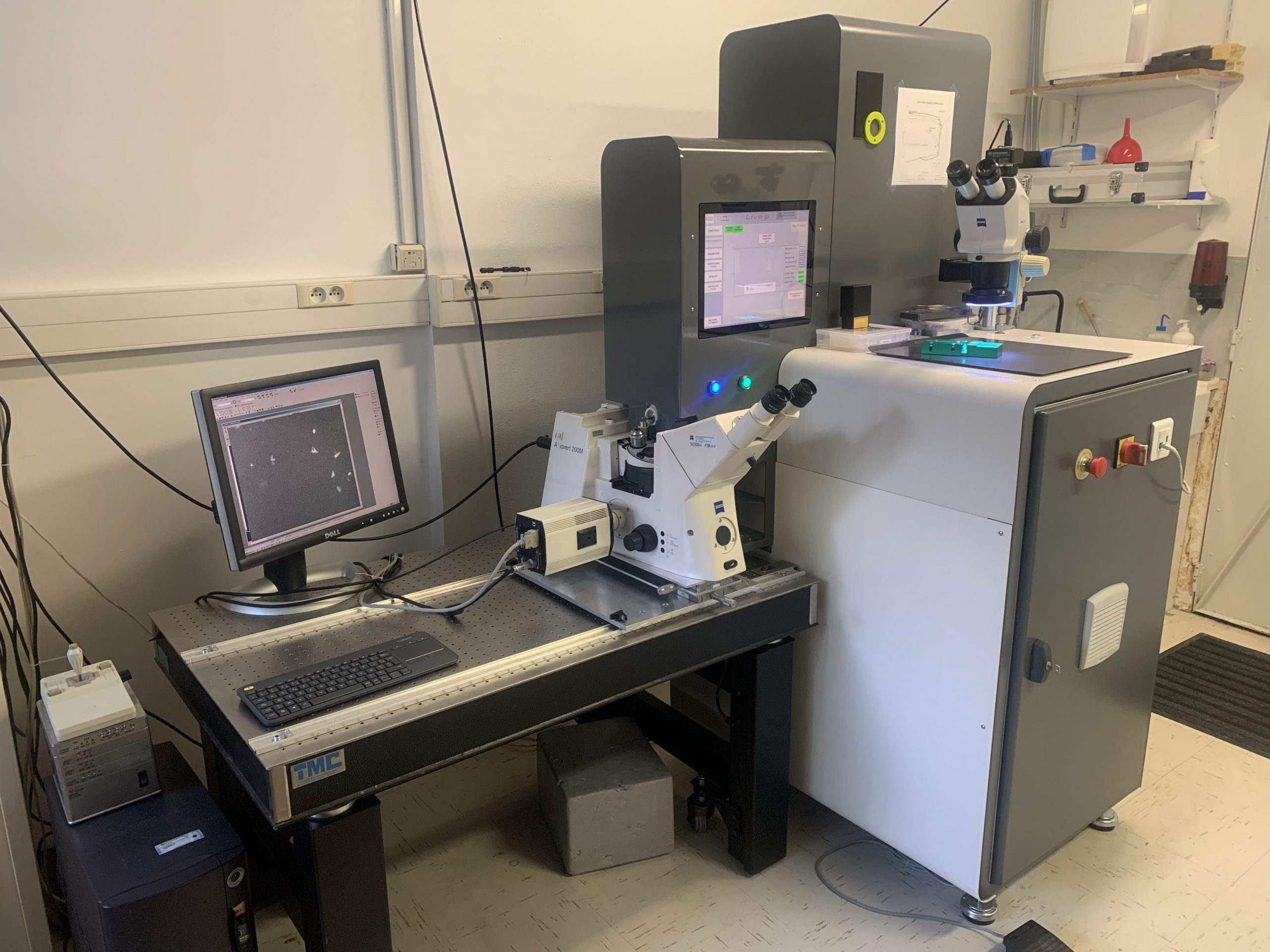
The Peterson (1957) Nanotechnology Materials Core Facility in the Robert A. Swanson (1969) Biotechnology Center at the Koch Institute for Integrative Cancer Research at MIT recently acquired a high pressure freezer called the Live μ that will let researchers do just that. This instrument allows scientists to image the same biological sample using fluorescent light microscopy and electron microscopy in close succession. These two techniques are usually performed on separate samples, but with the Live μ, researchers will be able to identify fleeting sub-cellular events using light microscopy, then preserve cells and observe the same events in high resolution using electron microscopy — a combination that was not previously available to researchers at MIT. In fact, the Live μ, which is sold by the Paris-based company CryoCapCell, will be the first instrument of its kind in the country.
Although high-pressure freezers like the Live μ have been around for decades, integration with a light microscope is what makes the Live μ special. The instrument itself is a washing machine-sized freezing instrument, equipped with an arm to hold a biological sample under a nearby light microscope. When researchers observe an interesting biological event using the light microscope, they can quickly retract the arm and insert the sample into the Live μ’s inner chamber, exposing it to low temperature and high pressure and freezing it in less than two seconds. Cells must be preserved before they can be observed using an electron microscope, and by freezing samples faster than ice crystals can form, the Live μ creates pristine samples that accurately represent the state of cells before preservation. Superimposing pictures taken using the light microscope on top of images from an electron microscope allows researchers to use the fluorescent signals like a “treasure map,” says Abigail Lytton-Jean, the director of the Peterson Facility.
Exocytosis is a vital sub-cellular event that could be studied using the Live μ. In this cellular process, cells use bubble-like vesicles to ferry proteins from the internal compartments where they’re made to the cell’s surface, where they can sense the external environment, attach cells to one another, or carry information to other cells. Exocytosis is important for many aspects of biology, and a variety of scientists, from ecologists to cancer researchers to microbiologists, would benefit from a greater understanding of this process. With the Live μ, researchers may be able to use light microscopy to catch the vesicles that mediate exocytosis when they dock with the cell’s surface, then use electron microscopy to understand the details of this association.
Researchers creating artificial materials to replace human tissues could also benefit from the Live μ, Lytton-Jean says. These materials are thick and contain a lot of water, but the Live μ is capable of freezing them without generating ice crystals that change their structure. Using this instrument, scientists can examine the internal structure of these synthetic materials and assess their similarities to live tissue.
“People who want to use the Live μ are coming from all sorts of labs,” Lytton-Jean says.
The world of biology and electron microscopy is wildly exciting right now, she adds, thanks in part to instruments like this. “People who have worked with electron microscopes for decades have told me that this is the most exciting time they’ve ever lived in.”
The Live μ recently took its place in the back of the Peterson Facility, under a picture of the Eiffel Tower that Lytton-Jean brought back from Paris when she first went to test the Live μ at CryoCapCell’s headquarters years ago. The Live μ is only the latest addition to a vast suite of instrumentation focused on cutting-edge cryo-electron microscopy and CLEM workflows, expanding the facility’s unusually large portfolio of workflows.
“There aren’t many places in the country that can do all of the different workflows we offer, and all in one place,” said Lytton-Jean. “High pressure freezing is the first step in the preservation process, so having this instrument in our lab will further enable many new workflows with our existing instrumentation. Although these workflows are challenging and sophisticated, our team of dedicated scientists are familiar with conducting this work.”