Investigating how cell orientation drives tissue growth during development

In a new study published July 7, 2022 in eLife, Adam Martin’s lab at the MIT Department of Biology identified the mechanical forces and molecular cues that help the spindles in cells located in the portion of the embryo destined to become the fly’s head to assume the same orientation.

Raleigh McElvery
July 7, 2022

Raleigh McElvery

During development, virtually all multicellular creatures must build themselves up from a ball of cells to a multilayered, fully-functioning organism. In the case of a fruit fly embryo, in order go from blob to organism, the cells must coordinate their divisions along the same axis to drive tissue growth in a specific direction — eventually going on to form the three germ layers known as the endoderm, ectoderm, and mesoderm.

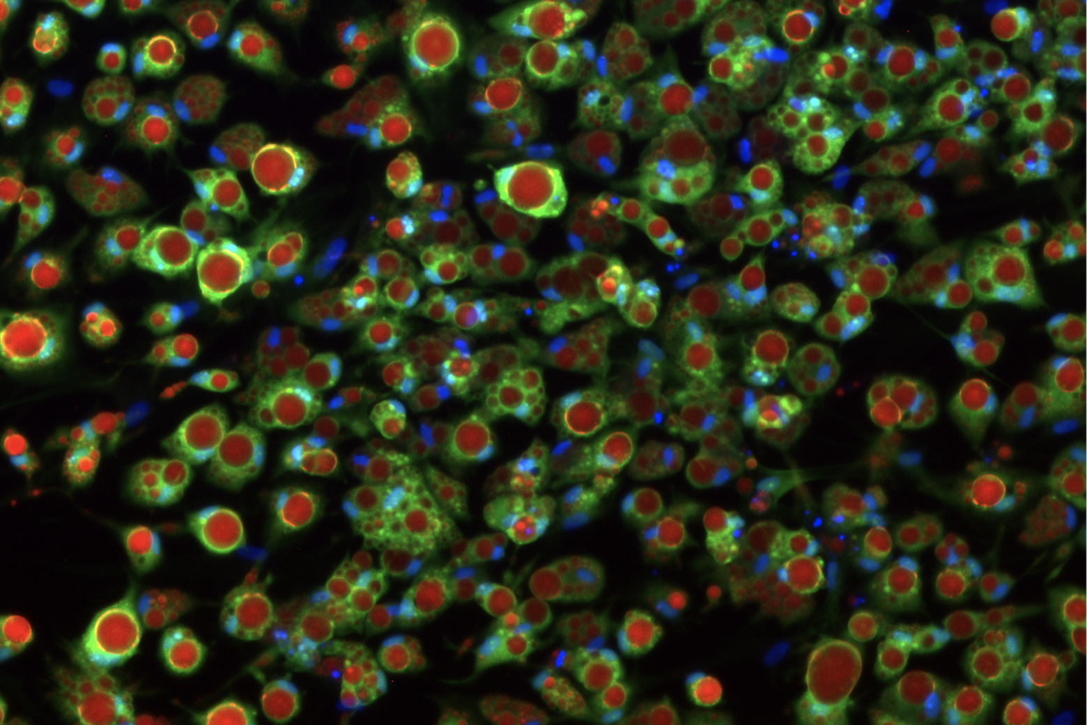
Scientists have long studied how cells orient themselves to divide in a specific direction. It’s clear that this orientation is determined by a bundle of tiny fibers inside the cell called the spindle, which helps segregate the chromosomes so they can be distributed between the two daughter cells as the parent cell splits. When the spindles in neighboring cells are parallel with one another, then the cells will divide along the same axis.

In a new study published in eLife on July 7, 2022, Adam Martin’s lab at the MIT Department of Biology identified the mechanical forces and molecular cues that help the spindles in cells located in the portion of the embryo destined to become the fly’s head to assume the same orientation.

According to Martin, an associate professor of biology and the study’s senior author, his lab was among a handful of labs to take interest in this coordinated spindle orientation in the early fruit fly embryo — and identified the molecular, mechanical, and molecular cues that orient the spindle in a living organism.

“Cell divisions in the fly embryo was thought to be regulated independently of cell shape changes and morphogenetic movements,” Martin says. “However, we found that forces associated with cell invagination actually oriented cell divisions through a novel mechanism that we describe.” First author Jaclyn Camuglia, he explains, spearheaded the project from start to finish.

Prior to Camuglia’s work, researchers knew that spindle orientation was controlled by a complex of multiple proteins, including one protein in fruit flies called Pins (known as LGN in vertebrates). The entire protein complex, including Pins, is coupled to motor proteins that help to rotate the spindle. However, it was still unclear what mechanical forces were orienting Pins to dictate spindle rotation.

“It’s a very striking phenomenon when you look into the microscope at a fly embryo and see all the spindles orienting together,” Camuglia says. “We wanted to know: How are these spindles oriented and why is that directionality important?”

The fly is an ideal organism for probing cell division, because it has a very consistent and predictable cell cycle. During development, cells divide a set number of times, pause briefly, and then start up again in very discrete pockets across the embryo. The researchers wanted to know how the cells in a subset of those pockets in the fly’s head were coordinating their divisions.

Camuglia found that, in healthy embryos, Pins was always recruited to the end of the cell along the anterior-posterior axis (from the fly’s head to rear). By cutting the embryo with a laser, treating it with chemical inhibitors, disrupting the connections between cells, and depleting certain transcription factor proteins, she was able to characterize the mechanical forces that directed Pins to the anterior-posterior axis and thus rotated the spindle in the proper direction. As it turns out, these forces stem from other large-scale tissue movements that occur at the same time to force the embryo to fold in on itself and eventually form the fly’s muscles.

The researchers don’t yet know the role that these coordinated divisions along the anterior-posterior axis play in the developing fruit fly head — or what might happen during development if the divisions were to go awry. But Martin and Camuglia suspect this process facilitates tissue elongation, and helps compensate for any cells that are lost from the tissue during the division process as the embryo folds in on itself.

“This study is unique because it connects mechanical forces to the specific molecular cues like Pins that coordinate cell division,” Camuglia explains. “The factors that shape a developing embryo are critical to understand, because all organisms — from fruit flies to humans — experience these driving forces.” This intersection between physics and biology, she says, is what makes the study so exciting.”

Video: Groups of cells divide in a coordinated and oriented manner in the fruit fly embryo.
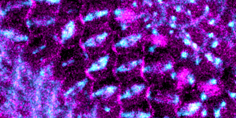
Top Image: Enrichment of cortical cues at one end of the cell coordinates the orientation of the mitotic spindle. Credit: Jaclyn Camuglia

Citation:
“Morphogenetic forces planar polarize LGN/Pins in the embryonic head during Drosophila gastrulation”
eLife, online 07/07/2022, DOI: https://doi.org/10.7554/eLife.78779
Jaclyn Camuglia, Soline Chanet, and Adam C Martin

Lab-grown fat cells help scientists understand type 2 diabetes
Eva Frederick | Whitehead Institute
June 16, 2022

In research published June 17 in the journal Science Advances, researchers in the lab of Whitehead Institute Founding Member Rudolf Jaenisch present a way to create fat cells that can be modified to display different levels of insulin sensitivity.

The cells accurately model healthy insulin metabolism, as well as insulin resistance, one of the key hallmarks of type 2 diabetes. “This system, I think, will be really useful for studying the mechanisms of this disease,” said Jaenisch, who is also a professor of biology at the Massachusetts Institute of Technology (MIT).

“It’s really exciting,” said Max Friesen, a postdoctoral researcher in Jaenisch’s lab and a first author of the study. “This is the first time that you can actually use a human stem cell-derived [fat cell] to show a real insulin response.”

Body fat — also known as adipose tissue — is essential for regulating your body’s metabolism and plays an important role in the storage and release of energy. When fat cells called adipocytes encounter the hormone insulin, they suck up sugar from the blood and store it for future use.

But over many years, factors such as genetics, stress, certain diets, or polluted air or water can cause this process to go awry, leading to type 2 diabetes. In this disease, adipocytes, as well as cells in the muscles and liver, become resistant to insulin and therefore unable to regulate the levels of sugar in the blood.

Tools to model diabetes in the lab generally rely on mice or on cells in a petri dish or test tube. Both these systems have their own problems. Mice, although they are comparable with humans in some respects, have a completely different metabolism and do not experience human diabetes comorbidities like heart attacks. And cell culture has, in the past, failed to replicate key markers of diabetes in a way that is comparable to human tissues.

That’s why Friesen and Andrew Khalil, another postdoc in Jaenisch’s lab, set out to create a new model. The researchers started with human pluripotent stem cells. These cells are the shapeshifters of the body — given the right conditions, they can assume the specific characteristics of almost any human cell type. The Jaenisch Lab has used them in the past to replicate liver cells, brain cells, and even cancerous tumors.

They decided to try to optimize an existing method for differentiating pluripotent cells into fat cells. The protocol created cells that looked like adipocytes, but these cells did not recreate the conditions of healthy insulin signaling or insulin resistance seen in the human body in type 2 diabetes. When healthy adipocytes encounter insulin in the human body, they respond by taking up glucose out of the bloodstream. These lab-made fat cells weren’t doing that, unless the researchers cranked up insulin levels to a thousand times higher than levels ever seen in humans. “Taking up glucose [in response to normal levels on insulin] is really the main function of an adipocyte, so if the model fails to do that, anything downstream in terms of disease research is not going to work either,” Friesen said.

Friesen and Khalil wondered if the lab-grown adipocytes’ low sensitivity to insulin could be a product of the conditions in which they grew. “We thought that maybe this happens because we’re feeding them an artificial culture medium, with all kinds of extra supplements that might be inhibiting their metabolic response,” Friesen said.

Friesen and Khalil decided to use a method called the Design of Experiments approach, which allows researchers to tease out the contributions of different factors to a specific outcome. Informed by this approach, they created nearly 30 different media compositions, each with slightly different levels of key ingredients such as glucose, insulin, the growth factor IGF-1, and albumin, a protein found in blood serum.

The medium that worked best had concentrations of insulin and glucose that were similar to the levels in the human body. When grown in this new medium, the cells responded to much lower concentrations of insulin, just like cells in the body. “So this is our healthy adipocyte,” Friesen said. “Next we wanted to see if we could make a disease model out of this — to make it an insulin-resistant adipocyte like you would see in the progression to type 2 diabetes.”

To desensitize the cells, they flooded the media with insulin for a short period of time. This caused the cells to become less sensitive to the hormone, and respond similarly to diabetic or pre-diabetic fat cells in a living person.

The researchers could then study how the cells responded to the change — such as what genes the insulin resistant cells expressed that healthy cells did not — in order to tease out the underlying genetics of insulin resistance. “We saw small changes in a lot of genes that are metabolism regulated, so that seems to be pointing to a deficiency of the metabolism or mitochondria of the insulin-resistant cells,” Friesen said. “That’s one thing we want to pursue in the future — figure out what is wrong with their metabolism, and then hopefully how to fix it.”

Now that they have created this new model for studying insulin resistance in fat cells, the researchers hope to develop similar procedures for other cells affected in diabetes.  “It seems that with some modifications, we can apply this method to other tissues as well,” Friesen said. “In the future, this will hopefully lead to a unified system for all stem cell-derived tissues, including liver, skeletal muscle, and other cell types, to get a really robust insulin response.”

Ankur Jain Named as Pew Scholar in Biomedical Sciences
Merrill Meadow | Whitehead Institute
June 13, 2022

The Pew Charitable Trusts has selected Whitehead Institute Member Ankur Jain to be a 2022 Pew Scholar in the Biomedical Sciences. The Pew program provides funding to young investigators of outstanding promise who work in areas of science relevant to the advancement of human health.

Jain, who joined the Whitehead Institute faculty in 2019, is one of 22 scientists selected to receive this year’s honor, chosen from among 197 nominations submitted by leading U.S. academic and research institutions. “I am grateful to the Pew Trusts for funding our work, and thrilled to be a part of the Pew community,” says Jain, who is also an assistant professor of biology and the Thomas D. and Virginia W. Cabot Career Development Professor at Massachusetts Institute of Technology.

The Pew award will provide research support for the next four years, enabling him to study the role of evolutionarily ancient metabolites called polyamines, which are essential for cell growth and survival.

“Polyamine concentrations within cells are carefully regulated, and disruptions in polyamine production are known to be associated with conditions ranging from cancer and aging to neurological disorders such as Parkinson’s disease” Jain explains. “But, despite being studied for more than a century, the specific role polyamines play in both healthy and diseased cells remains obscure. This is due, in part, to a lack of technologies effective in probing polyamines.”

Jain’s lab will harness the cell’s own polyamine detection machinery to build new tools to inspect polyamines. Those tools will allow his team to measure and track polyamines in individual cells, study how cells maintain their polyamine content, and explore how changing polyamine levels affect cellular functions. “Ultimately, this work could provide the basis for novel strategies for treating cancer or promoting healthy aging,” Jain observes.

Previously, Jain received a 2017 NIH Pathway to Independence Award and was named a 2019 Packard Fellow for Science and Engineering. He is the third current Whitehead Member to be named a Pew Scholar, following in the steps of Mary Gehring (2010) and Jing-Ke Weng (2014). Former Whitehead Fellow Fernando Camargo, now professor of stem cell and regenerative biology at Harvard University, also became a Pew Scholar in 2010.

Launched in 1985, the Pew Scholars in the Biomedical Sciences program supports top U.S. scientists at the assistant professor level and has, since inception, provided nearly 1000 young investigators with  funding for research projects that, though seemingly risky, have the potential to benefit human health. Pew Scholars are selected by a national advisory committee of eminent scientists, who evaluate candidates on the basis of proven creativity.

More information about Jain’s selection, the 2022 class of Pew Scholars, and the Pew Scholars program is available here.