Introducing the Amon Award Winners
MIT Koch Institute
October 25, 2022

Cheers to the inaugural winners of the Koch Institute’s Angelika Amon Young Scientist Award, Alejandro Aguilera and Melanie de Almeida. The new award recognizes graduate students in the life sciences or biomedical research from institutions outside the U.S. who embody Dr. Amon’s infectious enthusiasm for discovery science.

Aguilera, a student at the Weizmann Institute of Science in Israel, has developed a platform for studying mammalian embryogenesis. De Almeida, who recently completed her doctoral work at the Research Institute of Molecular Pathology in Austria, develops CRISPR screens to explore cancer vulnerabilities and gene regulatory networks.

Aguilera and de Almeida will visit the Koch Institute in November to deliver scientific presentations to the MIT community and Amon Lab alumni.

Unusual Labmates: How C. elegans Wormed Its Way into Science Stardom
Greta Friar | Whitehead Institute
September 20, 2022


Introduction

Michael Stubna, a graduate student in Whitehead Institute Member David Bartel’s lab, peers into his microscope at the Petri dish full of agar gel below. He spots one of his research specimens, a millimeter-long nematode worm known as Caenorhabditis elegans (C. elegans), slithering across the coating of bacteria–the worm’s food source–on the surface of the gel. The worm leaves sinuous tracks in its wake like a skier slaloming down a slope.


Michael looks up from the microscope and grabs his worm pick, a metal wire sticking out of a glass tube. He runs the end of the pick through a Bunsen burner flame until the wire glows red, using heat to sanitize the tool. Then he returns his attention to the microscope. He nudges the Petri dish to re-center the worm, and, once the pick has cooled, he coats the tip with some of the sticky bacterial food and uses it to skillfully pluck the worm from the surface of the gel. He puts a fresh dish of agar under the microscope, and presses the pick, with the worm still adhered, to the surface. Almost immediately, the worm sets off, carving fresh tracks into the pristine bacterial lawn.

Michael is cultivating C. elegans in order to use them to study microRNAs, tiny RNA molecules involved in gene regulation. Right now, Michael is the only researcher in the Bartel lab using the worms, but in the wider research world, C. elegans is a popular model organism. At first glance, C. elegans is a rather unassuming animal. Barely large enough to see with the naked eye, in nature the worms reside in soil and decomposing vegetation, feasting on bacteria. Except for their heads, their bodies can only bend up and down, so the animals crawl on their sides. The worms have simple tube bodies and are capable of a limited range of behaviors. Nevertheless, researchers frequently turn to C. elegans to learn about not only their biology, but our own. C. elegans is one of the most intimately understood species in biology—the first animal to have its complete genome sequenced or its neural circuitry completely mapped. How did this simple worm become so well studied and a fixture in laboratories around the world?

Making a model

The species C. elegans was first identified, and used in research, after being found in the soil in Algeria around 1900. However, its popularity as a research model skyrocketed in the 1970s, after biologist Sydney Brenner, then at the Medical Research Council  Laboratory of Molecular Biology and later the founder of the Molecular Sciences Institute, made the case for it as the best new model species for the field of molecular biology. [1], [2]

What makes C. elegans such a good model organism? The worm exists in a “just right” zone of biological complexity: it is complex enough to have many of the features that researchers want to study, but simple enough that those same features can be examined comprehensively. For example, each C. elegans has 302 neurons, which is enough to be a useful model for everything from questions about how brains form, to how they sense and respond to stimuli, to how neuronal pathways give rise to specific behaviors, to how different diseases cause neurodegeneration. At the same time, 302 neurons is a small enough number for researchers to be able to study each individual  neuron and its connections thoroughly. (In comparison, a fruit fly has around 100,000 neurons.)

In the same way, C. elegans has just enough complexity to be used to model other common aspects of animal biology, including muscle function, reproduction, digestion, wound healing, aging, and more. It shares many genes with humans and can even be used to model human disease. For example, researchers have used C. elegans to model neurodegenerative diseases such as Alzheimer’s and Parkinson’s.

Additionally, C. elegans has many advantages as a research subject. The worm’s skin is transparent, so researchers can easily observe and capture images of changes occurring inside of its body down to a cellular or even sub-cellular level.

C. elegans is small, hardy, and easy to rear in the laboratory. Fed a simple diet of bacteria and kept at 20 degrees Celsius, C. elegans will mature from an egg, through four larval stages, to a fertile adult in three days. It can then rapidly reproduce to provide researchers with thousands of specimens. C. elegans live for about three weeks, allowing for quick generational turnover, but if researchers want to keep the worms alive for longer, this is easy to do by putting the worms in stasis.

In nature, it is common to find the worms in a state of suspended animation, in which they can survive for months. [3] During poor environmental conditions, such as when food is scarce, instead of maturing into their usual third larval stage, the worms will enter what is known as a dauer stage, a hardier but inactive larval form. When environmental conditions improve, the worms exit the dauer stage and resume normal development. Researchers can recreate this process in the lab.

If a researcher is going out of town for a few days, they can keep their worms in a refrigerator at 4 degrees Celsius. They also have a simple solution for storing worms long term: freezing them. Worms put in a negative 80-degree Celsius freezer can survive for years and still be recovered. This makes the worms much easier to maintain than other common model organisms, which need constant maintenance to keep them alive, fed, and reproducing.

“With most model organisms, if you go away on vacation you need to find someone to look after your specimens while you are gone,” Stubna says. “That’s not necessary with worms.”

Another advantage of C. elegans is how easy it is to generate large numbers of them. Most C. elegans are self-fertilizing hermaphrodites. They cannot mate with each other, but each worm can generate hundreds of offspring on its own. The self-fertilizing nature of C. elegans generally works in researchers’ favor, as along with making it easy to generate lots of new worms, self-fertilization makes genetic inheritance simpler to predict and manipulate in order to maintain a desired mutation throughout the generations. However, sometimes researchers may want to cross-breed their specimens, for example to combine mutations found in separate adults in an offspring.
Fortunately for researchers, the rare C. elegans worm is male—around .1-.2% in the wild [4] — and males can fertilize hermaphrodites’ eggs, enabling cross-breeding. C. elegans males arise in the wild due to a deviation during sex cell division. Hermaphrodite C. elegans have two X chromosomes. When one of these fails to form correctly, the resulting worm with its one X chromosome will be male, having some key anatomical differences. If researchers need lots of males for their work, they can increase the percentage of a hermaphrodite’s offspring that are male by exposing the worm to heat before it reproduces or by using genetic manipulation. Then, researchers can selectively breed male-heavy populations to further increase the ratio over time.
Researchers have developed a variety of tools and approaches over the years with which to manipulate C. elegans genetically. These have enabled researchers to learn a lot about both the worm’s genes, and genes that it shares with humans and other animals. One useful approach is the development of marker strains.

Getting to know C. elegans inside and out

After Brenner landed on C. elegans as an ideal model, his research group began several ambitious projects to comprehensively understand the worm’s biology. In the following decades, the worm’s anatomy and genome would be detailed in unprecedented detail. The more that researchers learned about the worm, the better a model it became.

Creating a complete cell lineage and neural map

C. elegans is remarkable in that every worm has the same exact number of cells: 959 in the adult hermaphrodite (not counting the cells that will become eggs or sperm). 302 of these cells are neurons. Researchers in Brenner’s group created two first-of-their-kind resources documenting the details of this biology. First, they mapped the worms’ complete cell lineage, recording every cell division that occurs during the worms’ development from fertilized egg to adult. This resource makes it easy for researchers to study how different factors contribute to—or can alter—this development.

Then, the researchers created a wiring diagram, or connectome, of the hermaphrodite worm’s 302 neurons and their thousands of synapses—the junction points where neurons interact. Researchers have used this wiring diagram to identify neurons involved in many different behaviors in C. elegans, as well as to understand how brains form and function across animal species. [5,6] C. elegans was the first, and as of 2021 the only, animal to have had its brain completely mapped.

Capturing the complete genome

In 1998, C. elegans made the news as the first animal to have its complete genome sequenced. The completion of the 15-year-long sequencing project, helmed by the C. elegans Sequencing Consortium, was announced in a special issue of Science. Researchers had previously compiled complete genomes for a variety of single-celled species, but as an animal, C. elegans had a significantly larger genome. The complete genome provided many useful insights into individual genes, and the relationships between genes both within C. elegans and between species. The ambitious project also proved instructive for how to sequence large genomes. In fact, the Human Genome Project helped to fund the sequencing of C. elegans as a stepping stone to the ultimate goal of sequencing the human genome, which was achieved in 2003.

With C. elegans’ genetics, anatomy, and other biology so thoroughly documented, the worms became an even more potent model organism. Researchers now had a wealth of foundational knowledge about the worm that they could use to make and test hypotheses about specific questions.

Worm culture

As the use of C. elegans in science grew, a community formed among the worm’s researchers. The C. elegans community was quick to develop and share resources. The Worm Breeder’s Gazette is a semi-annual newsletter first published in 1975, which shares information of interest to the C. elegans community such as experimental techniques and new findings. The Caenorhabditis Genetics Center (CGC), founded in 1979, is a central repository from which researchers can order thousands of different strains of C. elegans for use in their own research.

From the early years, prominent researchers working with C. elegans believed strongly in sharing data both among researchers and with the public. This openness set the tone for the field of molecular biology more broadly; for example, open data sharing policies around the sequencing of the C. elegans genome encouraged the Human Genome Project to follow suit.

The worm community often had to build its own tools in order to share data on the scales its members desired. One big project was the creation of ACeDB (A C. elegans Database), a database management system capable of storing and displaying many different kinds of biological information about C. elegans, including its complete genome, in a user-friendly way. The current iteration of ACeDB, known as WormBase, contains the annotated genomes of C. elegans and related nematodes, information on every known C. elegans gene and its function, genetic maps, the C. elegans cell lineage and connectome, and much more. The ACeDB software was soon used to create similar databases for other model organisms. Such databases now exist for many different species, making detailed biological data widely available to everyone. These databases are also often used to share the latest research, maintain a consensus around scientific terminology and gene annotation, and provide educational resources on the model organism. Anyone looking for general information on C. elegans can also visit WormBook, an open access, online review of C. elegans biology.

With these resources and others, the C. elegans community fostered a culture of sharing and scientific openness that continues to this day.

A few of the many discoveries and further tool development

C. elegans researchwith its wealth of experimental tools and methods, pre-existing data with which to build and test hypotheses, and a worldwide community happy to share resources, has been the source of many important discoveries over the years. Many of these discoveries have also added to researchers’ toolkits, providing new ways to experiment with C. elegans and other research specimens. A few of these myriad impactful discoveries are highlighted below.

Insights into development and programmed cell death

Brenner and two researchers whom he had mentored, John Sulston, then at the The Wellcome Trust Sanger Institute, and Robert Horvitz, then at the Massachusetts Institute of Technology (MIT), were awarded the first Nobel Prize for work done in C. elegans in 2002. When the researchers were creating the C. elegans cell lineage map, they saw that some cells created during development died off at particular moments, and that this programmed destruction of cells, called apoptosis, was an essential part of creating the adult body. They identified key genes that regulate apoptosis, and their work led to insights into the role of apoptosis in human development, as well as in health and disease. For example, cancer cells are able to avoid apoptosis, and many modern cancer therapies work by reenabling apoptosis of cancer cells.

Andrew Fire at the Stanford University School of Medicine and Craig Mello at the University of Massachusetts Medical School used C. elegans to discover RNA interference (RNAi), a process that cells use to stop genes from being expressed. RNAi became an important research tool after researchers figured out how to tailor RNAi to turn off genes that they are interested in studying in different cells and species. Researchers turn off a gene and see what changes, which helps them figure out the gene’s function. People have also found uses for RNAi in medicine and industry. RNAi is easy to use in C. elegans — researchers can apply it to worms by simply feeding them modified bacteria — so this tool made the worms an even better model for genetics research. Fire and Mellow earned a Nobel Prize for their discovery in 2006.

Introducing a new visual tag

C. elegans also contributed to the development of another popular and powerful research tool, green fluorescent protein (GFP). GFP is a protein first found in jellyfish. It glows green under certain light waves. Martin Chalfie at Columbia University showed in C. elegans that the genetic code for GFP could be added as a tag to genes of interest, and then the products of those genes would glow, providing researchers with a great visual marker of where and when the genes were expressed. Chalfie shared the 2008 Nobel Prize in Chemistry for this work, and researchers now frequently use GFP and similar molecules as visual markers in experiments across species and cell types.

A model for aging

In 1993, Cynthia Kenyon at the University of California, San Francisco and colleagues discovered that mutations to a single gene, daf-2, along with the normal activity of a second gene, daf-16, could more than double the lifespan of C. elegans. Kenyon and others intrigued by this  discovery would go on to use C. elegans to ask questions about the molecular mechanisms governing aging. Researchers have also studied how equivalent genes affect aging in other animals, including humans.

A model for sex determination, reproduction, and development

C. elegans has been used to explore questions related to sex, reproduction, and development. Barbara Meyer, then at MIT, now at the University of California, Berkeley, discovered the mechanism of sex determination in the worm, and has uncovered mechanisms by which gene expression is regulated to compensate an animal having one or two X chromosomes. Other researchers have used C. elegans to make important discoveries about germ cells, the cells that give rise to eggs and sperm. Judith Kimble and John White, then at the MRC Laboratory of Molecular Biology, now at the University of Wisconsin–Madison discovered the first germline stem cell niche in C. elegans, which is the place where animals maintain a pool of stem cells with which to keep producing new germ cells over time. This finding had implications for fertility and regeneration research. Geraldine Seydoux at Johns Hopkins University has used C. elegans to investigate unique features of germ cells, as well as how sperm and egg interact and how the early embryo prepares to form a complex adult body.

Understanding sense of smell

Thanks in large part to C. elegans having such a well-mapped nervous system, the worm has been a common model for researchers studying how animals sense and respond to stimuli in their environments. Cori Bargmann, an alumna of Whitehead Institute, now at Rockefeller University, studies how C. elegans sense and process outside stimuli, how those stimuli can trigger changes in behavior, and how the brain can be rewired to modify behaviors over time. Bargmann’s research has particularly illuminated the worm’s sense of smell. She found the first evidence of a receptor for a specific smell, and her work more broadly shed light on how animals are able to recognize many different types of smells.

A rich history of discovery

This is just a small sampling of the important discoveries that have been made in C. elegans. WormBook has compiled a list of many such achievements, including the discovery of multiple key molecules and pathways present across animals.

Worms at Whitehead Institute

Michael uses C. elegans to study microRNAs.

C. elegans have long played an important role in microRNA research; in fact, microRNAs were first discovered in C. elegansVictor Ambros and colleagues, and Gary Ruvkun and colleagues, published papers describing the first identified microRNA, lin-4, and its target, in 1993. At first, researchers thought that the small gene-regulating molecule might be an oddity. However, in 2000, Ruvkun discovered a second microRNA, and by the next year researchers—including Whitehead Institute Member David Bartel—had identified many more microRNAs in C. elegans, as well as microRNAs in other species. [12] Collectively, this research implied that microRNAs were a common and important regulator of gene expression across species. The field of microRNA research exploded, and microRNAs became the focus of Bartel’s lab.

In spite of the importance of C. elegans in establishing microRNA research, both in the field at large and in the Bartel lab specifically, no one in the lab was using C. elegans as a research model when Michael joined. However, as Michael–who had previous experience working with C. elegans–began to plan his graduate research, he realized that the worm would be the perfect model in which to explore his topic of interest: how microRNAs are regulated. Bartel agreed, and so C. elegans made their triumphant return to Whitehead Institute.

“It’s great to return to C. elegans,” says Bartel, who is also a professor of biology at MIT and a HHMI investigator.  “Michael is working on sets of microRNAs that we discovered over 20 years ago. Since then, we and others have learned a lot about microRNAs, using a variety of research models. It will be fun to see what new things we will learn with C. elegans.

Michael is using C. elegans to better understand how microRNAs, which degrade messenger RNAs, are themselves degraded. In recent years, researchers discovered a surprising mechanism of microRNA degradation: in some instances, when a microRNA pairs with a messenger RNA, instead of this leading to the destruction of the messenger RNA, it leads to the destruction of the microRNA.

“The normal regulatory logic is completely flipped,” Michael says. “This was discovered in mammalian cells, and our lab and others have been working out the mechanism for how this happens.”

The Bartel lab found that a particular gene is necessary for this process. When Michael joined the lab, he wondered whether that gene’s equivalent in C. elegans serves the same role. He found that it does. However, there are differences between how this process works in C. elegans and mammals, in particular in the way that the RNAs pair to trigger destruction of the microRNA. Those differences, and what they reveal about how microRNAs are regulated, are what Michael is studying now.

“What is the underlying principle of what’s required for microRNAs to be degraded through this pathway in worms? It’s not known, and that’s what I’m trying to find out,” Michael says.

Michael hopes that what he learns from the worms will shed light on the logic for how microRNAs are regulated across animal species. This will in turn give researchers a better understanding of how cells are able to so precisely tailor their gene expression. The prospect of deciphering such a central facet of cell biology is exciting for Michael, but it’s just another day for C. elegans. Decades of results have shown that there’s no limit to what these simple worms can be used to discover.


New players in an essential pathway to destroy microRNAs

In a study from the lab of Whitehead Institute Member David Bartel, researchers have identified genetic sequences that can lead to the degradation of cellular regulators called microRNAs in the fruit fly Drosophila melanogaster.

Eva Frederick | Whitehead Institute
September 26, 2022

In a study from the lab of Whitehead Institute Member David Bartel, researchers have identified genetic sequences that can lead to the degradation of cellular regulators called microRNAs in the fruit fly Drosophila melanogaster. The findings were published September 22 in Molecular Cell.

“This is an exciting study that paves the way for a deeper understanding of the microRNA degradation pathway,” says Bartel, who is also a professor of biology at the Massachusetts Institute of Technology and investigator with the Howard Hughes Medical Institute. “Finding these ‘trigger’ sequences will allow us to more precisely probe the workings of this pathway in the lab, which is likely critical for flies — and possibly other species — to survive to adulthood.”

In order to produce new proteins, cells transcribe their DNA into messenger RNAs (or mRNAs), which provide information required to make the proteins . When a given mRNA has served its purpose, it’s degraded. The process of degradation is often led by tiny RNA sequences called microRNAs.

In previous work, researchers showed that certain mRNA or non coding RNA transcripts, rather than being degraded by microRNAs, can instead turn the tables on the microRNAs and lead to their destruction through a pathway called target-directed microRNA degradation, or TDMD. “This pathway leads to rapid turnover of certain microRNAs within the cell,” says former Bartel Lab graduate student Elena Kingston.

Kingston wanted to further understand the functions of the TDMD pathway in cells. “I wanted to get at the ‘why,’” she said. “Why are microRNAs regulated in this way, and why does it matter in an organism?”

Previous work on the TDMD pathway was primarily conducted in cultured cells. For the new study, the researchers decided to use the fruit fly Drosophila melanogaster.  A fly model could provide more insight into how the pathway worked in a live organism — including whether or not it had an effect on the organism’s fitness or was essential for survival.

The researchers created a model to study TDMD by using flies with mutations in an essential TDMD pathway gene called Dora (the equivalent human gene is called ZSWIM8, as detailed in this paper). Very few flies with mutations in Dora were seen to make it to adulthood. Most died early in development, suggesting the TDMD pathway was likely important for their embryonic viability.

Putting a finger on the triggers of the TDMD pathway

While microRNAs don’t need many complementary base pairs to bind and regulate their mRNA targets, the opposite is true in the TDMD pathway. In order to work properly, the TDMD pathway needs a highly specific trigger, which can either be a mRNA that codes for proteins, or a non-coding RNA. “What’s unique about a trigger is it has a site that the microRNA can bind to that has a lot of complementarity to the microRNA,” Kingston said.

During the isolation of the early Covid-19 pandemic, Kingston set out to write a program that could pick out probable triggers of microRNA degradation in Drosophila based on their sequencesThe program returned thousands of hits, and the researchers set to work narrowing down which sites were the best candidates to test in flies.

“As soon as we were able to get back into lab [after the lockdown], I took our top 10 or so candidates and tried perturbing them in flies,” she said. “Fortunately for me, about half of them ended up working out.”

These six new triggers more than double the list of known RNA sequences that can direct degradation of microRNAs. To take this finding a step further, the researchers conducted an analysis of what happened to the flies when a trigger was disrupted.

The researchers found that one of the triggers — a long non-coding RNA — plays a role in proper development of the cuticle, or the waterproof outer shell of a fly embryo. “We noticed that when we perturbed this trigger, the cuticles of fly embryos had altered elasticity,” Kingston said. “When we popped the embryos out of their egg shells, we could see these cuticles expand up and bloat.”

Because of the bloated phenotype, Kingston decided to name the long non-coding RNA marge after Aunt Marge, a character in the Harry Potter series. In “Harry Potter and the Prisoner of Azkaban”, Aunt Marge’s taunts lead Harry to accidentally perform magic on her, causing her to inflate and float away.

In the future, Kingston, who has since graduated and begun a career in the biotech industry, hopes researchers will pick up the torch on learning the roles of other TDMD triggers. “We still have several other triggers [from this paper] where there’s no known biological role for them in the fly,” she said. “I think this opens up the field for others to go in and to ask the questions, ‘Where are these triggers acting? What are they doing? And what’s the phenotype when you lose them?’”

Notes

Elena Kingston, Lianne Blodgett and David Bartel. “Endogenous transcripts direct microRNA degradation in Drosophila, and this targeted degradation is required for proper embryonic development.” Molecular Cell, September 22, 2022. DOI: https://doi.org/10.1016/j.molcel.2022.08.029

MIT biologist Richard Hynes wins Lasker Award

Hynes and two other scientists will share the prize for their discoveries of proteins critical for cellular adhesion.

Anne Trafton | MIT News Office
September 28, 2022

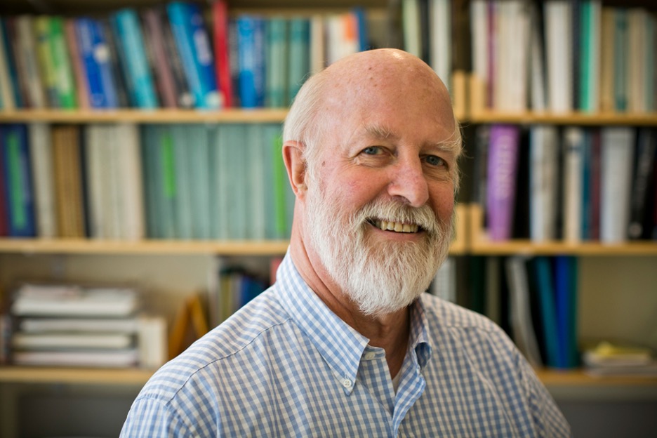
MIT Professor Richard Hynes, a pioneer in studying cellular adhesion, has been named a recipient of the 2022 Albert Lasker Basic Medical Research Award.

Hynes, the Daniel K. Ludwig Professor for Cancer Research and a member of MIT’s Koch Institute for Integrative Cancer Research, was honored for the discovery of integrins, proteins that are key to cell-cell and cell-matrix interactions in the body. He will share the prize with Erkki Ruoslahti of Sanford Burnham Prebys and Timothy Springer of Harvard University.

“I’m delighted, and it’s a pleasure to be sharing it with them,” Hynes says. “It’s great for the field, and for the trainees who did much of the work.”

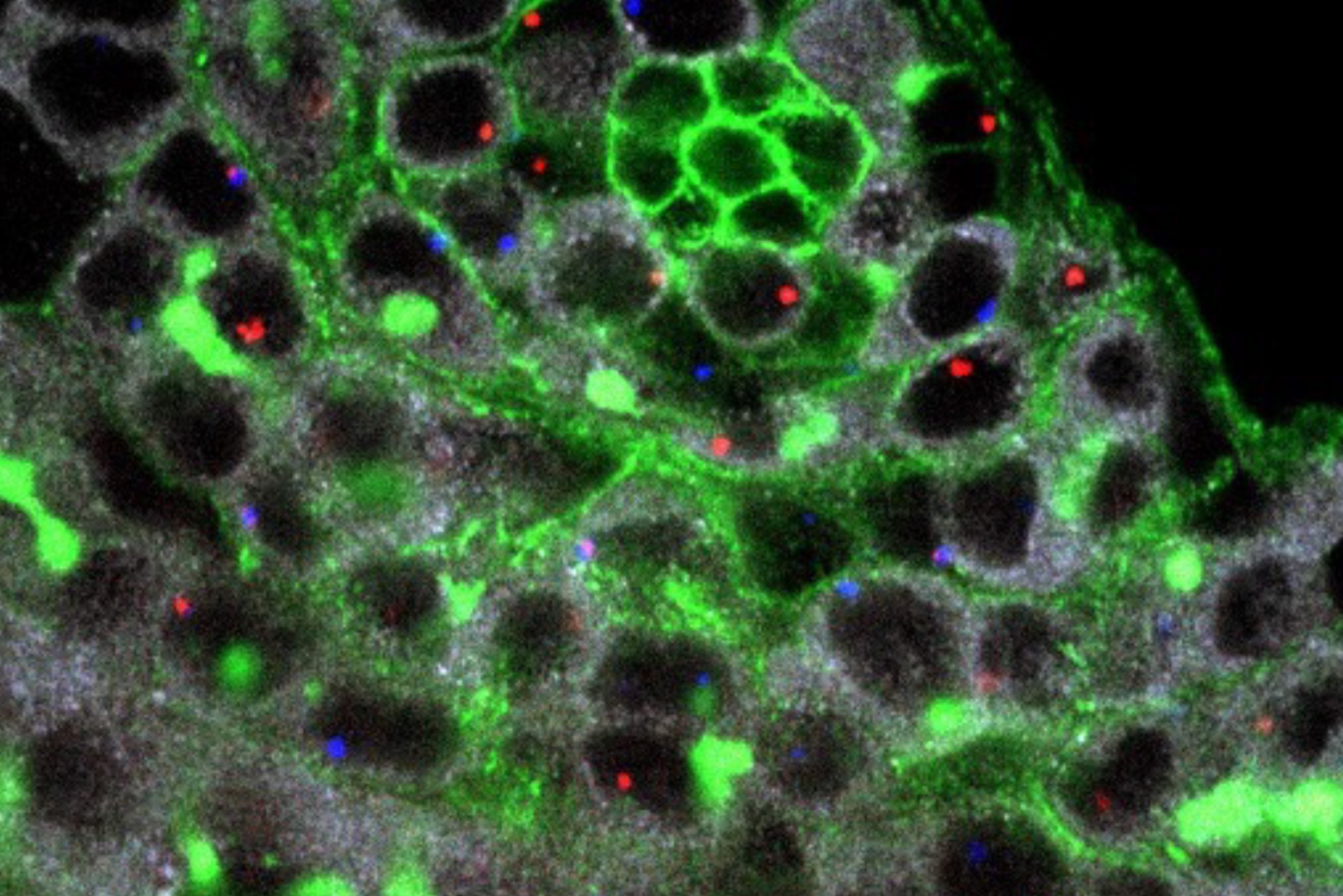
Hynes’ research focuses on proteins that allow cells to adhere to each other and to the extracellular matrix — a mesh-like network that provides structural support for cells. These proteins include integrins, a type of cell surface receptor, and fibronectins, a family of extracellular adhesive proteins. Integrins are the major adhesion receptors connecting the extracellular matrix to the intracellular cytoskeleton.

During embryonic development, cell adhesion is critical for cells to move to the correct locations in the embryo. Hynes’ work has also revealed that dysregulation of cell-to-matrix contact plays an important role in cancer cells’ ability to detach from a tumor and spread to other parts of the body, in a process known as metastasis.

“Professor Hynes’ contributions to the field of cancer biology, and more broadly, cellular biology, are numerous,” says Nergis Mavalvala, the Curtis and Kathleen Marble Professor of Astrophysics and the dean of the School of Science. “His investigations of fundamental biological questions — How do cells interact? How do they stick together? — changed how scientists approach cancer research and opened up avenues in developing potential therapeutics to disrupt metastatic disease.”

Born in Kenya, Hynes grew up in Liverpool, in the United Kingdom. Both of his parents were scientists: His father was a freshwater ecologist, and his mother a physics teacher. Hynes and all three of his siblings followed their parents into scientific fields.

“We talked science at home, and if we asked questions, we got questions back, not answers. So that conditioned me into being a scientist, for sure,” Hynes says.

After earning his bachelor’s and master’s degrees in biochemistry at Cambridge University, Hynes decided to head to the United States to continue graduate school. Colleagues at Cambridge suggested MIT, so he came to the Institute and earned his PhD in 1971. After doing a postdoc at the Imperial Cancer Research Fund Laboratories in London, he returned to MIT in 1975 as a faculty member in the Department of Biology and a founding member of MIT’s Center for Cancer Research (the predecessor of today’s Koch Institute).

Hynes began his career as a developmental biologist, studying how cells move to the correct locations during embryonic development. As a postdoc, he began studying the differences in the surface landscapes of healthy cells and tumor cells. This led to the discovery of a protein called fibronectin, which is often lost when cells become cancerous.

He and others found that fibronectin is part of the extracellular matrix, the network of proteins and other molecules that support cells and tissues in the body. When fibronectin is lost, cancer cells can more easily free themselves from their original location and metastasize to other sites in the body. Cells bind to the matrix through cell surface receptors known as integrins. In humans, 24 integrin proteins have been identified. These proteins help give tissues their structure, enable blood to clot, and are essential for embryonic development.

“These cell-matrix adhesion proteins hold us all together,” Hynes says. “If we didn’t have them, we’d be a pool of cells on the floor. And they’re contributors to lots of diseases: fibrosis, cancer, thrombosis, immune and autoimmune diseases. So, cell adhesion has become a huge field at both the basic science level and the therapeutic level.”

Since joining the MIT faculty, Hynes has also served as head and associate head of the Department of Biology, and as director of the Center for Cancer Research. He has also served as scientific governor of the Wellcome Trust in the United Kingdom, and as co-chair of National Academy committees establishing guidelines for stem cell and genome editing research.

His many awards include the Gairdner Foundation International Award, the Distinguished Investigator Award from the International Society for Matrix Biology, the Robert and Claire Pasarow Medical Research Award, the E.B. Wilson Medal from the American Society for Cell Biology and the Paget-Ewing Award, Metastasis Research Society. Hynes is also a member of the National Academy of Sciences, the National Academy of Medicine, the Royal Society of London, the American Association for the Advancement of Science, and the American Academy of Arts and Sciences.

The Lasker Award comes with a $250,000 prize, which will be shared between the three recipients.

Yami Acevedo ­Sánchez’s “go for it” attitude leads to MIT
Pamela Ferdinand
September 21, 2022

MIT PHD STUDENT YAMI ACEVEDO-SÁNCHEZ DISCOVERED SHE ENJOYED SCIENCE by watching television at home in Puerto Rico. While a strong student, encouraged by her mentors and parents to do well, she never imagined a science career would be in her future.

Acevedo-Sánchez is the second member of her extended family—her mother has 17 brothers and sisters; her father has 11—to earn a college degree. She didn’t learn about MIT until she began studying at the University of Puerto Rico, and attending the Institute felt like a very big step.

“I remember my thoughts were, ‘I’m never going to make it there.’ It felt really, really out of reach,” she says. “But I don’t say ‘no’ to myself. I just go for it.”

Today at MIT, Acevedo-Sánchez is pursuing her passion for biology, working to understand the basic processes that make all the complexity of life possible. “To me, it seems like a puzzle waiting for someone to assemble the pieces,” she says.

Her research focuses on a fundamental question: How do bacterial pathogens hijack a host? By studying how they travel between cells and spur infection, she hopes to discover more about the diseases they cause and potential therapies.

In particular, she is focused on Listeria monocytogenes, a widespread bacterium that can cause food poisoning. In high-risk populations, such as pregnant women or immunocompromised individuals, it can spread to the liver and then move through the bloodstream into the rest of the body. Listeria infection (listeriosis) has a high mortality rate, killing an estimated 20% to 30% of those infected, according to the US Food and Drug Administration.

Listeria hijacks molecular pathways as it spreads from cell to cell. It typically forces itself into neighboring cells by ramming into cell junctions (spots where cells connect). The force and speed Listeria uses to do this is about 0.2–1 microns per second—the equivalent of 50 feet per second if Listeria were the size of a submarine, Acevedo-Sánchez says: “It’s very impressive to watch!”

What is the mechanism of this action? Is it random, or programmed and regulated by the bacteria or our cells? Working with assistant professor of biology Rebecca Lamason and others in Lamason’s lab, Acevedo-Sánchez hopes to answer such questions through groundbreaking work that visualizes the cell membrane dynamics as Listeria spreads from cell to cell. To do this, the team uses a cellular line with a membrane marker (developed by Lamason) and a confocal microscope, which can capture high-resolution images deep inside cells.

Acevedo-Sánchez is especially interested in exploring how the mechanisms of two proteins, CAV1 and PACSIN2, promote cell-to-cell spread over long distances in a short amount of time. “These pathogens are constantly interacting with their host,” she says. “By understanding the key players that mediate those interactions from the bacteria side as well as the cell side, we can understand more about the microbiology of the bacteria and our own cell biology.”

Mentoring others

Outside the lab, Acevedo-Sánchez is working to support others like her who have not always believed they could pursue careers in science, technology, engineering, and mathematics. “There is tremendous power in having someone believe in your ability,” she says.

She has served as a mentor for the MIT Summer Research Program in Biology and supported first-year biology graduate students through the BioPals Program. Acevedo- Sánchez has also presented her work to middle school students around the world through the video series MIT Abstracts.

“Anyone can be a scientist, regardless of their background,” says Acevedo-Sánchez, who also has served as a graduate diversity ambassador at MIT. “You just need three things: be curious about the world that surrounds you, be willing to ask questions, and do the work yourself. Work smart and hard.”

Pamela Ferdinand is a 2003–2004 MIT Knight Science Journalism Fellow

Germ cells move like tiny bulldozers
Eva Frederick | Whitehead Institute
September 15, 2022

During fruit fly embryo formation, primordial germ cells — the stem cells that will later form eggs and sperm — must travel from the far end of the embryo to their final location in the gonads. Part of the primordial germ cell migration is passive; the cells are simply pushed into place by the movements of other cells. But at a certain point in development, the primordial germ cells must move on their own.

“A lot of the background in this field has been established by studying  how cells move in culture, and there’s this model that they move by using their cytoskeleton to push out their membranes to crawl,” said Benjamin Lin, a postdoctoral researcher in the lab of Whitehead Institute Director Ruth Lehmann. “We weren’t so sure they were actually moving that way in vivo.”

Now, in a new paper published September 14 in Science Advances, Lehmann who is also a professor of biology at the Massachusetts Institute of Technology, and researchers at Whitehead Institute and the Skirball Institute at New York University School of Medicine show that germ cells in growing fly embryos are in fact using a different method of movement which depends on a process called cortical flow, similar to the way bulldozers move on rotating treads. The research also reveals a new player in the pathway that governs this germ cell movement. “This work brings us one step closer to understanding the regulatory network that guides the germ cells on their long and complex journey across an ever-changing cellular landscape,” Lehmann said.

The research could also provide researchers with a new model for studying this type of cell movement in other situations — for example, cancer cells have been shown to move via cortical flow under certain conditions. ”We think there are more general implications for this mode of migratory behavior that go beyond primordial germ cells and apply to other migratory cells as well,” said Lin.

Balloon-shaped cells 

The first clue that Lehmann and Lin found that germ cells might not move the way scientists thought came from a simple observation. “When we began to study how these primordial germ cells move in the embryo, we saw that the cells actually remain shaped like a balloon while they’re moving and they don’t actually change their shape at all,” said Lin. “It’s really different from the crawling model.”

But if the cells weren’t moving by crawling, how were they moving through the embryo? To find out more, the researchers developed new techniques to image the germ cells in live fly embryos, and were able to watch clusters of a protein called actin moving backwards in each cell, as the cell itself was moving forward.

“There’s this thin layer of actin cytoskeleton just under the membrane of cells called the cortex, and they actually moved by making that cortex ‘flow,’” said Lin. “It’s like if you think of the tread of a bulldozer that’s moving backwards as the bulldozer is moving forward. The cells move that cortex backwards to generate friction to move the cell forward.”

Lin hypothesizes that this method of movement is especially well-suited to germ cells moving through a crowded embryo with many different cell types because instead of depending on recognizing specific proteins to “grab” in order to pull themselves through the embryo, it allows the germ cells to move independently. “Everything is pretty individualistic for primordial germ cells,” he said. “They don’t actually signal to each other at all, all the signaling is within each cell… And germ cells have to move through so many different tissues that they need a universal method of movement.”

A new role for a known protein

The researchers also found new information about how the cells control this form of motility. “We found that a protein called AMPK can control this pathway, which was really unexpected,” Lin said. “ Most people know it as a protein that senses energy. We found that this protein was important for helping these cells navigate. It’s one of these upstream players that can control how fast the cell goes, and in which direction.”

In the future, the researchers hope to map the entire pathway that allows germ cells to get to the right place at the right time in development. They also hope to learn more about the mechanisms behind cortical flow. “We want to figure out what is important for establishing these flows,” Lin said. “Our findings here could have implications not just for germ cells, but for other migrating cells as well.”

Notes

Benjamin Lin, Jonathan Luo, Ruth Lehmann. “An AMPK phosphoregulated RhoGEF feedback loop tunes cortical flow–driven amoeboid migration in vivo.” Science Advances, September 14, 2022. DOI: 10.1126/sciadv.abo0323

Hot off the press: parasite researchers melt down proteins to understand their roles in infection
Eva Frederick | Whitehead Institute
August 31, 2022

Much like humans, plants, and bacteria, the single-celled parasite Toxoplasma gondii (T. gondii) uses calcium as a messenger to coordinate important cellular processes. But while the messenger is the same, the communication pathways that form around calcium differ significantly between organisms.

“Since Toxoplasma parasites are so divergent from us, they have evolved their own sets of proteins that are involved in calcium signaling pathways,”  said Alice Herneisen, a graduate student in the lab of Whitehead Institute Member Sebastian Lourido.

Lourido and his lab study the molecular mechanisms that allow the single-celled parasite T. gondii and related pathogens to be so widespread and potentially deadly — and calcium signaling is an important part of the parasite’s process of invading its hosts. “Calcium governs this very important transition from the parasites replicating inside of host cells to parasites leaving those cells and seeking out new ones to infect,” said Lourido. “We’ve been really interested in how calcium plays into the regulation of proteins inside the parasite.”

A paper published August 17 in eLife provides some insight. In the paper, Herneisen, Lourido and collaborators used an approach called thermal profiling to broadly survey which parasite proteins are involved in calcium signaling in T. gondii. The new work reveals that an unexpected protein plays a role in parasite calcium pathways, and provides new targets that scientists could potentially use to stop the spread of the parasite. The data will also serve as a resource that other Toxoplasma researchers can use to find out whether their own proteins of interest interact with calcium pathways in parasite cells.

The heat is on

When studying calcium pathways in humans, researchers can often draw parallels from work in mice. “But parasites are very different from us,” said Lourido. “All of the principles that we’ve learned about calcium signaling in humans or mice can’t be readily translated over to parasites.”

So to study these mechanisms in Toxoplasma, the researchers had to start from scratch to determine which proteins were involved. That’s where the thermal profiling method came in. The method is based on the observation that proteins are designed to work well at specific temperatures, and when it becomes too hot for them, they melt. Consider eggs: when the proteins in egg whites and egg yolks are heated in a frying pan, the proteins begin to melt and congeal. “When we think about a protein melting, what we mean is the proteins unraveling,” said Lourido. “When proteins unravel, they expose side chains that bind to each other. They stop being individual proteins that are well-folded, and become a mesh.”

Small changes to the chemical structure of a protein — such as the changes resulting from binding a small molecule such as calcium — can alter the melting point of a protein. Researchers can then trace these alterations using proteomic methods. “Proteins that are binding calcium are changing in response to calcium, and are ultimately changing their thermal stability,” Herneisen said. “That’s kind of the language of proteins, alterations in their thermal stability.”

The thermal profiling method works by applying heat to parasite cells and graphing how each of the parasite’s proteins responds to changes in temperature under different conditions (for example, the presence or absence of calcium). In a 2020 paper, the researchers used the thermal profiling method to investigate the role of a protein called ENH1 in calcium signaling.

In their new paper, Lourido and Herneisen investigated the effect of calcium on all proteins in the parasite using two approaches. The researchers combined parasites with specific amounts of calcium, applied heat, and then performed proteomics techniques to track how the calcium affected the melting behavior of each protein. If a protein’s melting point was higher or lower than usual, the researchers could deduce that that protein was changed either by calcium itself or by another player in a calcium signaling pathway.

They then treated the parasites with a chemical that caused them to release stored calcium in a controlled manner and measured how a protein modification called phosphorylation changed over time. Together, these methods allowed them to infer how proteins might sense and respond to calcium within the signaling network.

Their approach provided data on nearly every expressed protein in the parasite cells, but the researchers zeroed in on one particular protein called Protein Phosphatase 1 (or PP1). The protein is ubiquitous across many species, but has never previously been implicated in calcium signaling pathways. They found that the protein was concentrated at the front end of the parasite. This region of the parasite cell is involved in motility and host invasion.

The protein’s role in the parasites — and in the other organisms in which it appears — is to remove the small molecules called phosphates from phosphorylated proteins. “This is a modification that can often change the activity of individual proteins, because it’s this big charge that’s been covalently stuck onto the surface of the protein,” Lourido said. “This ends up being a principle through which many, many different biological processes are regulated.”

How exactly PP1 interacts with calcium remains to be seen. When the researchers depleted PP1 in parasite cells, they found that the protein is somehow involved in helping the parasite take in calcium necessary for movement. It’s unclear whether or not it actually binds calcium or is involved in the pathway through another mechanism.

Because parasites use calcium signaling to coordinate life cycle changes such as entering or leaving  host cells, insights into the key players in calcium pathways could be a boon to public health. “These are kind of the pressure points or the hubs that would be ideal to target in order to prevent the spread and pathogenesis of these parasites,” Herneisen said.

Herneisen and collaborators focused primarily on PP1, but there are many other proteins to investigate using the data from this project. “I think part of the reason why I wanted to release this paper is so that the field could take the next steps,” she said. “I’m just one person — it would be great if 20 other people find that the protein that they were studying is calcium responsive, and they can chase down the exact reason for that or how it is involved in this greater calcium signaling network. This was exciting for us with regards to PP1, and I’m sure other researchers will make their own connections.”

Notes

Alice L. Herneisen,  Zhu-Hong Li, Alex W. Chan, Silvia NJ Moreno, and Sebastian Lourido. “Temporal and thermal profiling of the Toxoplasma proteome implicates parasite Protein Phosphatase 1 in the regulation of Ca2+-responsive pathways”. eLife, August 17, 2022. DOI: https://doi.org/10.7554/eLife.80336

The blueprint of a body
Eva Frederick | Whitehead Institute
July 20, 2022

Multicellular organisms evolved over millennia into a dazzling array of differently adapted creatures. With each generation, tiny worms, lavishly plumed birds, and even humans must create themselves anew from a single cell. To do so, they require a plan.

“How that multifunctional body plan is created is one of the deepest questions in developmental biology,” said Zak Swartz, until recently a postdoctoral researcher in the lab of Whitehead Institute Member Iain Cheeseman. “How do you take a single cell and pattern into a body that has different functions and features along it?”

Whitehead Institute researchers are tackling this question through a variety of different lenses. Researchers in Iain Cheeseman’s lab, including Swartz, have delved into the mysterious forces that underlie the polarity of an organism’s first cell. For the lab led by Pulin Li, research comes in at a later stage of development, when multiple cells combine to form a tissue and must communicate with each other to become an organized whole. Work on regeneration in Peter Reddien’s lab shows how some creatures can access their body blueprint throughout their lives to repair nearly any injury, and Yukiko Yamashita’s group studies how organisms pass on their body blueprints to their offspring through germ cells. Jonathan Weissman and his lab have created a “map” which researchers can use to find the function of a given gene, allowing them access to an organism’s most fundamental plans. Read on to learn about these scientists’ work, and more.

Laying out the plan

All multicellular organisms begin with a single cell, the fertilized egg. This cell has an essential role in setting out the body plan for the rest of an organism. It all starts with establishing polarity — in other words, figuring out which side of the cell is the top, and which is the bottom. This polarity establishes an axis of symmetry for the growing organism, and sets the stage for other developmental processes to come.

In a 2021 study, Cheeseman and postdoctoral researcher Zak Swartz investigated how one protein in specific, called Disheveled, localizes in a cell to help create this polarity in sea star embryos. Swartz found that Disheveled started out uniformly distributed in small aggregations throughout the egg cell, or oocyte. As the cell prepared to divide, Disheveled aggregations dissolved and then reformed at what would become the “bottom” of the oocyte.

Once the initial polarity is established, the oocyte can divide, creating a bilaterally symmetric sea star larvae. The burgeoning cluster of cells must then undergo other processes to define the several axes of symmetry that adult sea stars are known for.

Talking through it 

If an organism’s developmental blueprints are to be followed as development progresses, cells must be able to effectively communicate with each other. That cell to cell communication is the area of expertise of Whitehead Institute Member Pulin Li.

During her postdoctoral fellowship at the California Institute of Technology, Li studied tissue patterning — the mechanisms by which an organism’s newly forming tissues are laid out. Specifically, she investigated a developmental mechanism called morphogen gradient formation.

These gradients, composed of chemicals present in developing embryos, function as spatial coordinate systems and help determine how various cell types will be arranged in the organism — for example which groups of cells will form the liver, or the bones, or the brain, and where they will be within the body.

Li was able to recreate these gradients in the lab, in a Petri dish, and then interpret their signals using time lapse imaging and mathematical modeling. Here at Whitehead Institute, she follows a “bottom-up” approach to studying these complex systems. The best way to understand how something works, she says, is to build it yourself.

A key process in asymmetric cell division preserves the immortality of the germline
Eva Frederick | Whitehead Institute
July 27, 2022

During cell division, chromosomes are replicated into two copies — one for each daughter cell. These copies, called sister chromatids, are usually considered identical. In fact, it’s the two pairs of sister chromatids that make up the symmetrical X shape usually shown when visualizing chromosomes.

A 2013 paper from the lab of Whitehead Institute Member Yukiko Yamashita showed that in the case of asymmetric cell division — such as when a stem cell is dividing into two different kinds of daughter cells (i.e. a stem cell and a differentiating daughter)  — sister chromatids of sex chromosomes actually may carry distinct information, and the dividing cell “chooses” which of the daughters receive a specific copy.

What that “choice” means, and how it’s executed, has been a mystery — until now. A new paper from Yamashita, who is also a professor of biology at the Massachusetts Institute of Technology and an investigator with the Howard Hughes Medical Institute, published in Science Advances on July 27, illuminates the mechanisms that underlie nonrandom sister chromatid segregation, and suggests that the whole process may serve as a way to maintain the amount of ribosomal DNA (or rDNA) that is passed on to subsequent generations. “Tying together these two processes — rDNA copy number maintenance and nonrandom chromatid segregation — is an unexpected and exciting advance in our understanding of how germ cells are able to maintain their immortality,” said Yamashita.

George Watase, a postdoctoral scholar in the Yamashita Lab, led the study. Watase began his research intent on discovering the genetic underpinnings of nonrandom segregation of X and Y chromosomes in the fruit fly Drosophila melanogaster. As he surveyed the genome for genes that were essential to nonrandom segregation, it became apparent that ribosomal DNA was key for the process.

When rDNA was left intact, the sister chromatid  with more rDNA was preferentially chosen by the daughter stem cell instead of the differentiating daughter cell. When rDNA was removed from X and Y chromosomes, however, Watase found that the sister chromatids segregated randomly to the daughter cells.

Ribosomal DNA, or rDNA, is composed of a long stretch of repeats of certain base pairs. The rDNA provides the instructions and material to make ribosomes, which are essential for cells to create proteins. “Most genes exist only as a single copy, but in the case of rDNA we have hundreds of copies in our genome,” Watase said. “The reason for this is that we need a massive amount of ribosomes to synthesize proteins to maintain our cells’ viability.”

As organisms age, most of their cells naturally lose some of those rDNA repeats, including germline stem cells.  However, germline cells are sometimes called “immortal” — while all other cells in the body are made anew with each generation and die when an organism dies, germline cells such as sperm and eggs must carry DNA between generations. THerefore, the stem cells that produce sperm and eggs thus cannot keep losing rDNA repeats, and must bypass the mortality of other cells, by maintaining  a high number of rDNA repeats over time.

By isolating proteins that bind to rDNA, Watase discovered one specific gene, the protein product of which bound to rDNA and somehow assigned the sister chromatid with more rDNA repeats to the daughter cell that was destined to remain a germline stem cell.

This particular gene had not been described before, and Watase and Yamashita were now tasked with naming it. Fruit fly genes are named after what happens to the animal when the gene is removed. When this new gene was knocked down, the germ cells of subsequent generations gradually lost the immortality that separates germline stem cells from their differentiated counterparts.

Watase wasn’t sure how to convey the intricacies of this outcome. In the end, it was Watase’s wife who came up with the perfect name: Indra. In Hindu scriptures, Indra, the lord of all deities, was given a garland of fragrant flowers by a sage called Durvasa. Indra placed the garland on the trunk of his elephant, but the animal was irritated by the smell of the flowers and threw the garland down, trampling it underfoot. When Durvasa saw this, he became enraged and cursed Indra, taking away his immortality.

The name also opened up a world of possibilities for naming future genes that are important in nonrandom sister chromatid segregation. “People sometimes pull from Roman or Greek myths when naming genes, but not as many people use names from Hindu myths,” he said. “And since this is new biology, if we identify additional related genes in the future, we can use names from Hindu myths again.”

Watase and Yamashita’s study opens new avenues for future research. For example, the paper focused primarily on male fruit flies and the production of sperm via asymmetric division. Indra is expressed in the female germline as well, and when the gene is knocked down in females, the resulting phenotype is much more severe. “There must be some mechanism in female germ cells to avoid rDNA copy number reduction,” said Watase. “We just don’t know what that mechanism is.”

In the future, Watase and Yamashita also hope to elucidate how exactly Indra is interacting with cell division machinery to influence which chromatid ends up in the stem cell and which in the differentiating cell, and beyond this mechanism, how the stem cell “selects” the longer chromatid.

“Many biologists study germ cells, but few specifically study how they maintain their immortality,” said Yamashita. “This study is a step towards understanding this fascinating property of germ cells. It’s a really fascinating area and we really have to keep digging deeper into this phenomenon.”

Yiyin Erin Chen and Sam Chunte Peng named as core members of Broad Institute and MIT
Broad Communications
July 12, 2022

Broad Institute of MIT and Harvard has named Erin Chen, a dermatologist and microbiologist, and Sam Peng, a biophysicist and physical chemist with expertise in single-molecule imaging, as core institute members.

Chen will join in January 2023 and will also serve as an assistant professor in the Department of Biology at MIT and an attending dermatologist at Massachusetts General Hospital. Peng joined in July 2022 and will serve as an assistant professor in the Department of Chemistry at MIT.

Chen’s lab will study the communication between the immune system and the diverse microbes that colonize every surface of the human body, with a focus on the human body’s largest organ, the skin.

Peng’s lab will develop novel probes and microscopy techniques to visualize the dynamics of individual molecules in living cells, which will improve the understanding of molecular mechanisms underlying human diseases.

“We are delighted to welcome Sam and Erin to the Broad community,” said Todd Golub, director of the Broad. “These creative scientists are each taking inventive approaches to understand the molecular signals and interactions that underlie biological processes in health and disease. These insights will help further the Broad’s mission of advancing the understanding and treatment of human disease.”

Erin Chen.
Erin Chen

Erin Chen earned her BA in biology from the University of Chicago, her PhD from MIT, and her MD from Harvard Medical School. Prior to joining the Broad, Chen was a Howard Hughes Medical Institute Hanna Gray Postdoctoral Fellow at Stanford University, in the lab of Michael Fischbach. She was also an attending dermatologist at the University of California San Francisco and at the San Francisco VA Medical Center. During her postdoctoral research, Chen developed genetic methods to study harmless commensal skin bacteria. She engineered these bacteria to generate anti-tumor immunity, pioneering a novel approach to vaccination and cancer immunotherapy.

At the Broad, members of the Chen lab will continue to employ microbial genetics, immunologic approaches, and mouse models to dissect the molecular signals used by commensal microbes to educate the immune system. Ultimately, Chen aims to harness these microbe-host interactions to engineer novel therapeutics for human disease.

“I’m excited to join the collaborative scientific community at the Broad and MIT, including those who have pioneered novel tools for examining biological mechanisms at higher spatial resolution,” said Chen. “The biology I study is quite basic, but I’m motivated by the potential impact it could have on patients. Figuring out how commensal skin bacteria are captured by the immune system could unlock a whole new therapeutic toolbox.”

Sam Peng
Sam Peng

Sam Peng earned his BS in chemistry from the University of California, Berkeley, and his PhD from MIT in physical chemistry. He completed his postdoctoral research at Stanford University as an NIH K99 Pathway to Independence scholar in the lab of Steve Chu. During his postdoctoral research, he developed long-term single molecule imaging in live cells using a novel class of nanoprobes. He applied this new technique to study axonal transport in neurons and the molecular dynamics of dynein — a motor protein involved in transporting cargo in cells.

At the Broad, the Peng group will aim to elucidate the molecular mechanisms underlying human diseases. Lab members will develop and integrate a diverse toolbox spanning single-molecule microscopy, super-resolution microscopy, spectroscopy, nanomaterial engineering, biophysics, chemical biology, and quantitative modeling to uncover previously unexplored biological processes. With bright and photostable probes, lab members will have unprecedented capability to record ultra-long-term “molecular movies” in living systems with high spatiotemporal resolutions and to reveal molecular interactions that drive biological functions. Peng’s group will focus on studying molecular dynamics, protein-protein interactions, and cellular heterogeneity involved in neurobiology and cancer biology. Their long-term goal is to translate these mechanistic insights into drug discovery.

“Because my research is so multi-disciplinary, joining the Broad and MIT communities allows us to integrate a range of experimental tools and to collaborate with colleagues and students from diverse backgrounds,” said Peng. “I’m excited to see how our techniques can enable discoveries for a variety of cellular processes, including those underlying complex brain functions and dysfunctions. Many problems that previously seemed inaccessible now appear to be within reach in the foreseeable future.”