Siniša Hrvatin Named a Searle Scholar

Biology Professor and Whitehead Institute Member Siniša Hrvatin has been named as one of the 15 researchers to be selected as 2023 Searle Scholars. The Searle Scholars Program supports the research of exceptional young faculty in the biomedical sciences and chemistry.

Merrill Meadow | Whitehead Institute
May 12, 2023

Whitehead Institute Member Siniša Hrvatin has been named as one of the 15 researchers to be selected as 2023 Searle Scholars. The Searle Scholars Program supports the research of exceptional young faculty in the biomedical sciences and chemistry.

Chosen by an advisory board of eminent scientists, Searle Scholars are considered among the most creative researchers pursuing careers in academic research. Their investigations address challenging research questions and can lead to new insights that fundamentally change their fields—and to opportunities for translating discoveries into new therapeutics and diagnostics.

“I am truly grateful for the support of the Searle Scholar Program as we embark on this ambitious project,” says Hrvatin, who joined the Institute in 2021 and is also an assistant professor of biology at Massachusetts Institute of Technology. The three-year grant accompanying the award will support his work developing a new animal model for the study of hibernation.

“The ability to maintain nearly constant body temperature is a defining feature of mammalian and avian evolution; but, when challenged by harsh environments, many species decrease body temperature and metabolic rate and initiate energy-conserving states of torpor and hibernation,” Hrvatin notes. “Science has not yet answered the fundamental questions of how mammals initiate, regulate, and survive these extraordinary hypometabolic and hypothermic states.

“However, those answers could have profound medical applications,” he explains. “For example, harnessing the mechanisms behind hibernation might provide new approaches to protect neurons from ischemic injury and to preserve tissues and organs for transplantation.”

In the Searle-supported study, Hrvatin aims to discover a control center in the brain that regulates distinct stages of hibernation in the Syrian hamster. His lab will start by identifying the brain regions active during the deep torpor stage of hibernation and, using molecular profiling techniques, will then identify the specific neuronal populations and molecular pathways involved. Finally, the team will develop new tools to determine specific activities in those neural populations that are necessary for natural hibernation—and that may be sufficient to induce a synthetic state of hibernation.

“Taken together,” Hrvatin says, “I believe that our discoveries and the tools we build will help establish the first controllable animal model of hibernation.”

Since 1981, 677 scientists have been named Searle Scholars and the Program has awarded more than $152 million in support for Scholars’ research. To date, 85 Searle Scholars have been inducted into the National Academy of Sciences, 20 have been recognized with a MacArthur Fellowship, and two have been awarded the Nobel Prize for Chemistry.

The Human Genome Project Turns 20: Here’s How It Altered the World

"It simply changed the way that people thought that biology could be done."

Ed Cara | Gizmodo
April 11, 2023

On April 14 2003, scientists announced the end to one of the most remarkable achievements in history: the first (nearly) complete sequencing of a human genome. It was the culmination of a decade-plus endeavor that involved thousands of scientists across the globe. Many people hoped the accomplishment would change the world for the better.

For the 20-year anniversary of this historic event, we took a look back at the Human Genome Project and its impact. How did it shape science moving forward? How many of the expected goals have been reached since? And what lies ahead for the study of genetics?

A genome is the entire set of genetic information that makes up an organism. This information is packaged into sequences of DNA we call genes, which in humans are spread along 23 pairs of chromosomes. Only a small portion of these genes contain the instructions for coding the many proteins essential for life, but much of the rest is still thought to be important to our functioning. As scientists would eventually confirm, one copy of the human genome has around 3 billion base pairs of DNA. The sheer magnitude of the effort needed to map all this wasn’t lost on the researchers involved in the project, especially given the technology available decades ago.

“There’s been lots of analogies that people have put forward—like us being Lewis and Clark. We didn’t really have a map,” said Richard Gibbs, founder and director of the Baylor College of Medicine Human Genome Sequencing Center in Texas, one of the major institutions involved in the project.

Gibbs and his many colleagues knew they had to make compromises. Despite the advancements in sequencing technology since the official start of the project in 1990, they couldn’t fill in every gap with their current tools. And the first human genome was a composite of several blood donors in the U.S., not a single person. Along the way, private company Celera entered the picture, promising that it would complete a separate genome project using its own techniques even faster. Ultimately, both groups finished ahead of schedule around the same time, with the first draft sequences released in 2000, though Celera announced its success a few months earlier.

Regardless of the victor, the feat certainly did usher in a new era of genetics research—one that has seen great leaps in speed and efficiency since 2003.

“I think the very most important accomplishment in the past 20 years has been the advent of next-generation sequencing. The ability to perform sequencing in a massively parallel way, so that you could do it far more quickly and cheaply,” said Stacey Gabriel, director of the Genomics Platform at the Broad Institute of MIT and Harvard, another major research site involved in the Human Genome Project. “And that has come with all of the associated advancement in our computational abilities, too, to really be able to take that data and analyze it at a massive scale as well.”

The original project cost $2.7 billion, with most of the genome being mapped over a two-year span. Nowadays, the current speed record for sequencing a genome is around five hours (more often, though, it takes weeks), and this past fall, the company Illumina unveiled a machine that it claims will cost as little as $200 per sequence, down from the recently typical $600 cost.

Greg Findlay leads the Genome Function Laboratory at the Francis Crick Institute in the UK. His team is one of many around the world that is building on the work of the original project. They’re currently trying to identify and understand how certain variants in tumor-suppressor genes can raise our risk of cancer.

“So what my lab tries to do is actually understand what variants do to the genome. That is, we want to know exactly which variants cause disease and why they cause disease. Right now, we’re focused on certain genetic changes that lead to cancer, and I think this particular field has really been revolutionized by the Human Genome Project,” Findlay said. “We now know there are many, many different genetic paths by which cells can turn into cancer. And we know this, because we’ve been able to actually sequence the DNA in so many different tumors repeatedly, across all different types of cancer, to see what are the mutations that actually lead to cancer forming.”

Perhaps equally important was the project’s impact on scientific collaboration. The effort directly led to an international agreement meant to ensure open access to DNA sequences. It also made clear that great things could be possible when large groups of scientists worked together, according to Gibbs.

“It simply changed the way that people thought that biology could be done,” he said. “It built a model for team science that was not there before.”

Even at the time, though, the project knowingly left some things unfinished. They had mapped roughly 92% of the genome by 2003, but it would take almost 20 more years for other scientists to track down the remaining 8%. This missing “dark matter” of our genome could very well provide new clues about how humans evolved or our susceptibility to various diseases.

Much of the genetic information collected and analyzed since the project ended has come from white and European populations—a disparity that hampers our ability to truly understand the impact of genetics on everyone’s health. But scientists today are working on bridging that gap through initiatives like the Human Pangenome Project, which will sequence and make available the full genomes of over 300 people intended to represent the breadth of human diversity around the globe.

“There’s genetic variation that exists across all the world’s populations. And if you only use variations from a sliver of the world’s populations, and you try to apply that to everybody else, it just doesn’t work very well, because we all have different backgrounds,” said Lucinda Antonacci-Fulton, one of the project’s coordinators and director of project development & new initiatives at Washington University in St. Louis’s McDonnell Genome Institute. “So the more inclusive you can be, the better off you are in terms of treatments that you want to bring into the clinic.”

As important as genetics research has been, some of the expectations fueled by the Human Genome Project likely were too lofty. In 2000, for instance, President Bill Clinton claimed that the project would “revolutionize the diagnosis, prevention and treatment of most, if not all, human diseases.” While we do continue to discover new gene variants that strongly predict the odds of developing a specific disease or trait, there are countless others that we’re still in the dark about. Elsewhere, it’s become clear that our genome often only plays a small or negligible role in why we get sick or experience something in a particular manner. So although the project has helped unlock some of the mysteries of the world, there are so many more questions out there about why we are the way we are, and our genes are probably not going to provide a neat answer to many of them.

“I think where things are oversold, sometimes, is with this notion that just because the human genome is done, you’re going to be able to read it off for some sort of deterministic answer—where finding genes for disease becomes like falling off a log,” Gabriel notes. “But often human disease, especially the diseases that impact us the most, these are not simple genetic diseases. They’re multifactorial. They’re combinations of your genes, your behavior, exposure to the environment, sometimes just bad luck.”

None of this is to sell short the potential of genetics. Hans Lehrach, a former director at the Max Planck Institute for Molecular Genetics in Germany, was one of the first researchers involved in the Human Genome Project. He’s also one of many scientists who believe that we’ll someday be able to cheaply and easily scan a person’s genome at a moment’s notice and that this information, along with other aspects of our molecular make-up, will help guide the specific drugs or interventions doctors prescribe—a concept known as personalized medicine. Notably, the treatment of some cancers is already influenced by the variants that underlie their growth. Some experts even argue that widespread whole genome sequencing should start as early as birth, and there are already small-scale programs in the U.S., UK, and elsewhere testing out its potential benefits and risks.

“Not knowing about your genome is a bit like crossing the street while closing your eyes because you don’t want to see a bus coming. If we don’t sequence our genomes, the buses keep coming anyway—it just lets us open our eyes and maybe see the kind of danger that we can escape or do something about,” Lehrach said.

The Human Genome Project truly has changed the scientific landscape, but we’re still only at the very beginning of seeing the world that it’s made possible.

Balance between proteins keeps sperm swimming swiftly

Developing sperm cells swap out histones for proteins called protamines to coil DNA tightly enough to fit inside the hydrodynamic shape ideal for the task of swimming swiftly to an egg in order to fertilize it. If the balance of protamines in the sperm is wrong, however, the sperm may become misshapen and die, making the animal infertile. Whitehead Institute Member Yukiko Yamashita and former graduate student Jun Park have discovered why this imbalance causes infertility in the fruit fly.

Greta Friar | Whitehead Institute
April 10, 2023

Sperm must swim swiftly to an egg in order to fertilize it, and so they have evolved hydrodynamic shapes. Most of the space in the head of sperm cells is taken up by the DNA they carry, so the cells coil up their DNA super tightly to stay small and streamlined. In most cell types, DNA is coiled around proteins called histones. These do not package DNA tightly enough for sperm, so when a sperm cell is developing, it swaps out histones for another type of protein called protamines that coil DNA very tightly.

Many species, including humans, mice, and flies, have multiple types of protamines. If the balance between the different types is wrong, then the sperm’s DNA may not be packaged correctly and it may become misshapen and die, making the animal infertile. Whitehead Institute Member Yukiko Yamashita and former graduate student Jun Park have discovered why this imbalance causes infertility in the fruit fly (Drosophila melanogaster). The finding, published in the Proceedings of the National Academy of Sciences on April 10, showed a mechanism that balances different types of protamines to ensure male fertility.

Mst77F is a major fruit fly protamine. Yamashita and Park determined that the fruit fly protamine Mst77Y, which is related to Mst77F, can interfere with the function of Mst77F. Fruit flies usually make a lot of Mst77F and a little of Mst77Y. The researchers found that when expression of the Mst77Y gene is too high, especially when expression of Mst77F is low, it disrupts the process of DNA packaging, leading to infertility.

How does Mst77Y interfere with Mst77F? The researchers discovered that this is because the Mst77Y gene makes faulty protamines. There are multiple copies of Mst77Y on the fly’s Y chromosome. They likely evolved from a copy of Mst77F, which is not on a sex chromosome. However, the different versions of Mst77Y have lost or altered parts that they need in order to function, so unlike the Mst77F protamine, Mst77Y protamines likely cannot coil DNA tightly around themselves. In spite of the fact that the Mst77Y protamines do not work correctly, they are dominant: when they are present, the sperm cell will use them over the functional Mst77F protamines.

“Mst77Y is a half-broken tool,” says Yamashita, who is also a professor of biology at the Massachusetts Institute of Technology and a Howard Hughes Medical Institute investigator. “It is able to take the place of the working tool, Mst77F, but not to do its job, so when too much Mst77Y is present, the sperm cell does not have enough working tools in place to compact its DNA.”

The researchers also figured out how sperm cells keep expression of Mst77F high and Mst77Y low: with the help of a protein called Modulo. In order for an RNA read from a gene to be made into a protein, it needs to have a tail added to it made up of a string of adenines—one of the four building blocks that make up RNA. Modulo makes sure that the cell preferentially adds this tail to the RNA coding for Mst77F. Although Yamashita and Park did not determine the exact mechanism by which Modulo ensures this preferential treatment, they did observe that Modulo and the Mst77F RNA group together in the same part of the cell, the nucleolus, whereas Mst77Y does not.

Altogether, these findings explain why and how fruit fly sperm cells carefully balance the levels of these two protamines. However, the research raises the question, what are sperm cells using the non-functional Mst77Y protamines for? Yamashita and Park can only speculate, but the answer may have to do with their observation that high levels of Mst77Y killed off more X-chromosome bearing sperm than Y-chromosome bearing sperm. Past research has suggested that protamines may be involved in a process called meiotic drive, which animals can use to skew the sex ratio of their offspring. This new work is not only consistent with that hypothesis, but provides a possible mechanism to explain how protamines contribute. The researchers note that they did not see a strong effect on the sex ratio of offspring in this experiment, but hope that this work could set the stage to understand the role of non-functional protamines in meiotic drive.

“At the cell level, we were able to show that there’s some basis for this protamine to be involved in biasing whether X or Y chromosome bearing sperm survive,” Park says. “An interesting next question would be to see if there are certain conditions in which this mechanism more clearly acts as a driver at the level of offspring’s sex ratio.”

Notes

Park, Jun I., George W. Bell, and Yukiko M. Yamashita. 2023. “Derepression of Y-linked multicopy protamine-like genes interferes with sperm nuclear compaction in D. melanogaster,” PNAS 120 (16). https://www.pnas.org/doi/10.1073/pnas.2220576120

New research supports finding explaining why some patients may test positive for COVID-19 long after recovery

SARS-CoV-2, the virus that causes COVID-19, seems to have become a permanent presence in our lives. Research from Whitehead Institute Founding Member Rudolf Jaenisch’s lab reveals that this may be true on multiple levels.

Greta Friar | Whitehead Institute
February 28, 2023

SARS-CoV-2, the virus that causes COVID-19, seems to have become a permanent presence in our lives. Research from Whitehead Institute Founding Member Rudolf Jaenisch’s lab reveals that this may be true on multiple levels. Jaenisch, postdoc Liguo Zhang, and colleagues have shown that when the virus infects people, it is capable of integrating parts of its genetic code into the human genome through a process called reverse transcription. This genomic integration is rare, but due to how many hundreds of millions of people have been infected, it has likely occurred many times.

In a paper published in the journal Viruses on February 25, the researchers use and compare multiple methods to show that SARS-CoV-2 can integrate into host cells’ genomes. The paper is a follow up to Jaenisch and Zhang’s 2021 paper in the Proceedings of the National Academy of Sciences, which provided initial evidence of SARS-CoV-2 genomic integration. The original paper intended to solve the puzzle of why some people who had had COVID-19 were still testing positive long after recovering from the disease. The answer the researchers found was that parts of the viral genome were reverse transcribed into the human genome, meaning the viral RNA was transcribed or “read” into DNA (a reverse of the usual process) and then that DNA was stitched into the cell’s DNA. Then, when the cells’ genomes were transcribed into RNA, the portion of the virus’ genome that had been incorporated would be included and could be recognized by a PCR test, leading to a positive result.

In order to further substantiate the findings described in the previous paper, Jaenisch and Zhang have now performed additional experiments and analyses. The new paper explains why some experiments testing for viral genomic integration would come up with a negative result, and how this is consistent with Jaenisch and Zhang’s conclusion. Additionally, Jaenisch and Zhang examine whether viral RNA put into cells, as a model of the COVID-19 mRNA vaccines, can also integrate into the human genome, and find initial evidence that it cannot.

“This paper puts our data on a very firm footing,” Jaenisch says. “Hopefully, it will clarify some of the issues raised in the discussion that followed the first paper, and provide some reassurance to people who were worried about the implications for the vaccine.”

Hunting for a needle in a haystack

The main challenge in finding evidence of SARS-CoV-2 integrating into the human genome is that this event appears to be very rare. In the new paper, Jaenisch and Zhang used digital PCR, an approach that can sensitively detect specific DNA sequences in cells, to see how commonly the sequence that they would find in instances of viral RNA being read into DNA appeared in infected cells. Specifically, they looked for reverse transcribed SARS-CoV-2 complementary DNA (cDNA), DNA that is made from the virus’ original mRNA. Digital PCR revealed that for every one thousand cells, reverse transcribed viral cDNA was only present in around four to twenty cells. This number includes all detected instances of viral cDNA, whether integrated into the genome or not, so genomic integration is likely even rarer—indeed, the new research suggests that only a fraction of the total cDNA identified is from genomic integration.

Because genomic viral integration is so rare, Jaenisch and Zhang needed to use multiple complementary methods to test for it. One approach, called whole genome sequencing (WGS), is able to search cells’ genomes in great detail. When it does come across an instance of viral genomic integration, it can identify not only the reverse transcribed viral sequence, but also two sequences near the viral sequence that are added when it is integrated into the genome by a common reverse transcription complex called LINE1, which is encoded in the host cells. The combination of viral cDNA plus the two nearby cellular host sequences provides very strong evidence that viral cDNA is not only present but has been incorporated into the cell’s genome. However, WGS can only search the equivalent of a few cells’ genomes, and so when searching for a rare event, like SARS-CoV-2 integration, it often comes up empty. People skeptical of the first paper performed this type of experiment and came up with a negative result; Jaenisch and Zhang were not surprised by that, and it is consistent with their own findings when using this approach.

“Because the human cell genome coverage by whole genome sequencing is very limited, you would need to run the sequencing experiment many times in order to have a good chance of detecting one viral genome copy,” Zhang says.

In order to make the most of WGS, Jaenisch and Zhang induced their cells to overexpress LINE1, the cellular machinery that reverse transcribes viral RNA into the human genome. This exponentially increases the amount of viral cDNA that gets made; when the researchers performed digital PCR on their cells with overexpression, it detected fourteen to twenty thousand cDNA copies per thousand cells. Consequently, WGS was able to detect instances of viral cDNA plus the two nearby sequences that are the telltale signature of genomic integration in these cells.

“This is unambiguous proof of viral genomic integration,” Zhang says.

This type of experiment is called a positive control. Researchers use it to prove that, in ideal circumstances, the biological phenomenon they are curious about can occur. The question then becomes: does the phenomenon happen in normal circumstances? This was a criticism raised by some researchers in response to the first paper: they were not convinced that viral genomic integration happens in the cells of an infected person, which do not have the same levels of LINE1.

The search gets shallower but wider

Jaenisch and Zhang used another approach to hunt for evidence of viral genomic integration in cells without LINE1 overexpression. The approach, called an enrichment method and performed with the tool TagMap, can analyze thousands of cells—enough cells to reliably find evidence of a rare event. However, it cannot get the same detail as whole genome sequencing; TagMap enriches and captures shorter sequences of DNA, so it can only capture one of the two nearby sequences that act as a signature alongside viral cDNA. However, the smaller stretch of DNA that the researchers focused on still has features that can be used as evidence of integration. With this approach, Jaenisch and Zhang detected many instances of viral cDNA linked to the nearby cellular sequence.

Jaenisch and Zhang argue that the combined results of these experiments show strong proof of viral integration. Whole genome sequencing provides very strong proof that viral genomic integration can occur in the right conditions. Enrichment with TagMap provides reasonably strong proof that viral genomic integration occurs in normal cells.

“Each of these methods has advantages and disadvantages. You have to combine them to get the complete picture,” Jaenisch says.

Turning to the vaccine

After reaffirming their results that genomic integration of SARS-CoV-2 happens following viral infection, the researchers wanted to know whether the same thing happens with mRNA from the COVID-19 vaccines—which had been a concern expressed by many in the wake of the first paper. Jaenisch and Zhang could not get access to the actual vaccine RNA, packaged into a lipid coat, which is used for vaccination. Instead, they created a model of vaccine injection, inserting a bit of SARS-CoV-2 genetic material (mRNA) into cells through transfection, or non-infection “delivery” of genetic content into cells.

The researchers found that transfection of SARS-CoV-2 mRNA did not lead to genomic integration in the same way that infection did. Infection naturally produces a large amount of viral RNA and causes an inflammatory response in cells. Such cellular stresses increase the level of the reverse transcription machinery. Transfection does not do this, and correspondingly, the researchers found no evidence with TagMap that it led to viral genomic integration by LINE1 in normal cells.

The researchers’ model of vaccine injection is missing several key features of the actual vaccine. In the future, Jaenisch hopes to follow up on this research using the actual vaccine RNA sequence, and testing in an animal model to more closely match what happens during vaccine injection. In the meantime, the researchers hope that these initial results are reassuring.

“We need to do further testing, but our results are consistent with vaccine RNA not integrating,” Jaenisch says.

Notes

Zhang, Liguo, Punam Bisht, Anthony Flamier, M. Inmaculada Barrasa, Max Friesen, Alexsia Richards, Stephen H. Hughes, and Rudolf Jaenisch. 2023. “LINE1-Mediated Reverse Transcription and Genomic Integration of SARS-CoV-2 mRNA Detected in Virus-Infected but Not in Viral mRNA-Transfected Cells” Viruses 15, no. 3: 629. https://doi.org/10.3390/v15030629

MIT vs. poop

When Greater Boston faced a sewage crisis, the MIT biology department was there to help.

Saima May Sidik, SM ’21
February 21, 2023

View of Neponset River in Mattapan.

In the 1800s, Boston had a problem: it stank, and there was no question why. Modern sewage treatment had not yet been invented, so human waste ran through makeshift drainage systems into the surrounding rivers and bays. “Large territories have been at once, and frequently, enveloped in an atmosphere of stench so strong as to arouse the sleeping, terrify the weak, and nauseate and exasperate everybody,” an official from the City Board of Health wrote in an annual report in 1872.

Nausea and exasperation were the least of Bostonians’ worries: sewage spreads bacteria that cause diseases such as cholera and typhoid fever. In 1849, a major cholera outbreak killed more than 600 Boston residents, prompting city officials to take action. By the 20th century, Boston had a system for pumping waste away from the city and into the ocean.

Hughes House
When heiress Sarah Hughes married, her father gave her a mansion, abutting the Neponset River. She had just one complaint: the river “smells so horribly that you can’t sit near it.” Her anonymous support of MIT’s sewage treatment research helped clean up the river and significantly reduced rates of typhoid fever.
MILTON HISTORICAL SOCIETY

The system improved conditions downtown, but for people living along certain sewage-laden waterways, the problem was far from solved. One of these was Sarah Hughes, daughter of the merchant and railroad stockholder John Murray Forbes, who was known as “the richest man in New England.” In 1889, Forbes gave Hughes an elaborate wedding present: a mansion in Milton, near the Neponset River, that would become known as the Hughes House. There was just one problem: “Neponset River smells so horribly that you can’t sit near it,” Hughes wrote in a letter she sent to MIT president Henry Smith Pritchett in 1902.

Hughes had the means to do more than just complain. So for about 15 years, she anonymously donated thousands of dollars to MIT each year for research on treating human waste. By the 1920s, sewage purification systems—developed partly thanks to Hughes’s support—had reduced rates of typhoid fever significantly. In 1908, the disease struck at least 15 people per 100,000 in 35 US cities. Ten years later, only two or three cities were experiencing such dire problems. Techniques developed during this period underlie today’s methods for treating human waste.

Wryly suggesting that few Bostonians would believe in germs smaller than lobsters, Hughes pushed the researchers to describe their findings in plain language.

At the helm of MIT’s sewage treatment research was William Sedgwick, the founding head of the biology department, who had been working with the Massachusetts State Department of Health to improve water safety since 1888. At an experiment station in Lawrence, he and his colleagues built on European research to devise a method of removing pathogenic microbes by filtering sewage through sand. Harmless microbes that grew in the sand added to the filtering effect and prevented the harmful bacteria from passing through.

William Sedgwick
William Sedgwick, the founding head of the biology department, led MIT’s sewage treatment research.
MIT MUSEUM

Filtering an entire city’s waste would require an unfeasibly large bed of sand, however. Using Hughes’s funding, Sedgwick and his colleagues—including MIT student Anne Rogers, Class of 1904, and recent graduates Charles-Edward Amory Winslow, Class of 1898, SM 1899, and Earle Phelps, Class of 1899—aimed to devise more scalable methods. The work took place on the corner of Albany Street and Massachusetts Avenue in Boston, where they built a station consisting of a series of holding tanks positioned above 25 test tanks and filters divided between two levels. The researchers pumped sewage from a sewer that ran below the street into the holding tanks. They could then control the rate at which sewage ran into the test tanks on the upper level. After being subjected to treatment there, sewage ran into the lower level of tanks, where it underwent one or more additional experimental treatments. In this way, the researchers could test how sewage fared under different combinations of sequential treatments.

After a few years they noted some general trends. Four-foot-deep beds were better than six-foot-deep ones, but the improvement wasn’t enough to offset the smaller capacity and diminished filtration rate. The material filling the beds was less critical—coke, brick, and broken stone all worked. The size of the pieces was important, however; half-inch stone filtered much more effectively than coarser material.

Sewage treatment was an international field of study in the early 1900s, and many of the experiments performed at MIT involved fine-tuning techniques that had been developed elsewhere. For example, other groups had discovered that sewage could be reduced to an odorless liquid if it was left to trickle through a bed of coarse rocks, but this liquid still contained pathogenic bacteria. At the MIT station on Albany Street, researchers tested whether chlorine could kill these bacteria and render the liquid harmless. The experiment was a success, and the researchers estimated that 85 cents’ worth of chlorine could sterilize a million gallons of sewage. Today, chlorine is still part of the strategy at many wastewater facilities, including Boston’s Deer Island plant.

Hughes insisted on remaining anonymous, but rumors circulated about who was funding the station. In 1903, the philanthropist George Foster Peabody offered to pay for printing and distribution of 100,000 copies of an article about it, leading many to speculate that he was the donor. Behind the scenes, meanwhile, Hughes offered advice to the researchers through letters she wrote to Pritchett. Coauthor of the textbook Lessons on Practical Subjects, she believed strongly in the importance of educating the populace on essential topics and pushed the researchers to release pamphlets explaining their findings in plain language so that members of the state board of health, politicians, and the public could understand them. She had very little faith in the ability of the average Bostonian to comprehend the importance of sewage treatment otherwise. “Their slow and bewildered minds must be taught by the plainest language,” she wrote. “Unless a typhoid or diphtheria germ were the size of a lobster they would not believe in it.”

filter house diagram
MIT researchers tested sewage treatment options at its Albany Street station, which had a series of holding tanks positioned above 25 test tanks and filters divided between two levels.
MIT CONTRIBUTIONS FROM THE SANITARY RESEARCH LABORATORY AND SEWAGE EXPERIMENT STATION, VOLUME II

Hughes and Pritchett also discussed the business side of research and grappled with the slow, incremental process of scientific discovery. “We must admit quite frankly that they have not solved the problem which you had in mind, although I think they have made some important contributions toward it. Whether you care to have the work go on further is for you to say,” Pritchett wrote in 1905. Hughes was patient, and she kept sending money.

By 1909, the researchers had outgrown the Albany Street station, and Hughes donated money to erect a new one abutting the Calf Pasture Pumping Station in Dorchester, on what’s now the campus of the University of Massachusetts Boston. At this new location, the researchers could obtain sewage directly from an outflow—a more realistic scenario than using pumps to raise it from below the street. Here they began to move beyond studying how large cities could handle sewage, considering, for example, how rural communities lacking sewers could dispose of their waste. As effective sewage treatment systems became widespread, the MIT biology department shifted its focus toward other important research questions, like how to incorporate pasteurization into the burgeoning ice cream industry.

After Hughes’s death in January of 1917, Sedgwick wrote in a report to MIT’s president that many people would be surprised to learn that the donor was a woman. He went on to say, “We shall long cherish the memory of her alert, original, incisive, and powerful personality; of her determination to uphold whatsoever things are lovely and of good report; and her eagerness to put down all evil, to do away with filth, and to cleanse and purify the dirty places of this too often unclean world.”

Not so inactive X chromosome

Whitehead Institute Member David Page has spent his career understanding how the differences between X and Y contribute to these sex differences, but a recent project is taking his lab in a new direction: understanding how the differences between X chromosomes contribute to sex differences.

Greta Friar | Whitehead Institute
February 7, 2023

Nearly every cell in our body contains pairs of each of our chromosomes, and these pairs are identical in all but one case: that of our sex chromosomes. Males typically have one X and one Y sex chromosome, while females typically have two X chromosomes. In recent years, research has suggested that these different chromosomes can influence far more than sex determination. Gene expression from the sex chromosomes appears to contribute to sex differences in health and disease, which males and females experience in everything from the incidence of getting certain diseases, to the symptoms of diseases, to responses to drugs, and more. For example, women are more likely to develop autoimmune disorders, while men are more likely to develop heart conditions.

Whitehead Institute Member David Page has spent his career understanding how the differences between X and Y contribute to these sex differences, but a recent project is taking his lab in a new direction: understanding how the differences between X chromosomes contribute to sex differences. Although females’ pair of X chromosomes contain the same genes, they have different patterns of gene expression. New research from Page and postdoc Adrianna San Roman reveals just how different the two types of X chromosomes are. The findings, published in the journal Cell Genomics on February 8, show that one type of X chromosome, known as the inactive X chromosome, can modulate the gene expression of the other type of X chromosome, known as the active X chromosome. Their work indicates that inactive X chromosomes have underappreciated roles in gene regulation and, most likely, in sex differences in health and disease.

Difference rooted in history

Females’ two X chromosomes have different gene expression activity because of the sex chromosomes’ evolutionary history. The X and Y sex chromosomes evolved from a pair of identical non-sex chromosomes. Because of this ancestry, the sex chromosomes still contain genes that are important outside of regulating sex differences, such as genes that contribute to our immune system or regulate gene expression throughout the body. However, over time the Y chromosome shrank and lost most of its genes. Researchers think that in order to make up for the loss of necessary genes on the Y, expression of the corresponding genes on the X chromosome increased. This ensured that males still had the necessary levels of gene expression from their sex chromosomes, but now females, with two copies of X both working overtime, had levels of gene expression that were too high. To solve this problem, our bodies developed a process called X chromosome inactivation, by which the majority of genes on all but one copy of the X chromosome in each cell are silenced, or turned off. This means that everyone, male and female alike, has one copy of the X chromosome working at full strength–the active X chromosome. In males, the active X chromosome is paired with a Y chromosome, and in females, it is paired with a so-called inactive X chromosome, on which most of the genes are turned off.

In spite of the evolution of X chromosome inactivation, some percentage of genes on the inactive X chromosome are still expressed, such as genes that have an active counterpart on the Y chromosome. Previous research has indicated that about a quarter of the genes on the inactive X are, in fact, active, so researchers have long been aware that the chromosome is not completely silent. However, it’s still often painted as a passive copy playing backup for its more active partner. San Roman’s work shows that the inactive X chromosome’s gene expression is much more potent and complex than that.

A spectrum of sex chromosomes

In order to understand the inactive X chromosome’s contributions to gene expression, San Roman and colleagues in the Page lab collected blood and skin samples from people born with unusual combinations of sex chromosomes—everything from X0 (one X chromosome) to XXXXY. People with these different sets of chromosomes often have health issues; for example, X0 females have Turner syndrome, which can cause heart defects, hearing impairment, and more; and XXY males have Klinefelter syndrome, which can cause infertility, weak muscles, and more. Page and San Roman hope their research could provide useful insights into these health issues as well as into sex differences between XY males and XX females.

In people with more than one X chromosome, every X but one is an inactive X. The researchers graphed sex chromosome gene expression, measuring the change in expression level of each gene with the addition of each inactive X, for people with anything from zero to three inactive X chromosomes, as well as different numbers of Y chromosomes. They also looked at the relative contribution to overall expression from the active versus inactive X chromosomes. One might expect the graphs they made to be relatively straightforward: for genes that are turned off on the inactive X chromosome, the gene expression level would not change at all as the number of copies of the inactive X increased. For genes that are turned on, the gene expression level would double with two X chromosomes, triple with three X chromosomes, and so on. When the researchers looked at chromosomes other than X with extra copies—namely, Y and chromosome 21—this is essentially the pattern they observed. Gene expression from additional X chromosomes, however, was not so straightforward.

Each additional inactive X chromosome changes gene expression by the same amount. However, the researchers found a surprising diversity in expression levels across X chromosome genes. The presence of each additional inactive X might increase one gene’s expression by 20 percent and another’s by 70 percent. Then the results grew more surprising: for some genes, the addition of an inactive X decreased their expression. For some genes that are only expressed on the active X chromosome, and completely silent on the inactive X, additional inactive X chromosomes nonetheless changed their expression level.

These discrepancies led the researchers to a startling finding. The X chromosomes do not function independently of each other. Instead, the inactive X chromosome can modulate expression of genes on the active X chromosome. In other words, some genes on the inactive X chromosome regulate genes on the active X chromosome, dialing their expression up or down. Altogether, the researchers found that 38% of the X chromosome genes in the two cell types that they tested are affected by the presence of inactive X chromosomes, either because the genes are expressed on the inactive X, or because the inactive X regulates their expression on the active X, or through some combination of the two mechanisms.

These findings show that the inactive X plays a much more active role in gene expression and regulation than was previously appreciated. Rather than just playing second fiddle to the active X chromosome, the inactive X is sometimes harmonizing with and sometimes even conducting its partner.

Rethinking the role of the inactive X in health and disease

Page and San Roman hope that their findings will help refocus research into sex differences. Previous research into the mechanisms behind these differences has focused on the effects of having X versus Y chromosomes. Page and San Roman’s work show that researchers also need to consider how the presence (in females) or absence (in males) of an inactive X chromosome contributes to sex differences.

“Everybody on the planet carries one active X chromosome, so that first X chromosome really does not contribute, we think, to differences between males and females,” says Page, who is also a professor of biology at the Massachusetts Institute of Technology and Investigator with the Howard Hughes Medical Institute. “If we transition from saying that females are XX and males are XY, to saying that females are Xi [have an inactive X] and males are Y, that really focuses the question.”

Page lab researchers have already begun using their findings to identify X chromosome genes that are likely to be important for sex differences in health and disease. From their list of genes that change in expression based on the presence of an inactive X, the researchers narrowed in on a top ten list of genes that need to maintain a specific expression level or else there will be severe negative consequences. These genes are also likely to be responsible for causing the health issues associated with different atypical sex chromosome compositions, because changes in their expression level are most likely to have strong effects on cells.

“This is a new way of thinking about how the X chromosome is expressed and how it might be impacting our biology,” San Roman says. “This top ten list will be really interesting to consider in the future in terms of how the level of expression of these genes affects cells and tissues in very fundamental ways.”

Notes

Citation:

Adrianna K. San Roman, Alexander K. Godfrey, Helen Skaletsky, Daniel W. Bellott, Abigail F. Groff, Hannah L. Harris, Laura V. Blanton, Jennifer F. Hughes, Laura Brown, Sidaly Phou, Ashley Buscetta, Paul Kruszka, Nicole Banks, Amalia Dutra, Evgenia Pak, Patricia C. Lasutschinkow, Colleen Keen, Shanlee M. Davis, Nicole R. Tartaglia, Carole Samango-Sprouse, Maximilian Muenke, and David C. Page. (2023). The human inactive X chromosome modulates expression of the active X chromosome. Cell Genomics. https://doi.org/10.1016/j.xgen.2023.100259

New instrument lets MIT researchers combine previously disparate microscopy techniques

The first Live μ in the country will reveal fleeting sub-cellular events in high resolution

Saima Sidik
February 1, 2023

Inside cells, events can unfold quickly. Sub-cellular compartments constantly re-arrange while proteins move along structural fibers and membranes fuse and divide. By attaching fluorescent tags to sub-cellular structures, researchers can watch events unfold in real time using light microscopes. But to see the finest details of these processes, scientists need to shift from using light microscopy to using beams of electrons to generate even higher resolution images using a technique called electron microscopy. Using these techniques together is a powerful and rapidly growing strategy called correlative light electron microscopy (CLEM). In CLEM, light microscopy images are used to target regions of interest, and then the same sample is interrogated with electron microscopy to see the same areas at higher resolution.

The Peterson (1957) Nanotechnology Materials Core Facility in the Robert A. Swanson (1969) Biotechnology Center at the Koch Institute for Integrative Cancer Research at MIT recently acquired a high pressure freezer called the Live μ that will let researchers do just that. This instrument allows scientists to image the same biological sample using fluorescent light microscopy and electron microscopy in close succession. These two techniques are usually performed on separate samples, but with the Live μ, researchers will be able to identify fleeting sub-cellular events using light microscopy, then preserve cells and observe the same events in high resolution using electron microscopy — a combination that was not previously available to researchers at MIT. In fact, the Live μ, which is sold by the Paris-based company CryoCapCell, will be the first instrument of its kind in the country.

Although high-pressure freezers like the Live μ have been around for decades, integration with a light microscope is what makes the Live μ special. The instrument itself is a washing machine-sized freezing instrument, equipped with an arm to hold a biological sample under a nearby light microscope. When researchers observe an interesting biological event using the light microscope, they can quickly retract the arm and insert the sample into the Live μ’s inner chamber, exposing it to low temperature and high pressure and freezing it in less than two seconds. Cells must be preserved before they can be observed using an electron microscope, and by freezing samples faster than ice crystals can form, the Live μ creates pristine samples that accurately represent the state of cells before preservation. Superimposing pictures taken using the light microscope on top of images from an electron microscope allows researchers to use the fluorescent signals like a “treasure map,” says Abigail Lytton-Jean, the director of the Peterson Facility.

Exocytosis is a vital sub-cellular event that could be studied using the Live μ. In this cellular process, cells use bubble-like vesicles to ferry proteins from the internal compartments where they’re made to the cell’s surface, where they can sense the external environment, attach cells to one another, or carry information to other cells. Exocytosis is important for many aspects of biology, and a variety of scientists, from ecologists to cancer researchers to microbiologists, would benefit from a greater understanding of this process. With the Live μ, researchers may be able to use light microscopy to catch the vesicles that mediate exocytosis when they dock with the cell’s surface, then use electron microscopy to understand the details of this association.

Researchers creating artificial materials to replace human tissues could also benefit from the Live μ, Lytton-Jean says. These materials are thick and contain a lot of water, but the Live μ is capable of freezing them without generating ice crystals that change their structure. Using this instrument, scientists can examine the internal structure of these synthetic materials and assess their similarities to live tissue.

“People who want to use the Live μ are coming from all sorts of labs,” Lytton-Jean says.

The world of biology and electron microscopy is wildly exciting right now, she adds, thanks in part to instruments like this. “People who have worked with electron microscopes for decades have told me that this is the most exciting time they’ve ever lived in.”

The Live μ recently took its place in the back of the Peterson Facility, under a picture of the Eiffel Tower that Lytton-Jean brought back from Paris when she first went to test the Live μ at CryoCapCell’s headquarters years ago. The Live μ is only the latest addition to a vast suite of instrumentation focused on cutting-edge cryo-electron microscopy and CLEM workflows, expanding the facility’s unusually large portfolio of workflows.

“There aren’t many places in the country that can do all of the different workflows we offer, and all in one place,” said Lytton-Jean. “High pressure freezing is the first step in the preservation process, so having this instrument in our lab will further enable many new workflows with our existing instrumentation. Although these workflows are challenging and sophisticated, our team of dedicated scientists are familiar with conducting this work.”

The molecules behind metastasis
Greta Friar | Whitehead Institute
January 4, 2023

Many cancer cells never leave their original tumors. Some cancer cells evolve the ability to migrate to other tissues, but once there cannot manage to form new tumors, and so remain dormant. The deadliest cancer cells are those that can not only migrate to, but also thrive and multiply in distant tissues. These metastatic cancer cells are responsible for most of the deaths associated with cancer. Understanding what enables some cancer cells to metastasize—to spread and form new tumors—is an important goal for researchers, as it will help them develop therapies to prevent or reverse those deadly occurrences.

Past research from Whitehead Institute Member Robert Weinberg and others suggests that cancer cells are best able to form metastatic tumors when the cells are in a particular state called the quasi-mesenchymal (qM) state. New research from Weinberg and Arthur Lambert, once a postdoc in Weinberg’s lab and now an associate director of translational medicine at AstraZeneca, has identified two gene-regulating molecules as important for keeping cancer cells in the qM state. The research, published in the journal Developmental Cell on December 19, shows that these molecules, ΔNp63 and p73, enable breast cancer cells to form new tumors in mice, and illuminates important aspects of how they do so.

Most potent in the middle

Cells enter the qM state by undergoing the epithelial-mesenchymal transition (EMT), a developmental process that can be co-opted by cancer cells. In the EMT, cells transition from an epithelial state through a spectrum of more mesenchymal states, which allows them to become more mobile and aggressive. Cells in the qM state have only transitioned partway through the EMT, becoming more, but not fully, mesenchymal. This middle ground is perfect for metastasis, whereas cells on either end of the spectrum—cells that are excessively epithelial or excessively mesenchymal—lose their metastatic abilities.

Lambert and colleagues wanted to understand more about how cancer stem cells, which can seed metastases and recurrent tumors, remain in a metastasis-prone qM state. They analyzed how gene activity was regulated in those cells and identified two transcription factors—molecules that influence the activity of target genes—as important. One of the transcription factors, ΔNp63, appeared to most directly control cancer stem cells’ ability to maintain a qM state. The other molecule, p73, seemed to have a similar role because it can activate ΔNp63. When either transcription factor was inactivated, the cancer stem cells transitioned to the far end of the EMT spectrum and so were unable to metastasize.

Next, the researchers looked at what genes ΔNp63 regulates in cancer stem cells. They expected to find a pattern of gene regulation resembling what they would see in healthy breast stem cells. Instead they found a pattern closely resembling what one would see in cells involved in wound healing and regeneration. Notably, ΔNp63 stimulates EGFR signaling, which is used in wound healing to promote rapid multiplication of cells.

“Although this is not what we expected to see, it makes a lot of sense because the process of metastasis requires active proliferation,” Lambert says. “Metastatic cancer cells need both the properties of stem cells—such as the ability to self-renew and differentiate into different cell types—and the ability to multiply their numbers to grow new tumors.”

This finding may help to explain why qM cells are so uniquely good at metastasizing. Only in the qM state can the cells strongly stimulate EGFR signaling and so promote their own proliferation.

“This work gives us some mechanistic understanding of what it is about the quasi-mesenchymal state that drives metastatic tumor growth,” says Weinberg, who is also the Daniel K. Ludwig Professor for Cancer Research at the Massachusetts Institute of Technology.

The researchers hope that these insights could eventually contribute to therapies that prevent metastasis. They also hope to pursue further research into the role of ΔNp63. For example, this work illuminated a possible connection between ΔNp63 and the activation of dormant cancer cells, the cells that travel to new tissues but then cannot proliferate after they arrive there. Such dormant cells are viewed as ticking time bombs, as at any point they may reawaken. Lambert hopes that further research may reveal new insights into what causes dormant cancer cells to eventually gain the ability to grow tumors, adding to our understanding of the mechanisms of metastatic cancer.

Notes

Arthur W. Lambert, Christopher Fiore, Yogesh Chutake, Elisha R. Verhaar, Patrick C. Strasser, Mei Wei Chen, Daneyal Farouq, Sunny Das, Xin Li, Elinor Ng Eaton, Yun Zhang, Joana Liu Donaher, Ian Engstrom, Ferenc Reinhardt, Bingbing Yuan, Sumeet Gupta, Bruce Wollison, Matthew Eaton, Brian Bierie, John Carulli, Eric R. Olson, Matthew G. Guenther, Robert A. Weinberg. “ΔNp63/p73 drive metastatic colonization by controlling a regenerative epithelial stem cell program in quasi-mesenchymal cancer stem cells.” Developmental Cell, Volume 57, Issue 24,
2022, 2714-2730.e8, https://doi.org/10.1016/j.devcel.2022.11.015.

The Interview: MIT President Sally Kornbluth

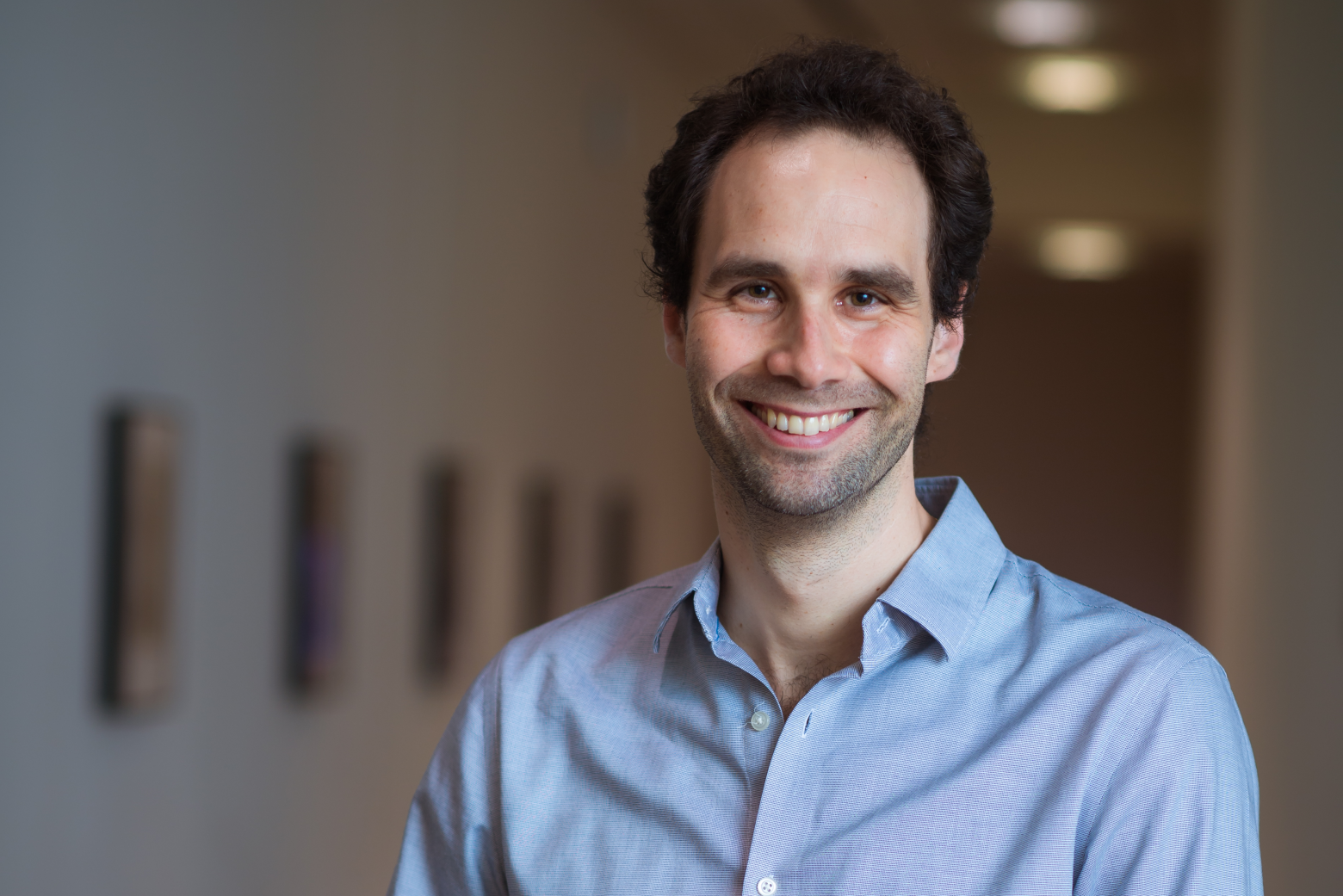
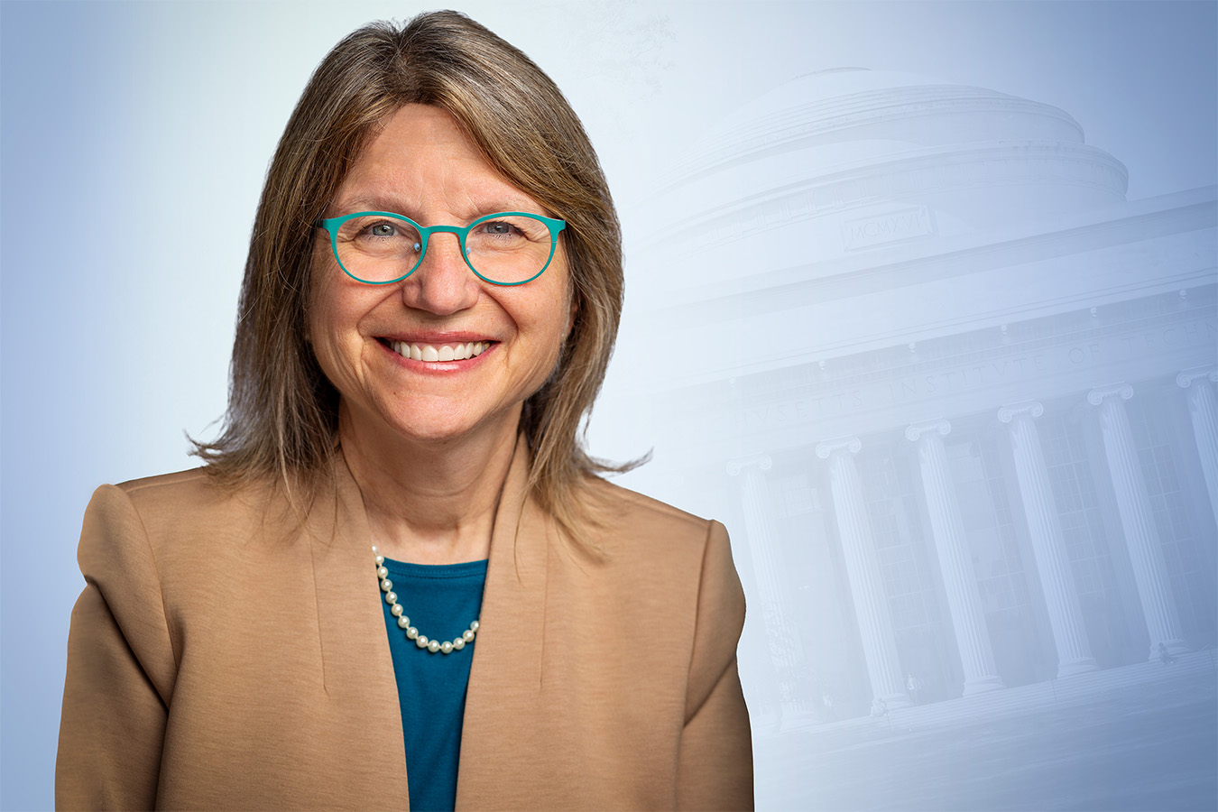
The incoming queen of Kendall Square talks Smoots, "cancel culture," and how to get more young women into STEM.

Jonathan Soroff | Boston Magazine
December 20, 2022

With renewed concerns about diversity, affordability, and censorship on campus—to say nothing about the future of space exploration and renewable energy—there’s a lot going on at MIT these days. For recently named president Sally Kornbluth, who is moving to the Bay State from North Carolina, where she’d served as provost at Duke University since 2014, it means the chance to shape one of the world’s most prestigious universities at a time of momentous change.

We caught up with her to discuss all of that, plus Smoots, the Sox, and how she plans to navigate the academic waters north of the Charles when she officially takes her post on January 1.

What do you anticipate being the best perk of your new job?

I’m a scientist by training, and I haven’t had a lab for some time. So I can live vicariously through the work of others, in a way, and really enjoy the discoveries that they’re making. I expect to learn about a lot of exciting projects, findings, discoveries, and inventions that I can help enable or support in a way that I could never do in my own work. I think it’s going to be like a candy store for the intellectually curious.

What percentage of what goes on academically or in research at MIT do you think will be comprehensible to you since—for most of us—the answer is zero?

Well, I’ll certainly understand what’s going on in the biology department, deeply. A lot of my colleagues, I follow their work. I have some understanding of what’s going on in engineering—although I’m not an engineer—particularly in the biomedical or biological engineering space. And, you know, I’ve closely followed a lot of different disciplines in my work as provost. I’m excited by what’s going on in the arts at MIT, the social sciences, and the humanities. A big part of the MIT ethos and culture is to try to make the work really accessible to others because it’s important to people’s lives on this planet. So, either I will understand it and help to translate it, or my faculty and colleagues will help translate it for me.

First thing you’ll do when you walk in the door?

The first thing I’ve got to do is get a map. I have never seen such a confusing welter of buildings that are numbered in a seemingly crazy manner. And then, honestly, just really get out there and meet everybody: the students, the faculty, the staff. It’s really going to be an exciting moment to get to know all these new people and all their exciting work.

So, offhand, do you know MIT’s Latin motto?

I believe it’s “Mind and hand.”

Yes! “Mens et Manus.” Well done. What do you think are the things you’ll miss most about North Carolina, and what are you most looking forward to in moving to New England?

I’ve been here [in North Carolina] a long time. I’m going to miss all my friends and colleagues. You know, my kids grew up here, there’s people here that I’ve known for years. Also, the weather’s pretty mild here. But it’s funny. I was up at MIT last weekend, and I was walking around with friends, and something really struck me, which is you don’t realize how much the foliage, plants, and trees that you were used to seeing growing up make you feel at home. I grew up in northern New Jersey, and I went to school at Williams College. I was with a friend who’s also from the Northeast, and she reached out and touched this shrub. She said, “You remember this?” I said, “Yes. I haven’t seen it in years.” I’m kind of excited about going back to this environment that’s so familiar.

But probably less excited about a long winter?

Well, I just bought myself a nice warm coat.

This next question is extremely important: Are you a sports fan, and if so, are you ready to swear fealty to Red Sox Nation, Patriots Nation, and Celtics Nation?

It’s so funny. I was thinking about that because when I was growing up, and my father would be watching sports on television, I’d say, “Dad, who are you rooting for?” And he’d say, “Nobody. I just find the game interesting.” I like watching sporting events, but I must admit, I’m not rabid for one team or another. I will be rooting for the MIT Engineers. But I have to say, I’ve gotten emails from people saying, “Don’t you dare root for the Red Sox!” Maybe I’ll maintain my neutrality for a bit, but then I might get sucked in.

But I assume you’ll always have a warm spot for the Blue Devils?

Yeah, of course. Plus, I have an extensive wardrobe of Duke stuff.

In a nutshell, what do you see as MIT’s greatest strength?

Honestly, it’s the ingenuity and brilliance of the faculty and students. If you believe that higher education is the talent development game, you can’t be anyplace better than MIT to help do that. It’s just brilliant people doing what they do best, and it’s amazing to me the amount of mind-bending work going on there.

Here’s another gotcha question: Do you know what a Smoot is?

I do. I know because my son is a graduate student at MIT, and we were walking across the bridge, like a year or two ago, and he explained it to my husband and me.

Are you prepared for, and what do you think of, the incredibly elaborate pranks MIT students are famous for, like taking apart and reassembling a police car on top of the dome?

I have to admit that I find those kinds of things incredibly amusing. I remember hearing about pranks like that throughout my career. My favorite was a sign on an elevator that said, “Elevator has now become voice activated. Please loudly announce the floor you wish to go to.” And there were all these people yelling, “Fourth floor!” It was hilarious. So, I’m familiar with them, and I think it’ll be fun.

On a more serious note, you’re joining a heavily female executive team: board chair, chancellor, provost, dean of science. Do you think that has particular significance?

I think we’ve reached a point, or I hope that we have, where we’re selecting the top talent and tapping into the full range of human talent. I think all of the leaders at MIT, and I hope I’m included, have been selected for their skills. It’s wonderful that they’re also women, but I believe that it’s a really strong team. My husband always says he thinks women should run the world.

How do we, as a society, get more young girls interested and involved with math and science?

One way is that I do think the presence of more women in these areas provides more role models, and it behooves women who have had success in these areas to reach down the pipeline and help others have the same success. The other thing is to have low barriers to entry into these areas. Because in some areas, girls may not have been traditionally encouraged to jump in. Girls, as well as boys, should be able to gravitate to their true interests and talent and not have to scale a wall to get into certain areas.

At this point, you’ve served in an administrative role for nearly nine years. Do you think you could go back to teaching an undergraduate course in your field of biology, or has that ship sailed?

I’d have to do a lot of reading, a lot of catch-up. But the basic skill set is still there. Could I understand what I read and learn to think about ways to teach it effectively? I think so. To go back and run a lab from scratch? That would be a bigger mountain to climb than teaching a course.

Any thoughts about the affirmative action question facing the Supreme Court?

Well, obviously, we’ll see how this plays out, and certainly, MIT will follow the law, whatever that is. But I think the bottom line is that institutions really, really benefit from a diversity of perspectives and a diversity of backgrounds, and regardless of the outcome of the Supreme Court decision, it’s going to be important for a place like MIT to still be able to hear truly diverse voices. A diverse team just comes up with much better ideas and discoveries. It’s not an echo chamber.

Do you think that in academics and society, too much emphasis is placed on sort of “brand name” schools?

There are many, many, many institutions in this country where you can get a fabulous education. So, do I divide the world in that way? Not necessarily. That said, what’s exciting to me about MIT and other institutions you might name is the high concentration of fabulous scholars. There are some institutions that can offer students exposure to that kind of scholarship as part of their experience.

Your predecessor had to navigate censorship and “cancel culture” on campus. How do you intend to handle that?

You’ve got to foster a culture where freedom of speech is strongly supported, even if that speech is maybe something someone doesn’t want to hear. That’s fine, as long as it doesn’t incite violence and doesn’t target individuals. That said, it can be difficult because people feel that words can hurt them. They don’t like to hear things they don’t want to hear. But I believe it’s the role of an educational institution to expose students to ideas or positions that they might not have otherwise entertained or heard.

Will it be weird to be president of a university where your son is a Ph.D. candidate?

[Laughs.] You might ask him that. I hope it won’t be weird for him. For me, it’s delightful because I’ll get to see him more often. And I’m not going to show up at his lab with a batch of cookies.

Thoughts on the idea of making tuition free to all?

You know, I can’t speak to that for MIT now, but I will say this: 85 percent of MIT graduates leave debt-free. There is a very robust financial aid program that’s both need-blind admissions-based and meeting the full needs of students financially. MIT is, no doubt, in a very privileged position in this way to have the resources to do that, but I don’t think that an MIT education is where these problems currently reside.

You were the chair of the trustees for the Duke Kunshan University partnership. China is so demonized these days; do you see it as an ally or a threat?

Well, let me just say up front that the partnership was really meant to bring liberal, American-style education to China, so it was not a deeply political play, nor was it a heavily research-based program. China is a place to approach with some balance. The open exchange of ideas has really fueled science, taking advantage of brilliant ideas from all over the world. But you have to balance that with national security threats and risks, which are very real. And greater minds than mine are grappling with that. I don’t demonize it as a country, but there are certainly thorny issues that have to be navigated.

What are your hobbies or pastimes?

I have two dogs that I like to walk all the time. People will see them walking around campus. I like to read. I have to admit that I like to watch those British mysteries. In fact, given the number I’ve watched, it’s surprising there’s a person left alive in the British Isles. I like to ride my bike. I like to hike. And during the pandemic, I took up needlepoint and felt flower making, which is a little odd. Some sort of latent craftiness that I never knew I had.

Any desire for a Nobel Prize?

No. I’ve never done anything that would merit a Nobel Prize. But I hope to be able to create, continue to create, I should say, fertile ground for future Nobel Prize winners.


New tool can assist with identifying carbohydrate-binding proteins

Groundbreaking research can help alleviate the challenges affiliated with studying carbohydrates.

Danielle Doughty | Department of Chemistry
December 19, 2022

One of the major obstacles that those conducting research on carbohydrates are constantly working to overcome is the limited array of tools available to decipher the role of sugars. As a workaround, most researchers utilize lectins (sugar-binding proteins) isolated from plants or fungi, but they are large, with weak binding, and they are limited in their specificity and in the scope of sugars that they detect. In a new study published in ACS Chemical Biology, researchers in Professor Barbara Imperiali’s group have developed a platform to address this shortcoming.

“The challenge with polymers of carbohydrates is that their biosynthesis is not template driven,” said Imperiali, the senior author of the study, and a Professor in the Departments of Chemistry and Biology. “Biology, medicine, and biotechnology have been fueled by technological advancements for proteins and nucleic acids. The carbohydrate field lags terribly behind, and is desperately seeking tools.”

Identifying carbohydrate-binding proteins

Biosynthesizing carbohydrates requires every link between individual sugar molecules to be made by a particular enzyme, and, there’s no ready way to decipher the structures and sequences of complex carbohydrates. Antibodies to carbohydrates can be generated,  but doing so is challenging, expensive, and results in a molecule that is far larger than what is really needed for the research. An ideal resource for this field plagued with limited mechanisms would be discovery of binding proteins, of limited size, that recognize small chunks of carbohydrates to piece together a structure by using those binders, or methods to detect and identify particular carbohydrates within complicated structures.

To achieve their breakthrough, the authors of this study used directed evolution and clever screen design to identify carbohydrate-binding proteins from proteins that have absolutely no ability to bind carbohydrates at all.  Their findings lay the groundwork for identifying carbohydrate-binding proteins with diverse and programmable specificity.

Streamlining for collaboration

This exciting breakthrough will allow researchers to go after a user-defined sugar target without being limited by what a lectin does, or challenged by the abilities of generating antibodies. These results could serve to inspire future collaborations with engineering communities to maximize the efficiency of glycobiology’s yeast surface display pipeline. As it is, this pipeline works well for proteins, but sugars are far more difficult targets and require the pipeline to be modified.

In terms of future applications, the potential for this innovation ranges from diagnostic to, in the longer term, therapeutic, and paves the way for collaborations with researchers at MIT and beyond. Chemistry Professor Laura Kiessling’s research group works with Mycobacterium tuberculosis (Mtb), which has an unusual cell wall composition with unique, distinct, and exclusive sugars. Using this method, a binder could potentially be evolved to that particular feature on Mtb. Chemical Engineering Professor Hadley Sikes develops paper-based diagnostic tools where the binding partner for a particular epitope or marker is laid down, and with the use of this discovery, in the longer term, a lateral flow assay device could be developed.

Laying the groundwork for future solutions

In cancer, certain sugars are over-represented on cell surfaces, so theoretically, researchers can utilize this finding, which is also amenable to labeling, to develop a tool out of the evolved glycan binder for detection.

This discovery also stands to contribute significantly to improving cell imaging. Researchers can modify binders with a fluorophore using a simple ligation strategy, and can then choose the best fluorophore for tissue or cell imaging. The Kiessling group, for example, could apply small protein binders labeled with fluorophore to detect bacterial sugars to initiate fluorescence-activated cell sorting to probe a complex mixture of microbes. This could in turn be used to determine how a patient’s microbiome has been disturbed. It also has the potential to screen the microbiome of a patient’s mouth or their upper or lower gastrointestinal tract to read out the imbalance within the community using these types of reagents. In the more distant future, the binders could potentially have therapeutic purposes like clearing the gastrointestinal tract or mouth of a particular bacterium based on the sugars that the bacterium displays.