How cells combat chromosome imbalance

Biologists discover the immune system can eliminate cells with too many or too few chromosomes.

Anne Trafton | MIT News Office
June 19, 2017

Most living cells have a defined number of chromosomes: Human cells, for example, have 23 pairs. As cells divide, they can make errors that lead to a gain or loss of chromosomes, which is usually very harmful.

For the first time, MIT biologists have now identified a mechanism that the immune system uses to eliminate these genetically imbalanced cells from the body. Almost immediately after gaining or losing chromosomes, cells send out signals that recruit immune cells called natural killer cells, which destroy the abnormal cells.

The findings raise the possibility of harnessing this system to kill cancer cells, which nearly always have too many or too few chromosomes.

“If we can re-activate this immune recognition system, that would be a really good way of getting rid of cancer cells,” says Angelika Amon, the Kathleen and Curtis Marble Professor in Cancer Research in MIT’s Department of Biology, a member of the Koch Institute for Integrative Cancer Research, and the senior author of the study.

Stefano Santaguida, a research scientist at the Koch Institute, is the lead author of the paper, which appears in the June 19 issue of Developmental Cell.

“A downward spiral”

Before a cell divides, its chromosomes replicate and then line up in the middle of the cell. As the cell divides into two daughter cells, half of the chromosomes are pulled into each cell. If these chromosomes fail to separate properly, the process leads to an imbalanced number of chromosomes in the daughter cells — a state known as aneuploidy.

When aneuploidy occurs in embryonic cells, it is almost always fatal to the organism. For human embryos, extra copies of any chromosome are lethal, with the exceptions of chromosome 21, which produces Down syndrome; chromosomes 13 and 18, which lead to developmental disorders known as Patau and Edwards syndromes; and the X and Y sex chromosomes, extra copies of which may cause various disorders but are not usually lethal.

In recent years, Amon’s lab has been exploring an apparent paradox of aneuploidy: When normal adult cells become aneuploid, it impairs their ability to survive and proliferate; however, cancer cells, which are nearly all aneuploid, can grow uncontrollably.

“Aneuploidy is highly detrimental in most cells. However, aneuploidy is highly associated with cancer, which is characterized by upregulated growth. So, a very important question is: If aneuploidy hampers cell proliferation, why are the vast majority of tumors aneuploid?” Santaguida says.

To try to answer that question, the researchers wanted to find out more about how aneuploidy affects cells. Over the past few years, Santaguida and Amon have been studying what happens to cells immediately after they experience a mis-segregation of chromosomes, leading to imbalanced daughter cells.

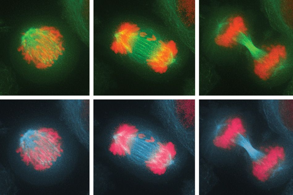
In the new study, they investigated the effects of this imbalance on the cell division cycle by interfering with the process of proper chromosome attachment to the spindle, the structure that holds chromosomes in place at the cell’s equator before division. This interference leads some chromosomes to lag behind and get shuffled into the two daughter cells.

The researchers found that after the cells underwent their first division, in which some of the chromosomes were unevenly distributed, they soon initiated another cell division, which produced even more chromosome imbalance, as well as significant DNA damage. Eventually, the cells stopped dividing altogether.

“These cells are in a downward spiral where they start out with a little bit of genomic mess, and it just gets worse and worse,” Amon says.

“This paper very convincingly and clearly shows that when chromosomes are lost or gained, initially cells can’t tell if their chromosomes have mis-segregated,” says David Pellman, a professor of pediatric oncology at Dana-Farber Cancer Institute who was not involved in the study. “Instead, the imbalance of chromosomes leads to cellular defects and an imbalance of proteins and genes that can significantly disrupt DNA replication and cause further damage to the chromosomes.”

Targeting aneuploidy

As genetic errors accumulate, aneuploid cells eventually become too unstable to keep dividing. In this senescent state, they start producing inflammation-inducing molecules such as cytokines. When the researchers exposed these cells to immune cells called natural killer cells, the natural killer cells destroyed most of the aneuploid cells.

“For the first time, we are witnessing a mechanism that might provide a clearance of cells with imbalanced chromosome numbers,” Santaguida says.

In future studies, the researchers hope to determine more precisely how aneuploid cells attract natural killer cells, and to find out whether other immune cells are involved in clearing aneuploid cells. They would also like to figure out how tumor cells are able to evade this immune clearance, and whether it may be possible to restart the process in patients with cancer, since about 90 percent of solid tumors and 75 percent of blood cancers are aneuploid.

“At some point, cancer cells, which are highly aneuploid, are able to evade this immune surveillance,” Amon says. “We have really no understanding of how that works. If we can figure this out, that probably has tremendous therapeutic implications, given the fact that virtually all cancers are aneuploid.”

The research was funded, in part, by the National Institutes of Health, the Kathy and Curt Marble Cancer Research Fund, the American Italian Cancer Foundation, a Fellowship in Cancer Research from Marie Curie Actions, the Italian Association for Cancer Research, and a Koch Institute Quinquennial Cancer Research Fellowship.

Biologists identify key step in lung cancer evolution

Blocking the transition to a more aggressive state could offer a new treatment strategy.

Anne Trafton | MIT News Office
May 10, 2017

Lung adenocarcinoma, an aggressive form of cancer that accounts for about 40 percent of U.S. lung cancer cases, is believed to arise from benign tumors known as adenomas.

MIT biologists have now identified a major switch that occurs as adenomas transition to adenocarcinomas in a mouse model of lung cancer. They’ve also discovered that blocking this switch prevents the tumors from becoming more aggressive. Drugs that interfere with this switch may thus be useful in treating early-stage lung cancers, the researchers say.

“Understanding the molecular pathways that get activated as a tumor transitions from a benign state to a malignant one has important implications for treatment. These findings also suggests methods to prevent or interfere with the onset of advanced disease,” says Tyler Jacks, director of MIT’s Koch Institute for Integrative Cancer Research and the study’s senior author.

The switch occurs when a small percentage of cells in the tumor begin acting like stem cells, allowing them to give rise to unlimited populations of new cancer cells.

“It seems that the stem cells are the engine of tumor growth. They’re endowed with very robust proliferative potential, and they give rise to other cancer cells and also to more stem-like cells,” says Tuomas Tammela, a postdoc at the Koch Institute and lead author of the paper, which appears in the May 10 online edition of Nature.

Tumor stem cells

In this study, the researchers focused on the role of a cell signaling pathway known as Wnt. This pathway is usually turned on only during embryonic development, but it is also active in small populations of adult stem cells that can regenerate specific tissues such as the lining of the intestine.

One of the Wnt pathway’s major roles is maintaining cells in a stem-cell-like state, so the MIT team suspected that Wnt might be involved in the rapid proliferation that occurs when early-stage tumors become adenocarcinomas.

The researchers explored this question in mice that are genetically programmed to develop lung adenomas that usually progress to adenocarcinoma. In these mice, they found that Wnt signaling is not active in adenomas, but during the transition, about 5 to 10 percent of the tumor cells turn on the Wnt pathway. These cells then act as an endless pool of new cancer cells.

In addition, about 30 to 40 percent of the tumor cells begin to produce chemical signals that create a “niche,” a local environment that is necessary to maintain cells in a stem-cell-like state.

“If you take a stem cell out of that microenvironment, it rapidly loses its properties of stem-ness,” Tammela says. “You have one cell type that forms the niche, and then you have another cell type that’s receiving the niche cues and behaves like a stem cell.”

While Wnt has been found to drive tumor formation in some other cancers, including colon cancer, this study points to a new kind of role for it in lung cancer and possibly other cancers such as pancreatic cancer.

“What’s new about this finding is that the pathway is not a driver, but it modifies the characteristics of the cancer cells. It qualitatively changes the way cancer cells behave,” Tammela says.

“It’s a very nice paper that points to the influence of the microenvironment in tumor growth and shows that the microenvironment includes factors secreted by a subset of tumor cells,” says Frederic de Sauvage, vice president for molecular oncology research at Genentech, who was not involved in the study.

Targeting Wnt

When the researchers gave the mice a drug that interferes with Wnt proteins, they found that the tumors stopped growing, and the mice lived 50 percent longer. Furthermore, when these treated tumor cells were implanted into another animal, they failed to generate new tumors.

The researchers also analyzed human lung adenocarcinoma samples and found that 70 percent of the tumors showed Wnt activation and 80 percent had niche cells that stimulate Wnt activity. These findings suggest it could be worthwhile to test Wnt inhibitors in early-stage lung cancer patients, the researchers say.

They are also working on ways to deliver Wnt inhibitors in a more targeted fashion, to avoid some of the side effects caused by the drugs. Another possible way to avoid side effects may be to develop more specific inhibitors that target only the Wnt proteins that are active in lung adenocarcinomas. The Wnt inhibitor that the researchers used in this study, which is now in clinical trials to treat other types of cancer, targets all 19 of the Wnt proteins.

The research was funded by the Janssen Pharmaceuticals-Koch Institute Transcend Program, the Lung Cancer Research Foundation, the Howard Hughes Medical Institute, and the Cancer Center Support grant from the National Cancer Institute.

New model could speed up colon cancer research

Introducing genetic mutations with CRISPR offers a fast and accurate way to simulate the disease.

Anne Trafton | MIT News Office
May 1, 2017

Using the gene-editing system known as CRISPR, MIT researchers have shown in mice that they can generate colon tumors that very closely resemble human tumors. This advance should help scientists learn more about how the disease progresses and allow them to test new therapies.

Once formed, many of these experimental tumors spread to the liver, just like human colon cancers often do. These metastases are the most common cause of death from colon cancer.

“That’s been a missing piece in the study of colon cancer. There is really no reliable method for recapitulating the metastatic progression from a primary tumor in the colon to the liver,” says Omer Yilmaz, an MIT assistant professor of biology, a member of MIT’s Koch Institute for Integrative Cancer Research, and the lead senior author of the study, which appears in the May 1 issue of Nature Biotechnology.

The study builds on recent work by Tyler Jacks, the director of the Koch Institute, who has also used CRISPR to generate lung and liver tumors in mice.

“CRISPR-based technologies have begun to revolutionize many aspects of cancer research, including building mouse models of the disease with greater speed and greater precision. This study is a good example of both,” says Jacks, who is also an author of the Nature Biotechnology paper.

The paper’s lead authors are Jatin Roper, a research affiliate at the Koch Institute and a gastroenterologist at Tufts Medical Center, and Tuomas Tammela, a research scientist at the Koch Institute.

Mimicking human tumors

For many years, cancer biologists have taken two distinct approaches to modeling cancer. One is to grow immortalized human cancer cells known as cancer cell lines in a lab dish. “We’ve learned a lot by studying these two-dimensional cell lines, but they have limitations,” Yilmaz says. “They don’t really reproduce the complex in vivo environment of a tumor.”

Another widely used technique is genetically engineering mice with mutations that predispose them to develop cancer. However, it can take years to breed such mice, especially if they have more than one cancer-linked mutation.

Recently, researchers have begun using CRISPR to generate cancer models. CRISPR, originally discovered by biologists studying the bacterial immune system, consists of a DNA-cutting enzyme called Cas9 and short RNA guide strands that target specific sequences of the genome, telling Cas9 where to make its cuts. Using this process, scientists can make targeted mutations in the genomes of living animals, either deleting genes or inserting new ones.

To induce cancer mutations, the investigators package the genes for Cas9 and the RNA guide strand into viruses called lentiviruses, which are then injected into the target organs of adult mice.

Yilmaz, who studies colon cancer and how it is influenced by genes, diet, and aging, decided to adapt this approach to generate colon tumors in mice. He and members of his lab were already working on a technique for growing miniature tissues known as organoids — three-dimensional growths that, in this case, accurately replicate the structure of the colon.

In the new paper, the researchers used CRISPR to introduce cancer-causing mutations into the organoids and then delivered them via colonoscopy to the colon, where they attached to the lining and formed tumors.

“We were able to transplant these 3-D mini-intestinal tumors into the colon of recipient mice and recapitulate many aspects of human disease,” Yilmaz says.

More accurate modeling

Once the tumors are established in the mice, the researchers can introduce additional mutations at any time, allowing them to study the influence of each mutation on tumor initiation, progression, and metastasis.

Almost 30 years ago, scientists discovered that colon tumors in humans usually acquire cancerous mutations in a particular order, but they haven’t been able to accurately model this in mice until now.

“In human patients, mutations never occur all at once,” Tammela says. “Mutations are acquired over time as the tumor progresses and becomes more aggressive, more invasive, and more metastatic. Now we can model this in mice.”

To demonstrate that ability, the MIT team delivered organoids with a mutated form of the APC gene, which is the cancer-initiating mutation in 80 percent of colon cancer patients. Once the tumors were established, they introduced a mutated form of KRAS, which is commonly found in colon and many other cancers.

The scientists also delivered components of the CRISPR system directly into the colon wall to quickly model colon cancer by editing the APC gene. They then added CRISPR components to also edit the gene for P53, which is commonly mutated in colon and other cancers.

“These new approaches reduce the time frame to develop genetically engineered mice from two years to just a few months, and involve very basic gene engineering with CRISPR,” Roper says. “We used P53 and KRAS to demonstrate the principle that the CRISPR editing approach and the organoid transplantation approach can be used to very quickly model any possible cancer-associated gene.”

In this study, the researchers also showed that they could grow tumor cells from patients into organoids that could be transplanted into mice. This could give doctors a way to perform “personalized medicine” in which they test various treatment options against a patient’s own tumor cells.

Fernando Camargo, a professor of stem cell and regenerative biology at Harvard University, says the study represents an important advance in colon cancer research.

“It allows investigators to have a very flexible model to look at multiple aspects of colorectal cancer, from basic biology of the genes involved in progression and metastasis, to testing potential drugs,” says Camargo, who was not involved in the research.

Yilmaz’ lab is now using these techniques to study how other factors such as metabolism, diet, and aging affect colon cancer development. The researchers are also using this approach to test potential new colon cancer drugs.

The research was funded by the Howard Hughes Medical Institute, the National Institutes of Health, the Department of Defense, the V Foundation V Scholar Award, the Sidney Kimmel Scholar Award, the Pew-Stewart Trust Scholar Award, the Koch Institute Frontier Research Program through the Kathy and Curt Marble Cancer Research Fund, the American Federation of Aging Research, and the Hope Funds for Cancer Research.