Five MIT students named 2019 Marshall Scholars

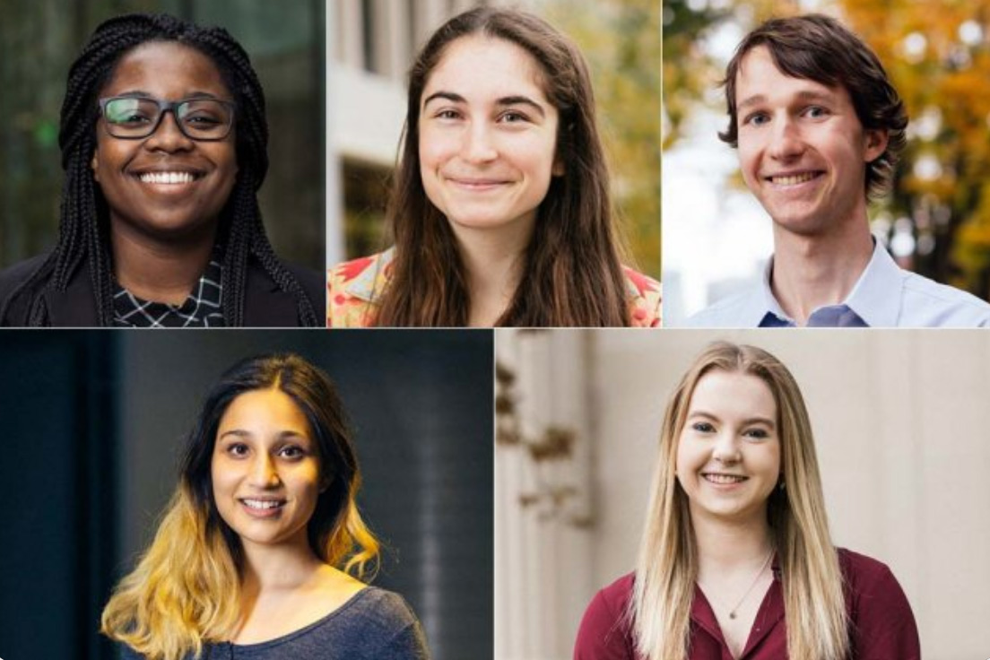
Radha Mastandrea, Katie O’Nell, Anna Sappington, Kyle Swanson, and Crystal Winston will begin graduate studies in the UK next fall.

Julia Mongo | Office of Distinguished Fellowships
December 3, 2018

Five MIT students — Radha Mastandrea, Kathryn O’Nell, Anna Sappington, Kyle Swanson ’18, and Crystal Winston — have been awarded Marshall Scholarships to pursue graduate studies in the United Kingdom. This class represents the largest number of Marshall Scholars from the Institute in a single year, and continues MIT students’ exceptional record of achievement in this elite fellowship program.

Funded by the British government, the Marshall Scholarship provides outstanding young Americans with the opportunity to earn advanced degrees in any academic subject at any university in the United Kingdom. Scholars are chosen through a rigorous national competition that assesses academic merit, leadership, and ambassadorial potential. Up to 40 Marshall Scholarships are granted each year.

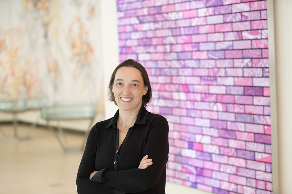
The MIT students were guided by Kimberly Benard, assistant dean of distinguished fellowships within MIT Career Advising and Professional Development, and by the Presidential Committee on Distinguished Fellowships co-chaired by professors Rebecca Saxe and Will Broadhead. “Working with students like our Marshall Scholars is one of the great pleasures and privileges of teaching at MIT,” says Saxe. “Every year I’m impressed by how hard all of the finalists work to develop and communicate their distinctive vision for the future and their place in it.”

“MIT’s five Marshall Scholars, Radha, Katie, Anna, Kyle, and Crystal, are extraordinary; they are intelligent, creative, and dedicated to making a better world,” says MIT Chancellor Cynthia Barnhart. “Together, they make up an eighth of the entire class of 2019 Marshall Scholars, a remarkable accomplishment that would not have been possible without the hard work and passion of these students; the incredible efforts of professors Broadhead and Saxe and the entire Presidential Committee for Distinguished Fellowships; and the staff who work around the clock to support all applicants through this demanding process. We are fortunate that our 2019 Marshall Scholars have such exciting opportunities to look forward to, and that they will be representing the MIT community to the world.”

Radha Mastandrea

Hailing from Westwood, Massachusetts, Mastandrea is an MIT senior double-majoring in physics and mathematics. She is headed to Cambridge, where she will study theoretical and experimental physics before returning to the U.S. to undertake a PhD in high-energy particle physics. She aims for a career in academia as a researcher and an advocate for open data.

As an undergraduate researcher with Professor Jesse Thaler at the MIT Center for Theoretical Physics, Mastandrea has been using machine learning to analyze a trove of open data from the CERN Large Hadron Collider to gain insight on quarks and gluons. Mastandrea has also conducted research on galaxy spectral emission data with Professor Michael McDonald at the MIT Kavli Institute for Astrophysics and Space Research, and neutrinoless double beta decays with Professor Lindley Winslow at the MIT Laboratory for Nuclear Science. During a summer internship at Caltech, she researched black holes with the Laser Interferometer Gravitational-wave Observatory (LIGO) Scientific Collaboration.

Mastandrea is committed to championing women in physics. As president of MIT’s Undergraduate Women in Physics, she has established connections between undergraduates and older role models to showcase women’s accomplishments in the physics field. She has mentored local high school girls by leading physics exploration days on the MIT campus, and she helped write the MIT physics department’s first community values statement to foster a culture of respect and support. Mastandrea is also co-captain of MIT Bhangra and has taught bhangra dance classes to members of the local community.

Kathryn “Katie” O’Nell

O’Nell, from San Diego, California, is an MIT senior majoring in brain and cognitive sciences. At Oxford University, she will pursue an MS in the Department of Experimental Psychology. She will then return to the U.S. to embark on doctoral studies in computational neuroscience with the goal of becoming a professor and researcher.

O’Nell is currently conducting studies on facial perception in the laboratories of Professor Rebecca Saxe in MIT’s Department of Brain and Cognitive Sciences and Professor Stefano Anzellotti of Boston College. O’Nell has also been examining the connection between gestures and speech with the MIT Speech Communication Group, and she has created calcium imaging processing pipelines in the laboratory of Professor Ann Graybiel at the McGovern Institute for Brain Research at MIT. O’Nell spent two summers interning at the J. Craig Venter Institute, which conducts genomic and bioinformatics research.

On campus, O’Nell is an associate advisor to first-year students and a member of the associate advisor steering committee. She is active with the MIT Model UN Conference for high school students and serves as special political and decolonization committee chair. She also mentors high school students as a Splash instructor for the MIT Educational Studies Program and as president of the MIT Academic Teaching Initiative. As campus engagement chair for the MIT Addir Interfaith Program, O’Nell plans and promotes campus-wide events, including an upcoming interfaith hackathon. O’Nell originated and oversees the popular murder mystery-themed puzzle hunt for her residence hall, MacGregor House.

Anna Sappington

A native of Riva, Maryland, Sappington will graduate this spring with a BS in computer science and molecular biology. She will earn master’s degrees in machine learning at University College London and medical sciences in oncology at the Cambridge University. She will then pursue an interdisciplinary career as a physician-scientist using computer science to improve precision medicine.

As an undergraduate researcher at the laboratory of Professor Aviv Regev in MIT’s Department of Biology and the Broad Institute of MIT and Harvard, Sappington has been developing machine-learning techniques to improve cell type classification for the Human Cell Atlas Initiative. She has also conducted research on hepatitis B virus infection in the laboratory of Professor Sangeeta Bhatia in the Department of Electrical Engineering and Computer Science and the Koch Institute for Integrative Cancer Research. Sappington has held summer research internships at the National Institutes of Health in Bethesda, Maryland, and as an Amgen Scholar at Kyoto University, Japan. In 2018, she was named a national Barry M. Goldwater Scholar and an MIT Burchard Scholar.

Sappington served as the Director of TechX, which hosts world-renowned events such as HackMIT. She is co-president of the MIT Biology Undergraduate Student Association, a teaching assistant for the biology department, and a peer health counselor and former residential director for MIT MedLinks. A talented dancer, Sappington has performed with MIT DanceTroupe and the MIT Asian Dance Team. For the past two years, she has volunteered for the Massachusetts General Hospital department of radiation oncology.

Kyle Swanson

Hailing from Bronxville, New York, Swanson graduated from MIT in 2018 with a BS in computer science and engineering, a BS in mathematics, and a minor in music. He will receive his MEng in computer science and engineering this spring. As a Marshall Scholar, Swanson will study mathematics and computer science at Cambridge. He intends to pursue a PhD in computer science to research applications of machine learning to the diagnosis and treatment of cancer.

As an undergraduate, Swanson worked with Professor Regina Barzilay in MIT’s Computer Science and Artificial Intelligence Laboratory (CSAIL) and Constance Lehman in the Breast Imaging Division of Massachusetts General Hospital on machine learning to improve breast cancer detection in mammography, and recently co-authored a paper published in Radiology.

Swanson’s master’s research with Barzilay focuses on machine learning for chemistry. He is currently developing machine-learning algorithms to predict molecular properties such as toxicity or solubility, with the hope of developing a property-prediction tool that can accelerate drug discovery. Swanson previously conducted research on cybersecurity at CSAIL. He has interned at Microsoft and other companies and taught machine learning to university students in Kazakhstan.

An accomplished musician, Swanson plays flute and piccolo with the MIT Symphony Orchestra where he has served as president. He is active with the music service club Ribotones, which performs concerts at nursing homes, and he has played with MIT’s Chamber Music Society and Wind Ensemble. Swanson has received multiple MIT awards recognizing his musical talent and scholarship.

Crystal Winston

Winston, from Charlotte, North Carolina, is an MIT senior majoring in mechanical engineering. At Imperial College, Winston will embark on a PhD in aerospace materials and structures to further develop her skills in redesigning transportation systems. Her goal is to start a company that transforms transportation technology.

As a sophomore, Winston co-invented a remote-controlled four-wheeled robot that converts into a flying drone, which won first place in a campus project exhibition. She has continued to refine this project and hopes it can eventually be used to reduce traffic congestion by transporting people on ground or through the air.

Winston has conducted research on a robot for detecting and repairing pipe leaks, at the MIT Mechatronics Research Laboratory. As a mechanical engineering intern at Google, she designed and programmed systems for the cameras on Google Maps Streetview cars, and at the NASA Jet Propulsion Laboratory she designed and prototyped an animal-inspired foot for the Europa Lander. She has been a mechanical team member for the MIT Solar Electric Vehicle Team, and helped design and manufacture an electric solar car for the 2017 Formula Sun Grand Prix.

Winston is dedicated to her work with the National Society of Black Engineers (NSBE) where she has held several executive positions. As an engineering immersion program mentor with NSBE, she helps local underrepresented minority high school students prepare for STEM careers. Winston is a member of the Tau Beta Pi and Pi Tau Sigma engineering honors societies.

The long and short of CDK12

A new study linking RNA processing to DNA repair may open new avenues to cancer therapy.

Bendta Schroeder | Koch Institute
December 3, 2018

Mutations in the BRCA1 and BRCA2 genes pose a serious risk for breast and ovarian cancer because they endanger the genomic stability of a cell by interfering with homologous recombination repair (HR), a key mechanism for accurately repairing harmful double-stranded breaks in DNA. Without the ability to use HR to fix double-stranded breaks, the cell is forced to resort to more error-prone — and thus more cancer-prone — forms of DNA repair.

The BRCA1 and BRCA2 genes are not the only genes whose mutations foster tumorigenesis by causing an inability to repair DNA double strand breaks by HR. Mutations in twenty-two genes are known to disrupt HR, giving rise to tumors with what researchers call “BRCAness” characteristics. All but one of these BRCAness genes are known to be directly involved in the HR pathway.

The one exception, CDK12, is thought to facilitate a set of different processes altogether, involving how RNA transcripts are elongated, spliced and cleaved into their mature forms. While the connection between this RNA-modulating gene to DNA repair remained poorly understood, the identification of CDK12 as a BRCAness gene piqued significant clinical interest.

The researchers who pinpointed this connection, Sara Dubbury and Paul Boutz, both work in the laboratory of Phillip Sharp, Institute Professor, professor of biology, and member of the Koch Institute for Integrative Cancer Research. In a study appearing online in Nature on Nov. 28, they describe how they discovered a previously unknown mechanism by which CDK12 enables the production of full-length RNA transcripts and that this mechanism was especially critical to maintain functional expression of the other BRCAness genes.

When the researchers knocked out expression of CDK12, mouse stem cells showed many signs of accumulating DNA damage that prevented DNA replication from going forward, classic indications of a BRCAness phenotype. To identify what roles CDK12 may play in regulating gene expression, the researchers turned to RNA sequencing to determine which genes had increased or decreased their overall expression.

To their surprise, only genes activated by p53 and early differentiation (side effects of accumulating unrepaired DNA damage and BRCAness in mouse stem cells) accounted for the lion’s share of changes to RNA transcription. However, when the researchers instead focused on the types of RNAs transcribed, they found that many genes produced unusually short transcripts when CDK12 was absent.

Not every stretch of DNA in a gene makes it into the final RNA transcript. The initial RNA from a gene often includes sections, which researchers call “introns,” that are cut out of transcript, the discovery that earned Sharp the 1993 Nobel Prize in Physiology or Medicine and the remaining sections. “Exons,” are spliced together to form a mature transcript (mRNA). Alternately, an intronic polyadenylation (IPA) site may be activated to cleave away the RNA sequence that follows it preventing intron removal and generating a prematurely shortened transcript. These processes allow the same gene to produce alternate forms of messenger RNA (mRNA), and thus be translated into different protein sequences.

Surprisingly CDK12 knockout cells produced significantly more IPA-truncated transcripts genome-wide, while full-length transcripts for the same genes were reduced. These shortened mRNAs can vary greatly in their stability, their ability to be translated into protein, and their protein function. Thus, even while a gene may be actively transcribed, its translation into functional proteins can be radically altered or depleted by IPA activation.

While this observation began to illuminate CDK12’s role in regulating mRNA processing, what remained puzzling was why CDK12 loss affected the HR pathway so disproportionately. In investigating this question, Dubbury and Boutz found that BRCAness genes were overrepresented as a group among those genes that have increased IPA activity upon CDK12 loss.

Additionally, while CDK12 suppresses IPA activity genome-wide, 13 of the other 21 BRCAness genes were found to be particularly vulnerable to CDK12 loss, in part, because they possess multiple high-sensitivity IPA sites, which have a compound effect in decreasing the total amount of full-length transcripts. Moreover, because multiple CDK12-senstive BRCAness genes operate in the same HR pathway, the researchers believe that the disruption to HR repair of double-stranded DNA breaks is amplified.

CDK12 mutations are found recurrently in prostate and ovarian cancer patients, making them an attractive diagnostic and therapeutic target for cancer. However, not enough is known about CDK12 to distinguish between true loss-of-function mutations and so-called “passenger mutations” with no functional consequence.

“The ability to identify patients with true loss-of-function mutations in CDK12 would enable clinicians to label a new cohort of patients with bona fide BRCAness tumors that could benefit from certain highly effective and targeted chemotherapeutics against BRCAness, such as PARP1 inhibitors,” says Dubbury, a former David H. Koch Fellow.

Dubbury and Boutz were able to confirm that IPA sites in key BRCAness genes were also used more frequently upon CDK12 loss in human tumor cells using RNA sequencing data from prostate and ovarian tumor patients with CDK12 mutations and by treating human prostate adenocarcinoma and ovarian carcinoma cells with a CDK12 inhibitor. This result suggests that the CDK12 mechanism observed in mouse cell lines is conserved in humans and that CDK12 mutations in human ovarian and prostate tumors may promote tumorigenesis by increasing IPA activity and thus functionally attenuating HR repair.

“These results not only give us a better understanding how CDK12 contributes to BRCAness, they also may have exciting potential impact in the clinic,” Dubbury says. “Currently available diagnostic techniques could be used to probe the usage of IPA sites found in this study to rapidly screen for patients with true loss-of-function CDK12 mutations, who would respond to BRCAness-targeted treatments.”

Paul Boutz, a research scientist in the Sharp Lab, is co-first author of the study, and has plans to follow-up many of these implications for ovarian and prostate cancer his lab at the University of Rochester School of Medicine and Dentistry.

“CDK12 provides a remarkable example of how factors that control the processing of RNA molecules can function as master regulators of gene networks, and thereby profoundly affect the physiology of both normal and cancerous cells,” he says.

Phil Sharp, the senior author on the work, says “Sara’s and Paul’s surprising discovery that CDK12 suppresses intronic polyadenylation has implications for fundamental new insights into gene structure as well as for control of cancer.”

Angelika Amon wins 2019 Breakthrough Prize in Life Sciences

Four other MIT researchers to receive New Horizons Prizes in math and physics; two alumni win Breakthrough Prize in Fundamental Physics.

Anne Trafton | MIT News Office
October 17, 2018

Angelika Amon, an MIT professor of biology, is one of five scientists who will receive a 2019 Breakthrough Prize in Life Sciences, given for transformative advances toward understanding living systems and extending human life.

Amon, the Kathleen and Curtis Marble Professor in Cancer Research and a member of MIT’s Koch Institute for Integrative Cancer Research, was honored for her work in determining the consequences of aneuploidy, an abnormal chromosome number that results from mis-segregation of chromosomes during cell division.

The award, announced this morning, comes with a $3 million prize.

“Angelika Amon is an outstanding choice to receive the Breakthrough Prize,” says Tyler Jacks, director of the Koch Institute and the David H. Koch Professor of Biology. “Her work on understanding how cells control the decisions to divide and the effects of imbalances in chromosome number has helped shape how we think about normal development and disease. Angelika is a fearless investigator and a true scientist’s scientist. All of us in the Koch Institute and across MIT are thrilled by this news.”

Two MIT alumni, Charles Kane PhD ’89 and Eugene Mele PhD ’78, both professors at the University of Pennsylvania, will share a Breakthrough Prize in Fundamental Physics. Kane and Mele are being recognized for their new ideas about topology and symmetry in physics, leading to the prediction of a new class of materials that conduct electricity only on their surface.

New Horizons winners

Also announced today, three MIT physics researchers will receive the $100,000 New Horizons in Physics Prize, awarded to promising junior researchers who have already produced important work.

Lisa Barsotti, a principal research scientist at MIT’s Kavli Institute, and Matthew Evans, an MIT associate professor of physics, will share the prize with Rana Adhikari of Caltech for their work on ground-based detectors of gravitational waves. Daniel Harlow, an MIT assistant professor of physics, will share the prize with Daniel Jafferis of Harvard University and Aron Wall of Stanford University for their work generating fundamental insights about quantum information, quantum field theory, and gravity.

Additionally, Chenyang Xu, an MIT professor of mathematics, will receive a 2019 New Horizons in Mathematics Prize for his work in the minimal model program and applications to the moduli of algebraic varieties.

“On behalf of the School of Science, I congratulate Angelika Amon for this extraordinary honor, in recognition of her brilliant work that expands our understanding of cellular mechanisms that may lead to cancer,” says Michael Sipser, dean of the MIT School of Science and the Donner Professor of Mathematics. “We celebrate all recipients of these prestigious awards, including MIT’s four researchers whose impressive early-career achievements in physics and mathematics are being recognized today. Our scientists pursue fundamental research that advances human knowledge, which in turn leads to a better world.”

Chromosome imbalance

Most living cells have a defined number of chromosomes. Human cells, for example, have 23 pairs of chromosomes. However, as cells divide, they can make errors that lead to a gain or loss of chromosomes.

Amon has spent much of her career studying how this condition affects cells. When aneuploidy occurs in embryonic cells, it is almost always fatal to the organism. For human embryos, extra copies of any chromosome are lethal, with the exceptions of chromosome 21, which produces Down syndrome; chromosomes 13 and 18, which lead to developmental disorders known as Patau and Edwards syndromes; and the X and Y sex chromosomes, extra copies of which may sometimes cause various disorders but are not usually lethal.

In recent years, Amon’s lab has been exploring an apparent paradox of aneuploidy: When normal adult cells become aneuploid, it impairs their ability to survive and proliferate; however, cancer cells, which are nearly all aneuploid, can grow uncontrollably. Amon has shown that aneuploidy disrupts cells’ usual error-repair systems, allowing genetic mutations to quickly accumulate.

A better understanding of the consequences of aneuploidy could shed light on how cancer cells evolve and help to identify new therapeutic targets for cancer. Last year, Amon discovered a mechanism that the immune system uses to eliminate aneuploid cells from the body, raising the possibility of harnessing this system, which relies on natural killer cells, to destroy cancer cells.

Amon, who was informed of the prize several weeks ago, was sworn to secrecy until today’s announcement.

“When I received the phone call, I was driving in the car with my daughter, and it was really hard to not be too excited and thereby spill the beans,” she says. “Of course I am thrilled that our work is recognized in this manner.”

Scientists Frank Bennett of Ionis Pharmaceuticals, Adrian Krainer of Cold Spring Harbor Laboratory, Xiaowei Zhuang of Harvard University, and Zhijian Chen of the University of Texas Southwestern Medical Center will also receive Breakthrough Prizes in Life Sciences.

The 2019 Breakthrough Prize and New Horizon Prize recipients will be recognized at the seventh annual Breakthrough Prize ceremony, hosted by actor, producer and philanthropist Pierce Brosnan, on Sunday, Nov. 4, at NASA Ames Research Center in Mountain View, California, and broadcast live on National Geographic.

Joining the resolution revolution

Department of Biology hosts a symposium to celebrate the launch of MIT.nano and its new Cryogenic Electron Microscopy Facility.

Raleigh McElvery | Department of Biology
October 16, 2018

It’s a time of small marvels and big ideas. Welcome to the Nano Age.

MIT’s preparations for this new era are in full swing, including the recent launch of MIT.nano, the Institute’s center for nanoscience and nanotechnology. And on the day after MIT.nano’s opening ceremonies, the Department of Biology hosted its Cryogenic Electron Microscopy (Cryo-EM) Symposium, which was co-organized by biology professor Thomas Schwartz and the director of the new facility, Edward Brignole.

“We organized the symposium to raise awareness of this new research capacity, and to celebrate the many people who worked to fund these instruments, design the space, build the suites, and set up the microscopes,” Brignole said of the Oct. 5 event. “We also wanted to bring together the various groups across MIT working on diverse technologies to improve Cryo-EM, from mathematicians, computer scientists, and electrical engineers to biologists, chemists, and biological engineers.”

The event featured pioneers leveraging Cryo-EM for various interdisciplinary applications both on campus and outside of MIT — from biology and machine learning to quantum mechanics.

The program included Ed Egelman from the University of Virginia, Mark Bathe from the MIT Department of Biological Engineering, Katya Heldwein from Tufts University’s School of Medicine, and Karl Berggren from the Department of Electrical Engineering and Computer Science. Also giving talks were computational and systems biology graduate student Tristan Bepler from MIT’s Computer Science and Artificial Intelligence Laboratory, Luke Chao from Harvard Medical School and Massachusetts General Hospital, postdoc Kuang Shen from the Whitehead Institute at MIT, and graduate student Jonathan Weed from the MIT Department of Mathematics. The talks were followed by a reception in Building 68 and guided tours of the Cryo-EM Facility.

Unlike other popular techniques for determining 3-D macromolecular structures, Cryo-EM permits researchers to visualize more diverse and complex molecular assemblies in multiple conformations. Cryo-EM is housed in precisely climate-controlled rooms in the basement of MIT.nano, built atop a 5 million pound slab of concrete to minimize vibrations. Two multimillion-dollar instruments are being installed that will enable scientists to analyze cellular machinery in near-atomic detail; the microscopes are the first instruments to be installed in MIT.nano.

As Schwartz explained to an audience of more than 100 people during his opening remarks, he and his colleagues realized they needed to bring this technology to the MIT community. Like many of the center’s other tools, they would be too costly to purchase and too onerous to maintain for a single researcher or lab.

“Microscopes are very special and expensive tools, so this endeavor turned out to be much more involved than anything else I have done during my 14 years at MIT,” he said. “But this was not an effort of one or two people, it really took a whole community. We have many people to thank today.”

Establishing the Cryogenic Electron Microscopy Facility at MIT has been a long-time dream for Catherine Drennan, a professor of chemistry and biology and a Howard Hughes Medical Institute investigator. At the symposium, Drennan spoke about her work using the microscopes to capture snapshots of enzymes in action.

She remembers it was a “happy coincidence” that the plans for MIT.nano and the Cryo-EM Facility unfolded around the same time and then merged together to become one multi-disciplinary effort. Drennan, Schwartz, and others worked closely with MIT.nano Founding Director Vladimir Bulović and Vice President for Research Maria Zuber to gain institutional support and jumpstart the project. It took six years to design and construct MIT.nano, and four to implement the Cryo-EM Facility.

“We had this vision that the Cryo-EM Facility would be a shared space that people from all around MIT would use,” Drennan said.

An anonymous donor offered $5 million to fund the first microscope, the Titan Krios, while the Arnold and Mabel Beckman Foundation contributed $2.5 million to purchase the second, the Talos Arctica.

“The Beckman Foundation is really pleased to be supporting this kind of technology,” said Anne Hultgren, the foundation’s executive director, who attended the symposium. “It was a win-win in terms of the timing and the need in the community. We are excited to be part of this effort, and to drive forward new innovations and experiments.”

The Beckman Foundation has made similar instrumentation grants to Johns Hopkins University School of Medicine, University of Pennsylvania’s Perelman School of Medicine, the University of Utah, and the University of Washington School of Medicine.

Drennan said that as the revolution in resolution continues to build, she hopes MIT’s new microscopes will bolster the resurging Cryo-EM community that’s slowly growing in and around Boston.

“Thanks to facilities like this, the Boston area went from being way behind the curve to right in front of it,” she said. “It’s an incredibly exciting time, and I can’t wait to see how we learn and grow as a research community.”

In pursuit of the elusive stem cell

New MIT initiative delves into the biology of stem cells and cancer stem cells, seeks ways to identify, purify, and propagate them.

Koch Institute
October 16, 2018

How does the body renew itself? How do cancer cells use the same or similar processes to form tumors and spread throughout the body? How might we use those processes to heal injuries or fight cancer?

A new research program at MIT is tackling fundamental biological questions about normal adult stem cells and their malignant counterparts, cancer stem cells. Launched last spring with support from Fondation MIT, the MIT Stem Cell Initiative is headed by Jacqueline Lees, the Virginia and D.K. Ludwig Professor of Cancer Research, professor of biology, and associate director of the Koch Institute for Integrative Cancer Research. Other founding members of the initiative are Robert Weinberg, a professor of biology, Whitehead Institute member, and director of the Ludwig Center at MIT; and Omer Yilmaz, an assistant professor of biology.

Rare power

Normal adult stem cells have been defined for more than a half-century. Relatively rare, they are undifferentiated cells within a tissue that divide to produce two daughter cells. One remains in the stem cell state to maintain the stem cell population, a process called self-renewal. The second daughter cell adopts a partially differentiated state, then goes on to divide and differentiate further to yield multiple cell types that form that tissue. In many fully formed adult tissues, normal stem cells divide periodically to replenish or repair the tissue. Importantly, this division is a carefully controlled process to ensure that tissues are restricted to the appropriate size and cell content.

Cancer stem cells are also of long-standing interest and share many similarities with normal adult stem cells. They perform the same division but, rather than differentiating, the additional cells produced by the second daughter cell amass to form the bulk of the tumor. Following surgery or treatment, cancer stem cells can regrow the tumor — and are frequently resistant to chemotherapy — making them especially dangerous. This unique ability of normal and cancer stem cells to both self-renew and form a tissue or tumor is referred to by researchers as “stemness,” and has important implications for biomedical applications.

Because of the key role they play in tissue maintenance and regeneration, normal stem cells hold great promise for use in repairing damaged tissues. Cancer stem cells, correspondingly, are the lifeblood of tumors. Although relatively rare within tumors, they are particularly important because they possess the ability to create tumors and are also chemotherapy-resistant. As a result, cancer stem cells are thought to be responsible for tumor recurrence after remission, and also for the formation of metastases, which account for the majority of cancer-associated deaths. Accordingly, an anti-cancer stem cell therapy that can target and kill cancer stem cells is one of the holy grails of cancer treatment — a means to suppress both tumor recurrence and metastatic disease.

Hiding in plain sight

One of the fundamental challenges to studying normal and cancer stem cells, and to ultimately harnessing that knowledge, is developing the ability to identify, purify, and propagate these cells. This has often proved tricky, as another key similarity between normal and cancer stem cells is that neither is visibly different from other cells in a tissue or tumor. Thus, a major goal in stem cell and cancer stem cell research is finding ways to distinguish these rare specimens from other cells, ideally by identifying unique surface markers that can be used to purify stem cell and cancer stem cell populations and enable their study.

The MIT Stem Cell Initiative is applying new technologies and approaches in pursuit of this goal. More specifically, the program aims to:

  • identify the stem cells and cancer stem cells in various tissues and tumor types;
  • determine how these cells change during aging (in the case of normal stem cells) or with disease progression (in the case of degenerative conditions and cancer); and
  • determine the similarities and differences between normal and cancer stem cells, with the goal of finding vulnerabilities in cancer stem cells that can be viable and specific targets for treatment.

Ultimately, the ability to identify, purify, and establish various populations of stem cells and cancer stem cells could help researchers better understand the biology of these cells, and learn how to utilize them more effectively in regenerative medicine applications and target them in cancer.

When biology meets technology

MIT Stem Cell Initiative studies focus on normal and cancer stem cells of epithelial tissues. Epithelia are one of four general tissue types in the body; they line most organs and are where the vast majority of cancers arise. Epithelial cells from different organs share some biological properties, but also have distinct differences reflecting the organ’s specific role and/or environment. In particular, the MIT Stem Cell Initiative has focused on the breast and colon, as these tissues are quite different from each other, yet each constitutes a major portion of cancer incidence.

New technologies are enabling the researchers to make significant headway in these investigations, progress that was not feasible just a few years ago. Specifically, they are using a combination of specially cultured cells, sophisticated and highly controllable mouse models of cancer, and single-cell RNA sequencing and computational analysis techniques that are uniquely suited to extracting a great deal of information from the relatively small number of stem cells.

While breast and colon work is ongoing, MIT Stem Cell Initiative members are planning studies of additional tissues and recruiting collaborators for pilot projects. The results of the researchers’ studies will advance understandings of stem cell regulation and may ultimately lead to advances in tissue regeneration and/or cancer analysis and treatment.

Progress against pancreatic cancer

Lustgarten Foundation names MIT laboratory to improve understanding and treatment of a deadly disease

Erika Reinfeld | Koch Institute
October 12, 2018

Genetically complex and hard to detect in its early stages, pancreatic cancer is the fourth leading cause of cancer mortality in the U.S. It is also a long-standing staple of the MIT cancer research portfolio, with multiple active projects at the Koch Institute and beyond seeking to transform the way the disease is studied and treated.

On Sept. 21, the Lustgarten Foundation, the nation’s largest private funder of pancreatic cancer research, honored MIT’s commitment to pancreatic cancer research with the naming of the Lustgarten Laboratory for Pancreatic Cancer Research at MIT. The Lustgarten Laboratory is headed by Tyler Jacks, director of the Koch Institute and the David H. Koch Professor of Biology.

The Lustgarten Foundation’s investment will support postdocs, graduate students, technicians and a senior scientist for its duration. The Lustgarten Lab’s goals are to better understand the immunological conditions and genetic events that contribute to the development of pancreatic cancer, to study the disease on a single-cell level in both humans and mouse models, and to develop novel high throughput tools for culture and drug testing using mini-organs known as organoids.

The Jacks lab is ideally suited for this massive undertaking, thanks to its solid portfolio of pancreatic cancer research, developed with the support of the Lustgarten Foundation and others, and its deep connections with both biology and engineering laboratories at the Koch Institute and across the MIT campus.

At the dedication ceremony, David Tuveson, the chief scientist of the Lustgarten Foundation, praised the Jacks lab’s “collective skills and talents” and its “highly collaborative approach” as the driving force behind many advances in pancreatic cancer research over the last two decades, a legacy of which Tuveson is part.

Of mice and mentorship

Jacks is widely considered a pioneer in the development of engineered mouse models of human cancers. It was in his laboratory, then a part of MIT’s Center for Cancer Research, the predecessor to the Koch Institute, that Tuveson began work on what would become the KPC mouse model of pancreatic ductal adenocarcinoma (PDAC). The model, which centers around the exploitation of commonly mutated genes Kras (a cancer driver) and p53 (a tumor suppressor) is now the gold standard for pre-clinical studies of the disease. With it, scientists can trace the development of tumors inside a living pancreas from a single mutated cell to metastatic invasion of distant organs.

In a way, the model presents a microcosm of the robust training environment within the Jacks Lab. Current postdoctoral researcher and designated Lustgarten Lab lead scientist Will Freed-Pastor praises his mentor’s willingness to step back and give his mentees space to make their own marks on the world. “Training future leaders,” he says, “is one of Tyler’s most valuable contributions to the field.”

With the new resources from Lustgarten, Jacks looks forward to bringing even more researchers into field and applying knowledge gained from his lab’s work in lung cancer, immunology, and gene editing to the unique challenges of pancreatic tumors.

“We have a formidable team and it is only going to get stronger,” Jacks says. “We are grateful for the Lustgarten Foundation’s investment in our work as it allows us to recruit new investigators from across MIT who have never worked in pancreatic cancer before but whose tools and approaches will help us develop new treatment paradigms for early diagnosis and intervention.”

A signature investment

The naming of the Lustgarten Laboratory for Pancreatic Cancer Research at MIT is happening side-by-side with that of Brian Wolpin’s lab across the river at Dana-Farber Cancer Institute. In addition to serving as the Jacks lab’s clinical liaison, Wolpin has collaborated on pancreatic cancer research with Matthew Vander Heiden, professor of biology and associate director of the Koch Institute.

The dual investment represents a milestone for the Lustgarten Foundation — the second and third lab spaces dedicated to pancreatic cancer research — in its 20th anniversary year. Tuveson heads the first, Cold Spring Harbor Laboratory, where he is now a professor and the director of the Cancer Center.

“We are so excited to usher in a new era of pancreatic cancer research,” says Kerri Kaplan, president and chief executive officer of the Lustgarten Foundation. “Twenty years ago, this was truly an ‘orphan’ disease, but thanks to the commitment and innovative approaches of these researchers at MIT and beyond, we are rapidly expanding our knowledge and ability to improve patient outcomes.”

As a researcher himself and the shepherd of MIT’s transition from the Center for Cancer Research to the Koch Institute, Jacks has long sought to balance basic science research with clinical applications. Pancreatic cancer was among the first disease areas identified as a priority for the Bridge Project, the Koch Institute’s signature collaboration between MIT and Dana-Farber/Harvard Cancer Center. The Lustgarten Foundation was among the primary supporters of the Bridge Project in its inaugural year. Even with these resources, however, pancreatic cancer continues to be a difficult disease to approach.

Jacks and his colleagues describe the Lustgarten investment as high-risk, high-reward — an innovation fund to move beyond incremental improvements at both the bench and the bedside.

“This gives us the freedom to ask very challenging questions about cancer cells themselves and the immune system,” Freed-Pastor says.

Jacks agrees. “This is exactly the right time for this work,” he says. “We understand so much more about this disease than we did two decades ago, and we now have the teams and technologies to transform that knowledge into actionable solutions for patients. We are honored by the Lustgarten Foundation’s trust in that endeavor.”

Immune cell variations contribute to malaria severity

Natural killer cells’ failure to respond to infection may explain why the disease is more grave in some patients.

Anne Trafton | MIT News Office
October 4, 2018

At least 250 million people are infected with malaria every year, and about half a million of those die from the disease. A new study from MIT offers a possible explanation for why some people are more likely to experience a more severe, and potentially fatal, form of the disease.

The researchers found that in some patients, immune cells called natural killer cells (NK cells) fail to turn on the genes necessary to effectively destroy malaria-infected red blood cells.

The researchers also showed that they could stimulate NK cells to do a better job of killing infected red blood cells grown in a lab dish. This suggests a possible approach for developing treatments that could help reduce the severity of malaria infections in some people, especially children, says Jianzhu Chen, one of the study’s senior authors.

“This is one approach to that problem,” says Chen, an MIT professor of biology and a member of MIT’s Koch Institute for Integrative Cancer Research. “Most of the malaria patients who die are children under the age of 5, and their immune system has not completely formed yet.”

Peter Preiser, a professor at Nanyang Technical University (NTU) in Singapore, is also a senior author of the study, which appears in the journal PLOS Pathogens on Oct. 4. The paper’s lead authors are NTU and Singapore-MIT Alliance for Research and Technology (SMART) graduate students Weijian Ye and Marvin Chew.

First line of defense

In 2010, Chen and his colleagues engineered strains of mice that produce several types of human immune cells and red blood cells. These “humanized” mice can be used to study the human immune response to pathogens that don’t normally infect mice, such as Plasmodium falciparum, the parasite that causes malaria.

A few years later, the researchers used those mice to investigate the roles of NK cells and macrophages in malaria infection. These two cell types are key players in the innate immune system, a nonspecific response that acts as the first line of defense against many microbes. Chen and his colleagues found that when they removed human NK cells from the mice and infected them with malaria, the quantity of parasites in the blood was much greater than in mice with NK cells. This did not happen when they removed human macrophages, suggesting that NK cells are the most important first-line defenders against malaria.

A natural killer (NK) cell binds to a malaria-infected red blood cell and destroys it. Credit: Weijian Ye

In that study, the researchers also found that in about 25 percent of the human blood samples they used, the NK cells did not respond to malaria at all. In the new paper, they set out to try to find out why that was the case. To do that, they sequenced the RNA of NK cells before and after they encountered malaria-infected red blood cells. This allowed the researchers to identify a small number of genes that get turned on in malaria-responsive NK cells but not in nonresponsive cells.

Among these genes was one that codes for a protein called MDA5, which was already known to be involved in helping immune cells such as NK cells and macrophages recognize foreign RNA. Further studies revealed that malaria-infected red blood cells secrete tiny bubbles called microvesicles that carry pieces of RNA from the malaria parasite. The studies also showed that NK cells absorb these microvesicles. If MDA5 is present, the NK cell is activated to kill the infected blood cell.

Nonresponsive NK cells, which have lower levels of MDA5, fail to recognize and kill the infected cells. NK cells are also responsible for secreting cytokines that summon T cells and other immune cells, so their failure to activate also hinders other elements of the immune response.

Boosting immunity

Chen and his colleagues also showed that they could activate the nonresponsive NK cells by treating them with a synthetic molecule called poly I:C, which is structurally similar to double-stranded RNA. For poly I:C to be effective, the researchers had to package it into tiny spheres called liposomes, which allow it to enter cells just like the RNA-carrying microvesicles do.

The researchers also found a correlation between the levels of MDA5 in the NK cells and the disease severity experienced by the patients who donated the blood samples. Next, they hope to take cells from human patients and use them to further examine this correlation in humanized mice, and also to explore whether treating the mice with poly I:C would have the same beneficial effect they saw in cells grown in a lab dish.

The research was funded by the National Research Foundation of Singapore through the SMART Interdisciplinary Research Group in Infectious Disease Research Program.

Regina Barzilay, James Collins, and Phil Sharp join leadership of new effort on machine learning in health

MacArthur “geniuses” in machine learning and synthetic biology to serve as faculty co-leads; Nobel laureate to chair advisory board of new research center.

School of Engineering
October 3, 2018

Regina Barzilay and James Collins have been named the faculty co-leads of the Abdul Latif Jameel Clinic for Machine Learning in Health, or J-Clinic, effective immediately, announced Anantha Chandrakasan, dean of the School of Engineering and chair of J-Clinic. Institute Professor Philip Sharp will also serve as the chair of J-Clinic’s advisory board.

Launched on Sept. 17, J-Clinic is the fourth major collaborative effort between MIT and Community Jameel, the social enterprise organization founded and chaired by Mohammed Abdul Latif Jameel ’78. A key part of the MIT Quest for Intelligence, J-Clinic will focus on developing machine learning technologies to revolutionize the prevention, detection, and treatment of disease. It will concentrate on creating and commercializing high-precision, affordable, and scalable machine learning technologies in areas of health care ranging from diagnostics to pharmaceuticals.

“J-Clinic will make a difference in patients’ lives everywhere from major hospitals to villages in the developing world. It will draw on MIT’s longstanding strengths in biomedical fields, on our decades of collaboration with the concentration of world-class teaching hospitals in Boston, and on our proximity to the world’s major biotech companies in Kendall Square,” says Chandrakasan.

Barzilay is the Delta Electronics Professor of Electrical Engineering and Computer Science at MIT and an investigator at the Computer Science and Artificial Intelligence Laboratory (CSAIL). She also co-directs a Machine Learning for Pharmaceutical Discovery and Synthesis Consortium that aims to develop AI algorithms for automation of drug design. Barzilay is a recipient of a MacArthur Fellowship, the National Science Foundation CAREER award, the MIT Technology Review TR35 Award, and a Microsoft Faculty Fellowship. She was also elected an Association of Computational Linguistics Fellow and an Association for the Advancement of Artificial Intelligence Fellow. Barzilay received her BS and MS from Ben-Gurion University of the Negev, Israel. She earned a PhD in computer science from Columbia University and did her postdoctoral work at Cornell University.

“Today almost every aspect of our life is driven by machine learning predictions — be it travel, banking or entertainment. The only area where we do not benefit from this powerful technology is the one which impacts us the most, our health care,” says Barzilay. “The goal of the center is to change it. We aim to bring the best of AI technology we develop in our labs at MIT to hospitals and clinics in the U.S. and around the world.”

Collins is the Termeer Professor of Medical Engineering and Science, a professor of biological engineering at MIT, and a member of the Harvard-MIT Health Sciences and Technology faculty. He is also a core founding faculty member of the Wyss Institute for Biologically Inspired Engineering at Harvard University and an Institute Member of the Broad Institute of MIT and Harvard. Collins’s numerous honors include a Rhodes Scholarship, a MacArthur Fellowship, and an NIH Director’s Pioneer Award. He is an elected member of all three national academies: the National Academy of Sciences, the National Academy of Engineering, and the National Academy of Medicine. He is also a member of the American Academy of Arts and Sciences, the National Academy of Inventors, and the World Academy of Sciences. Collins earned an BA in physics from the College of the Holy Cross and a PhD in medical engineering from the University of Oxford.

“Machine learning is the defining technology of this decade, though its impact on health care thus far has been meager. Through J-Clinic, we plan to train the next generation of scientists and engineers at the interface of machine learning and biomedicine, so as to enable the development of innovative AI-based technologies that can be used to improve lives of patients around the world,” says Collins. “I am honored to have the opportunity to work with Regina, Phil, and Anantha on this exciting new venture.”

Sharp is an Institute Professor at MIT in the Koch Institute for Integrative Cancer Research. In 1993 he shared the Nobel Prize in physiology or medicine for the discovery of split genes and in 2004 was awarded the National Medal of Science. He co-founded Biogen and served on its board for 29 years. In 2002, he co-founded Alnylam Pharmaceuticals and continues to serve on its board. He is chair of the scientific advisory committee of Stand Up to Cancer and a proponent of Convergence, the engagement of engineering, computational and physical sciences in biomedical science.

“The J-Clinic is an exciting opportunity for MIT scientists to bring machine learning to health care. I look forward to chairing its advisory group to accelerate its growth and impact,” says Sharp.

J-Clinic’s efforts will be global and multifaceted, says Chandrakasan. “J-Clinic’s remarkable leadership team will bring the world many exciting new healthcare solutions,” he says.

Chemists discover unexpected enzyme structure

Metal cluster in enzyme that breaks down carbon dioxide can switch between two different shapes.

Anne Trafton | MIT News Office
October 2, 2018

Many microbes have an enzyme that can convert carbon dioxide to carbon monoxide. This reaction is critical for building carbon compounds and generating energy, particularly for bacteria that live in oxygen-free environments.

This enzyme is also of great interest to researchers who want to find new ways to remove greenhouse gases from the atmosphere and turn them into useful carbon-containing compounds. Current industrial methods for transforming carbon dioxide are very energy-intensive.

“There are industrial processes that do these reactions at high temperatures and high pressures, and then there’s this enzyme that can do the same thing at room temperature,” says Catherine Drennan, an MIT professor of chemistry and biology and a Howard Hughes Medical Institute Investigator. “For a long time, people have been interested in understanding how nature performs this challenging chemistry with this assembly of metals.”

Drennan and her colleagues at MIT, Brandeis University, and Aix-Marseille University in France have now discovered a unique aspect of the structure of the “C-cluster” — the collection of metal and sulfur atoms that forms the heart of the enzyme carbon monoxide dehydrogenase (CODH). Instead of forming a rigid scaffold, as had been expected, the cluster can actually change its configuration.

“It was not what we expected to see,” says Elizabeth Wittenborn, a recent MIT PhD recipient and the lead author of the study, which appears in the Oct. 2 issue of the journal eLife.

A molecular cartwheel

Metal-containing clusters such as the C-cluster perform many other critical reactions in microbes, including splitting nitrogen gas, that are difficult to replicate industrially.

Drennan began studying the structure of carbon monoxide dehydrogenase and the C-cluster about 20 years ago, soon after she started her lab at MIT. She and another research group each came up with a structure for the enzyme using X-ray crystallography, but the structures weren’t quite the same. The differences were eventually resolved and the structure of CODH was thought to be well-established.

Wittenborn took up the project a few years ago, in hopes of nailing down why the enzyme is so sensitive to inactivation by oxygen and determining how the C-cluster gets put together.

To the researchers’ surprise, their analysis revealed two distinct structures for the C-cluster. The first was an arrangement they had expected to see — a cube consisting of four sulfur atoms, three iron atoms, and a nickel atom, with a fourth iron atom connected to the cube.

In the second structure, however, the nickel atom is removed from the cube-like structure and takes the place of the fourth iron atom. The displaced iron atom binds to a nearby amino acid, cysteine, which holds it in its new location. One of the sulfur atoms also moves out of the cube. All of these movements appear to occur in unison, in a movement the researchers describe as a “molecular cartwheel.”

“The sulfur, the iron, and the nickel all move to new locations,” Drennan says. “We were really shocked. We thought we understood this enzyme, but we found it is doing this unbelievably dramatic movement that we never anticipated. Then we came up with more evidence that this is actually something that’s relevant and important — it’s not just a fluke thing but part of the design of this cluster.”

The researchers believe that this movement, which occurs upon oxygen exposure, helps to protect the cluster from completely and irreversibly falling apart in response to oxygen.

“It seems like this is a safety net, allowing the metals to get moved to locations where they’re more secure on the protein,” Drennan says.

Douglas Rees, a professor of chemistry at Caltech, described the paper as “a beautiful study of a fascinating cluster conversion process.”

“These clusters have mineral-like features and it might be thought they would be ‘as stable as a rock,’” says Rees, who was not involved in the research. “Instead, the clusters can be dynamic, which confers upon them properties that are critical to their function in a biological setting.”

Not a rigid scaffold

This is the largest metal shift that has ever been seen in any enzyme cluster, but smaller-scale rearrangements have been seen in some others, including a metal cluster found in the enzyme nitrogenase, which converts nitrogen gas to ammonia.

“In the past, people thought of these clusters as really being these rigid scaffolds, but just within the last few years there’s more and more evidence coming up that they’re not really rigid,” Drennan says.

The researchers are now trying to figure out how cells assemble these clusters. Learning more about how these clusters work, how they are assembled, and how they are affected by oxygen could help scientists who are trying to copy their action for industrial use, Drennan says. There is a great deal of interest in coming up with ways to combat greenhouse gas accumulation by, for example, converting carbon dioxide to carbon monoxide and then to acetate, which can be used as a building block for many kinds of useful carbon-containing compounds.

“It’s more complicated than people thought. If we understand it, then we have a much better chance of really mimicking the biological system,” Drennan says.

The research was funded by the National Institutes of Health and the French National Research Agency.

A big new home for the ultrasmall

MIT.nano building, the largest of its kind, will usher in a new age of nanoscale advancements.

David L. Chandler | MIT News Office
September 24, 2018

Nanotechnology, the cutting-edge research field that explores ultrasmall materials, organisms, and devices, has now been graced with the largest, most sophisticated, and most accessible university research facility of its kind in the U.S.: It is the new $400 million MIT.nano building, which will have its official opening ceremonies next week.

The state-of-the-art facility includes two large floors of connected clean-room spaces that are open to view from the outside and available for use by an extraordinary number and variety of researchers across the Institute. It also features a whole floor of undergraduate chemistry teaching labs, and an ultrastable basement level dedicated to electron microscopes and other exquisitely sensitive imaging and measurement tools.

“In recent decades, we have gained the ability to see into the nanoscale with breathtaking precision. This insight has led to the development of tools and instruments that allow us to design and manipulate matter like nature does, atom by atom and molecule by molecule,” says Vladimir Bulović, the Fariborz Maseeh Professor in Emerging Technology and founding director of MIT.nano. “MIT.nano has arrived on campus at the dawn of the Nano Age. In the decades ahead, its open-access facilities for nanoscience and nanoengineering will equip our community with instruments and processes that can further harness the power of nanotechnology in service to humanity’s greatest challenges.”

“In terms of vibrations and electromagnetic noise, MIT.nano may be the quietest space on campus. But in a community where more than half of recently tenured faculty do work at the nanoscale, MIT.nano’s superb shared facilities guarantee that it will become a lively center of community and collaboration, says MIT President L. Rafael Reif. “I am grateful to the exceptional team — including Provost Martin Schmidt, Founding Director Vladimir Bulovic, and many others — that delivered this extraordinarily sophisticated building on an extraordinarily inaccessible construction site, making a better MIT so we can help to make a better world.”

Accessible and flexible

The 214,000-square-foot building, with its soaring glass facades, sophisticated design and instrumentation, and powerful air-exchange systems, lies at the heart of campus and just off the Infinite Corridor. It took shape during six years of design and construction, and was delivered exactly on schedule and on budget, a rare achievement for such a massive and technologically complex construction project.

“MIT.nano is a game-changer for the MIT research enterprise,” says Vice President for Research Maria Zuber. “It will provide measurement and imaging capabilities that will dramatically advance science and technology in disciplines across the Institute.”

At the heart of the building are two levels of clean rooms — research environments in which the air is continuously scrubbed and replaced to maintain a standard that allows no more than 100 particles of  0.5 microns or larger within a cubic foot of air. To achieve such cleanliness, work on the building has included strict filtration measures and access restrictions for more than a year, and at the moment, with the spaces not yet in full use, they far exceed that standard.

All of the lab and instrumentation spaces in the building will be used as shared facilities, accessible to any MIT researcher who needs the specialized tools that will be installed there over the coming months and years. The tools will be continually upgraded, as the building is designed to be flexible and ready for the latest advances in equipment for making, studying, measuring, and manipulating nanoscale objects — things measured in billionths of a meter, whether they be technological, biological, or chemical.

Many of the tools and instruments to be installed in MIT.nano are so costly and require so much support in services and operations that they would likely be out of reach for a single researcher or team. One of the instruments now installed and being calibrated in the basement imaging and metrology suites — sitting atop a 5-million-pound slab of concrete to provide the steadiest base possible — is a cryogenic transmission electron microscope. This multimillion dollar instrument is hosted in an equally costly room with fine-tuned control of temperature and humidity, specialized features to minimize the mechanical and electromagnetic interference, and a technical support team. The device, one of two currently being installed in MIT.nano, will enable detailed 3-D observations of cells or materials held at very low liquid-nitrogen temperatures, giving a glimpse into the exquisite nanoscale features of the soft-matter world.

Almost half of the MIT.nano’s footage is devoted to lab space — 100,000 square feet of it — which is about 100 times larger in size than the typical private lab space of a young experimental research group at MIT, Bulović says. Private labs typically take a few years to build out, and once in place often house valuable equipment that is idle for at least part of the time. It will similarly take a few years to fully build out MIT.nano’s shared labs, but Bulović expects that the growing collection of advanced instruments will rarely be idle. The instrument sets will be selected and designed to drastically improve a researcher’s ability to hit the ground running with access to the best tools from the start, he says.

Principal investigators often “find there’s a benefit to contributing tools to the community so they can be shared and perfected through their use,” Bulović says. “They recognize that as these tools are not needed for their own work 24/7, attracting additional instrument users can generate a revenue stream for the tool, which supports maintenance and future upgrades while also enhancing the research output of labs that would not have access to those tools otherwise.”

A facility sized to meet demand

Once MIT.nano is fully outfitted, over 2,000 MIT faculty and researchers are expected to use the new facilities every year, according to Bulović. Besides its clean-room floors, instrumentation floor, chemistry labs, and the top-floor prototyping labs, the new building also houses a unique facility at MIT: a two-story virtual-reality and visualization space called the Immersion Lab. It could be used by researchers studying subcellular-resolution images of biological tissues or complex computer simulations, or planetary scientists walking through a reproduced Martian surface looking for geologically interesting sites; it may even lend itself to artistic creations or performances, he says. “It’s a unique space. The beauty of it is it will connect to the huge datasets” coming from instruments such as the cryoelectron microscopes, or from simulations generated by artificial intelligence labs, or from other external datasets.

The chemistry labs on the building’s fifth floor, which can accommodate a dozen classes of a dozen students each, are already fully outfitted and in full use for this fall. The labs allow undergraduate chemistry students an exceptionally full and up-to-date experience of lab processes and tools.

“The Department of Chemistry is delighted to move into our new state-of-the-art Undergraduate Teaching Laboratories (UGTL) in MIT.nano,” says department head Timothy Jamison. “The synergy between our URIECA curriculum and this new space enables us to provide an even stronger educational foundation in experimental chemistry to our students. Vladimir Bulović and the MIT.nano team have been wonderful partners at all stages — throughout the design, construction, and move — and we look forward to other opportunities resulting from this collaboration and the presence of our UGTL in MIT.nano.”

The building itself was designed to be far more open and accessible than any comparable clean-room facility in the world. Those outside the labs can watch through MIT.nano’s many windows and see the use of these specialized devices and how such labs work. Meanwhile, researchers themselves can more easily interact with each other and see the sunshine and the gently waving bamboo plants outdoors as a reminder of the outside world that they are working to benefit.

A courtyard path on the south side of the building is named the Improbability Walk, in honor of the late MIT Institute Professor Emerita Mildred “Millie” Dresselhaus. The name is a nod to a statement by the beloved mentor, collaborator, teacher, and world-renowned pioneer in solid-state physics and nanoscale engineering, who once said, “My background is so improbable — that I’d be here from where I started.”

Those who walk through the building’s sunlight-soaked corridors and galleries will notice walls surfaced with panels of limestone from the Yangtze Platform of southwestern China. The limestone’s delicate patterns of fine horizontal lines are made up of tiny microparticles, such as bits of ancient microorganisms, laid down at the bottom of primeval waters before dinosaurs roamed the Earth. The very newest marvels to emerge in nanotechnology will thus be coming into existence right within view of some of their most ancient minuscule precursors.