Myth-busting on YouTube

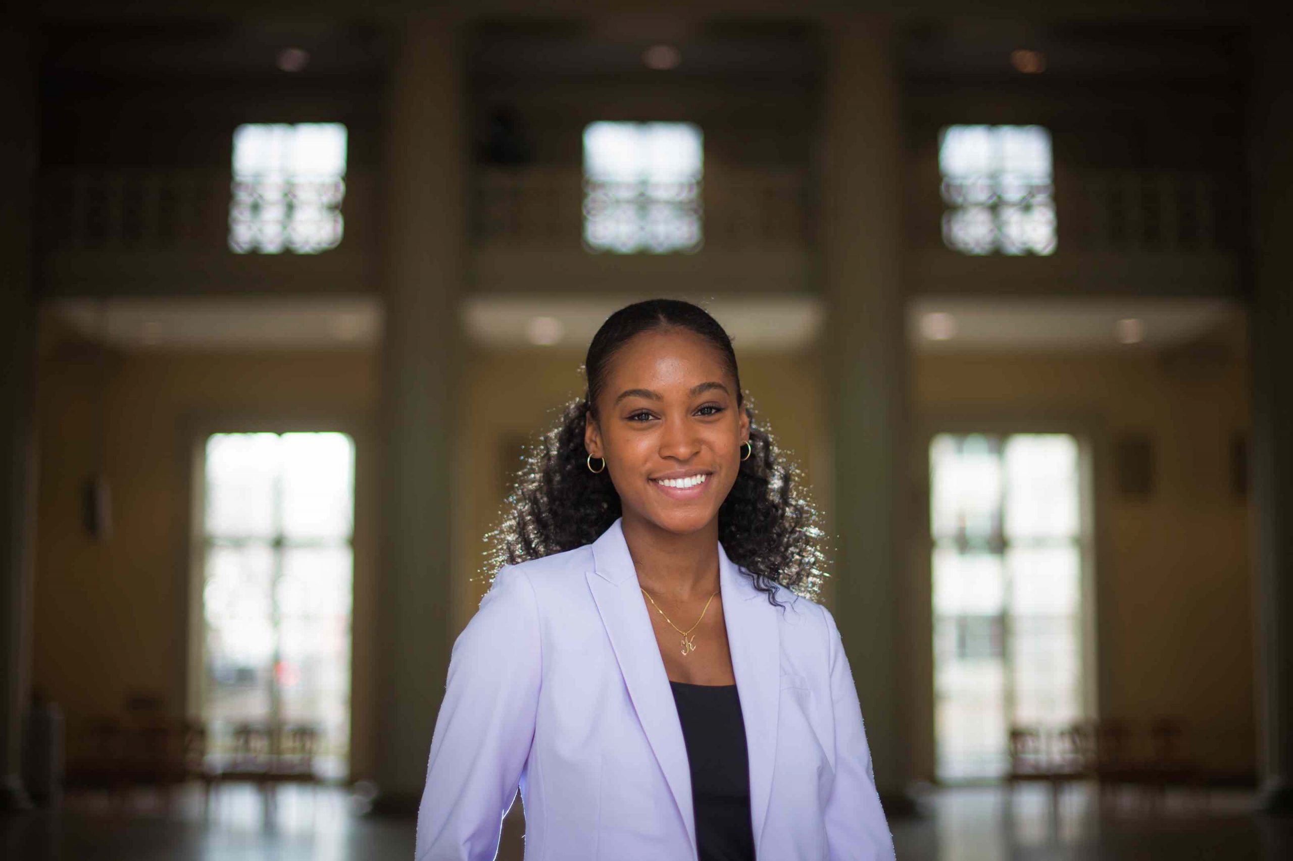
Postdoc Izabella Pena uses social media to combat the infodemic about the Covid-19 pandemic.

Fernanda Ferreira | School of Science
May 4, 2020

In mid-March, Izabella Pena received a WhatsApp text from a friend in Indianapolis, Indiana. “He said, ‘Oh, I got your audio message from a priest in rural São Paulo,’” remembers Pena, a postdoc in Department of Biology Professor David Sabatini’s lab at the Whitehead Institute for Biomedical Research.

Pena had recorded the five-minute audio message about risk groups and the novel coronavirus SARS-CoV-2 for her family’s text thread after she heard one-too-many comments about how only the elderly caught the more severe forms of Covid-19. She never imagined it would spread like wildfire. “I realized the power of these tools,” says Pena of WhatsApp. “You can really reach people and share your information.”

While Pena’s message was fact-checked and scientifically correct, a lot of the information being shared on these platforms isn’t. In Pena’s native Brazil, the messaging platform WhatsApp has played an outsized role in the spread of fake news concerning SARS-CoV-2. Seeing the onslaught of misinformation, Pena first panicked. Then she fought back, choosing to use the vehicles of fake news to spread facts. “We scientists need to learn how to use WhatsApp, YouTube, and Twitter to communicate,” says Pena. “Because that’s how people are getting their information.”

At first, Pena’s misinformation-busting efforts were focused on friends and family. She recorded short audio messages in Portuguese to answer their questions and try to convince them that Covid-19 isn’t just another cold. The rapid spread of her audio messages, which alerted listeners about the importance of physical isolation and risk groups, sparked an idea: to take her science communication efforts from WhatsApp to YouTube, where she could reach a larger audience. Video also has the benefit of being a visual medium, where there’s a face attached to the information being shared. “I think that if people see you, there’s more reliability,” says Pena.

Pena uploaded her first video in late March, answering questions she had received via WhatsApp about Covid-19. Since then, she’s uploaded another five videos and is aiming to release one a week while the pandemic lasts. Many of these videos are in direct response to the messages she gets from viewers. “For example, everybody is asking when is life going to go back to normal, and I think life is only going to go back to ‘normal’ when there’s a vaccine,” says Pena. On April 10, she uploaded a video focused on vaccines, explaining what exactly a vaccine is and how they are made.

On camera, Pena is warm and inviting, delivering updated information about the coronavirus’s biology and epidemiology without clunky jargon and with an abundance of analogies. In a recent video that delved into the biology of SARS-CoV-2 and the different treatments being explored for the virus, she compared the human protein TMPRSS2, which primes the virus’ spike protein to enable the fusion of the virion to a cell’s membrane, to the scissors you use to open a tough plastic snack bag.

In using analogies, Pena is following the advice of Paulo Freire, a famed Brazilian educator and one of her personal idols. “Freire says that the best way to teach something very complicated to someone is to try to bring that concept close to their lives,” says Pena.

Trying to make complex and novel science digestible requires time. According to Pena, just writing the script and developing the analogies takes a couple of hours. “I collect all the information I need before I write the script,” says Pena, whose videos include a long list of references in the description, an unexpected sight on YouTube. “Then I film and edit the video. It all takes a few hours.”

Pena’s videos are filmed late at night because she continues to perform research during the pandemic, mostly virtually. But, she explains, “I’m part of the essential personnel in my lab.” Pena’s work in the Sabatini Lab focuses on the lysosome, the garbage disposal unit of cells that breaks down old cell parts and waste to recycle nutrients. It’s the perfect organelle for someone who has always enjoyed cell metabolism.

“I’ve always liked how chemicals in the cells are made and broken down,” says Pena. Her PhD research at the University of Campinas in Brazil investigated how metabolic problems in the brain could cause epilepsy. Since joining the Sabatini lab in 2018, Pena studies neurodegenerative disorders, like Parkinson’s and Huntington’s, and what role the lysosome plays in them. “For neurodegenerative diseases, there’s a lot of evidence that there’s lysosome influence,” she says. “There are many lysosome gene mutations associated to these disorders, so it’s a nice target to look at.”

Mostly working from home in Cambridge, Massachusetts, Pena is analyzing data and writing grants and papers, balancing her research with her “after-hours job” as a science communicator. “It’s a lot of commitment and dedication, but I believe this is very important, so I’ll keep doing it,” she says. “We are living a hard time, where science and education are constantly under attack. As scientists, we need to help inform people with accurate and life-saving information”.

Recently, Pena added another job title to her resumé: vice-president of ContraCovid, an initiative to make coronavirus information accessible to Latino and immigrant individuals. “We are sharing information in four languages: English, Portuguese, Spanish, and Haitian Creole, to benefit the community here in the U.S. and abroad,” says Pena. But ContraCovid wants to do more, including creating videos like Pena’s in other languages and recruiting more scientists, so that their materials can reach more and more people.

Accessibility of information is at the front of Pena’s mind when she sits down to make a new video. “If you look at how scientists communicate with each other, it’s a bit intimidating,” says Pena. The jargon and the excess of data make it hard for the general public to locate the main takeaways. Pena focuses on stripping away the excess and delivering the message, such as the importance of flattening the curve, in an easily digestible manner.

When imagining her viewers, Pena thinks of her mother. “My mom is not a scientist, but she’s super into technology like YouTube and WhatsApp,” says Pena, who usually sends her audio clips and videos to her mom first, only uploading them once her mom gives the go-ahead. “My mom helps a lot with sharing the videos because she has lots of followers,” Pena laughs. That’s actually how her involvement in Covid-19 outreach started: with her mom wildly sharing Pena’s audio message about risk groups with her numerous followers.

3 Questions: Michael Yaffe on treating Covid-19 patients with acute respiratory distress

MIT professor and intensivist/trauma surgeon explains the new challenges that Covid-19 brings to treating patients in acute respiratory distress.

Bendta Schroeder | Koch Institute
April 30, 2020

During the Covid-19 pandemic, frontline health care workers have had to adapt rapidly to treating patients with lung failure, not only because of shortages of equipment such as ventilators often used to treat severe cases, but also because such approaches are not always effective due to the unique and still imperfectly understood pathology of Covid-19 infections.

Michael Yaffe, the David H. Koch Professor in Science, normally divides his time among his roles as a researcher and professor of biology and biological engineering at MIT, an intensivist/trauma surgeon at Beth Israel Deaconess Medical Center (BIDMC), and a colonel in the U.S. Army Reserve Medical Corps. Currently, he is developing treatments for Covid-19 infections in his laboratory at the Koch Institute for Integrative Cancer Research at MIT. Additionally, he runs one of the Covid-19 Intensive Care Units at BIDMC and serves as co-director of the acute care and ICU section of Boston Hope, the 500-bed pop-up hospital organized by the City of Boston, Massachusetts in the Boston Convention and Exposition Center. Yaffe shares how he is working to improve outcomes for Covid-19 patients and offers his perspective on how emergency care for acute respiratory distress will need to evolve during this crisis and beyond.

Q: What are the special considerations for Covid-19 patients receiving treatment for respiratory failure?

A: We have known about acute respiratory distress syndrome (ARDS) for decades. It was first recognized in battlefield casualties during the Vietnam War, and was initially called “Da-Nang Lung,” but later was understood to be the result of many different diseases. In ARDS, fluid builds up in the tiny air sacs, or alveoli, preventing the lungs from filling up with enough air, and in severe cases is treated by putting patients on ventilators or other devices that support breathing.

The type of lung injury we are seeing in Covid-19 patients behaves very differently from the traditional type of ARDS, and seems to involve early damage to the cells that line the lungs, followed by intense inflammation. The inflammation leads to a massive increase in blood clotting that affects all of the blood vessels in the body, but particularly the blood vessels in the lungs. As a consequence, even if we can force air into the lungs, it does not get delivered very efficiently into the bloodstream.

In ICUs in Boston, New York, and Colorado, we have started a clinical trial using a clot-busting drug called tPA that we think will help rescue patients whose lungs are failing despite maximal support with a mechanical ventilator. This approach has gathered a lot of attention from other hospitals, both nationally and internationally, who are also trying this approach. The work has now led to FDA approval for this drug as an Investigational New Drug, meaning that it is now approved for use in Covid-19 ARDS in the setting of clinical trials.

Q: How has your wide-ranging expertise equipped you to address new challenges that you face in the ICU?

A: I have been very fortunate to be well-prepared to help out in this crisis. First, my training as an intensive care physician and trauma surgeon makes me comfortable in a crisis situation. The clinical problems that we are dealing with here  — ARDS, kidney failure, etc. — are exactly within the scope of my regular clinical practice. Second, my Army deployment experience as a surgeon and critical care doctor in Afghanistan and in Central America has made me very comfortable having to make decisions in resource-limited situations. Finally, it has been incredibly fortuitous that much of my lab’s work has been in the area of cell injury, particularly cancer treatment-related cell injury, but also in the setting of a condition called systemic inflammatory response syndrome, which is essentially exactly what Covid-19 is. In this area, my lab has been studying the link between inflammation and blood clotting for over a decade, and the basic science insights from that work have now become central to our understanding of Covid-19 lung failure, which no one could have foreseen when we first started that research.

Q: What implications do you think the Covid-19 pandemic will have for emergency care after it is over?

A: I think the implications of Covid-19 for the future are immense. First, I hope the lessons learned from this pandemic lead to a complete re-thinking of our national public health policy (or lack of one, really) and a re-engagement with World Health Organization officials for monitoring the outbreak of emerging diseases.

Second, I think that this crisis may fuel additional research funding in the area of critical care medicine. Before the Covid-19 crisis, very few people had heard of ARDS, or even critical care as a field of medicine, since it does not have the glamour of conditions like cancer medicine or cardiovascular disease. Historically, research in this area has been underfunded, but now that ARDS has taken the spotlight in the news, I am hopeful that the recognition that some patients with Covid-19 are dying because of critical illness and lung failure will lead to new efforts to better understand the link between inflammation, lung function, and innate immunity, including blood coagulation. The Covid-19 crisis will not end when this first wave subsides, but will re-visit us again in the fall. Additionally, other coronavirus diseases as well as viral epidemics are likely to continue to plague us in the future.

One final lesson we are learning from this terrible pandemic is how important it is to treat all of the different parts of the body as a complex interacting unit, and to apply what we know from systems biology and other fields of study to understand how those parts are integrated into one coherent system. The lung failure, kidney failure, and inflammation of the heart that are the hallmarks of Covid-19 critical illness directly reflect how different inflammatory molecules in the blood alter the function of each of these different organ systems. Our traditional medical approach of having separate specialists in infectious disease, pulmonary medicine, renal medicine, and hematology does not work well when all the organ systems are cross-talking to each other. The job of the intensive care physician is to integrate all of the relevant basic biology and pathology of these organs into a comprehensive holistic treatment approach for the patient. Covid-19 has made that need to think across multiple disciplines and connect basic science to clinical care even more apparent.

Engineers develop precision injection system for plants

Microneedles made of silk-based material can target plant tissues for delivery of micronutrients, hormones, or genes.

David L. Chandler | MIT News Office
April 27, 2020

While the human world is reeling from one pandemic, there are several ongoing epidemics that affect crops and put global food production at risk. Oranges, olives, and bananas are already under threat in many areas due to diseases that affect plants’ circulatory systems and that cannot be treated by applying pesticides.

A new method developed by engineers at MIT may offer a starting point for delivering life-saving treatments to plants ravaged by such diseases.

These diseases are difficult to detect early and to treat, given the lack of precision tools to access plant vasculature to treat pathogens and to sample biomarkers. The MIT team decided to take some of the principles involved in precision medicine for humans and adapt them to develop plant-specific biomaterials and drug-delivery devices.

The method uses an array of microneedles made of a silk-based biomaterial to deliver nutrients, drugs, or other molecules to specific parts of the plant. The findings are described in the journal Advanced Science, in a paper by MIT professors Benedetto Marelli and Jing-Ke-Weng, graduate student Yunteng Cao, postdoc Eugene Lim at MIT, and postdoc Menglong Xu at the Whitehead Institute for Biomedical Research.

The microneedles, which the researchers call phytoinjectors, can be made in a variety of sizes and shapes, and can deliver material specifically to a plant’s roots, stems, or leaves, or into its xylem (the vascular tissue involved in water transportation from roots to canopy) or phloem (the vascular tissue that circulates metabolites throughout the plant). In lab tests, the team used tomato and tobacco plants, but the system could be adapted to almost any crop, they say. The microneedles can not only deliver targeted payloads of molecules into the plant, but they can also be used to take samples from the plants for lab analysis.

The work started in response to a request from the U.S. Department of Agriculture for ideas on how to address the citrus greening crisis, which is threatening the collapse of a $9 billion industry, Marelli says. The disease is spread by an insect called the Asian citrus psyllid that carries a bacterium into the plant. There is as yet no cure for it, and millions of acres of U.S. orchards have already been devastated. In response, Marelli’s lab swung into gear to develop the novel microneedle technology, led by Cao as his thesis project.

The disease infects the phloem of the whole plant, including roots, which are very difficult to reach with any conventional treatment, Marelli explains. Most pesticides are simply sprayed or painted onto a plant’s leaves or stems, and little if any penetrates to the root system. Such treatments may appear to work for a short while, but then the bacteria bounce back and do their damage. What is needed is something that can target the phloem circulating through a plant’s tissues, which could carry an antibacterial compound down into the roots. That’s just what some version of the new microneedles could potentially accomplish, he says.

“We wanted to solve the technical problem of how you can have a precise access to the plant vasculature,” Cao adds. This would allow researchers to inject pesticides, for example, that would be transported between the root system and the leaves. Present approaches use “needles that are very large and very invasive, and that results in damaging the plant,” he says. To find a substitute, they built on previous work that had produced microneedles using silk-based material for injecting human vaccines.

“We found that adaptations of a material designed for drug delivery in humans to plants was not straightforward, due to differences not only in tissue vasculature, but also in fluid composition,” Lim says. The microneedles designed for human use were intended to biodegrade naturally in the body’s moisture, but plants have far less available water, so the material didn’t dissolve and was not useful for delivering the pesticide or other macromolecules into the phloem. The researchers had to design a new material, but they decided to stick with silk as its basis. That’s because of silk’s strength, its inertness in plants (preventing undesirable side effects), and the fact that it degrades into tiny particles that don’t risk clogging the plant’s internal vasculature systems.

They used biotechnology tools to increase silk’s hydrophilicity (making it attract water), while keeping the material strong enough to penetrate the plant’s epidermis and degradable enough to then get out of the way.

Sure enough, they tested the material on their lab tomato and tobacco plants, and were able to observe injected materials, in this case fluorescent molecules, moving all they way through the plant, from roots to leaves.

“We think this is a new tool that can be used by plant biologists and bioengineers to better understand transport phenomena in plants,” Cao says. In addition, it can be used “to deliver payloads into plants, and this can solve several problems. For example, you can think about delivering micronutrients, or you can think about delivering genes, to change the gene expression of the plant or to basically engineer a plant.”

“Now, the interests of the lab for the phytoinjectors have expanded beyond antibiotic delivery to genetic engineering and point-of-care diagnostics,” Lim adds.

For example, in their experiments with tobacco plants, they were able to inject an organism called Agrobacterium to alter the plant’s DNA – a typical bioengineering tool, but delivered in a new and precise way.

So far, this is a lab technique using precision equipment, so in its present form it would not be useful for agricultural-scale applications, but the hope is that it can be used, for example, to bioengineer disease-resistant varieties of important crop plants. The team has also done tests using a modified toy dart gun mounted to a small drone, which was able to fire microneedles into plants in the field. Ultimately, such a process might be automated using autonomous vehicles, Marelli says, for agricultural-scale use.

Meanwhile, the team continues to work on adapting the system to the varied needs and conditions of different kinds of plants and their tissues. “There’s a lot of variation among them, really,” Marelli says, so you need to think about having devices that are plant-specific. For the future, our research interests will go beyond antibiotic delivery to genetic engineering and point-of-care diagnostics based on metabolite sampling.”

The work was supported by the Office of Naval Research, the National Science Foundation, and the Keck Foundation.

Katie Collins, Vaishnavi Phadnis, and Vaibhavi Shah named 2020-21 Goldwater Scholars

Three MIT undergraduates who use computer science to explore human biology and health honored for their academic achievements.

Fernanda Ferreira | School of Science
April 10, 2020

MIT students Katie Collins, Vaishnavi Phadnis, and Vaibhavi Shah have  been selected to receive a Barry Goldwater Scholarship for the 2020-21 academic year. Over 5,000 college students from across the United States were nominated for the scholarships, from which only 396 recipients were selected based on academic merit.

The Goldwater scholarships have been conferred since 1989 by the Barry Goldwater Scholarship and Excellence in Education Foundation. These scholarships have supported undergraduates who go on to become leading scientists, engineers, and mathematicians in their respective fields. All of the 2020-21 Goldwater Scholars intend to obtain a doctorate in their area of research, including the three MIT recipients.

Katie Collins, a third-year majoring in brain and cognitive sciences with minors in computer science and biomedical engineering, got involved with research in high school, when she worked on computational models of metabolic networks and synthetic gene networks in the lab of Department of Electrical Engineering and Computer Science Professor Timothy Lu at MIT. It was this project that led her to realize how challenging it is to model and analyze complex biological networks. She also learned that machine learning can provide a path for exploring these networks and understanding human diseases. This realization has coursed a scientific path for Collins that is equally steeped in computer science and human biology.

Over the past few years, Collins has become increasingly interested in the human brain, particularly what machine learning can learn from human common-sense reasoning and the way brains process sparse, noisy data. “I aim to develop novel computational algorithms to analyze complex, high-dimensional data in biomedicine, as well as advance modelling paradigms to improve our understanding of human cognition,” explains Collins. In his letter of recommendation, Professor Tomaso Poggio, the Eugene McDermott Professor in the Department of Brain and Cognitive Sciences and one of Collins’ mentors, wrote, “It is very difficult to imagine a better candidate for the Goldwater fellowship.” Collins plans to pursue a PhD studying machine learning or computational neuroscience and to one day run her own lab. “I hope to become a professor, leading a research program at the interface of computer science and cognitive neuroscience.”

Vaishnavi Phadnis, a second-year majoring in computer science and molecular biology, sees molecular and cellular biology as the bridge between chemistry and life, and she’s been enthralled with understanding that bridge since 7th grade, when she learned about the chemical basis of the cell. Phadnis spent two years working in a cancer research lab while still in high school, an experience which convinced her that research was not just her passion but also her future. “In my first week at MIT, I approached Professor Robert Weinberg, and I’ve been grateful to do research in his lab ever since,” she says.

“Vaishnavi’s exuberance makes her a joy to have in the lab,” wrote Weinberg, who is the Daniel Ludwig Professor in the Department of Biology. Phadnis is investigating ferroptosis, a recently discovered, iron-dependent form of cell death that may be relevant in neurodegeneration and also a potential strategy for targeting highly aggressive cancer cells. “She is a phenomenon who has vastly exceeded our expectations of the powers of someone her age,” Weinberg says. Phadnis is thankful to Weinberg and all the scientific mentors, both past and present, that have inspired her along her research path. Deciphering the mechanisms behind fundamental cellular processes and exploring their application in human diseases is something Phadnis plans to continue doing in her future as a physician-scientist after pursuing an MD/PhD. “I hope to devote most of my time to leading my own research group, while also practicing medicine,” she says.

Vaibhavi Shah, a third-year studying biological engineering with a minor in science, technology and society, spent a lot of time in high school theorizing ways to tackle major shortcomings in medicine and science with the help of technology. “When I came to college, I was able to bring some of these ideas to fruition,” she says, working with both the Big Data in Radiology Group at the University of California at San Francisco and the lab of Professor Mriganka Sur, the Newton Professor of Neuroscience in the Department of Brain and Cognitive Sciences.

Shah is particularly interested in integrating innovative research findings with traditional clinical practices. According to her, technology, like computer vision algorithms, can be adopted to diagnose diseases such as Alzheimer’s, allowing patients to start appropriate treatments earlier. “This is often harder to do at smaller, rural institutions that may not always have a specialist present,” says Shah, and algorithms can help fill that gap. One of aims of Shah’s research is to improve the efficiency and equitability of physician decision-making. “My ultimate goal is to improve patient outcomes, and I aim to do this by tackling emerging scientific questions in machine learning and artificial intelligence at the forefront of neurology,” she says. The clinic is a place Shah expects to be in the future after obtaining her physician-scientist training, saying, “I hope to a practicing neurosurgeon and clinical investigator.”

The Barry Goldwater Scholarship and Excellence in Education Program was established by Congress in 1986 to honor Senator Barry Goldwater, a soldier and statesman who served the country for 56 years. Awardees receive scholarships of up to $7,500 a year to cover costs related to tuition, room and board, fees, and books.

Neuroscientists find memory cells that help us interpret new situations

Neurons that store abstract representations of past experiences are activated when a new, similar event takes place.

Anne Trafton | MIT News Office
April 6, 2020

Imagine you are meeting a friend for dinner at a new restaurant. You may try dishes you haven’t had before, and your surroundings will be completely new to you. However, your brain knows that you have had similar experiences — perusing a menu, ordering appetizers, and splurging on dessert are all things that you have probably done when dining out.

MIT neuroscientists have now identified populations of cells that encode each of these distinctive segments of an overall experience. These chunks of memory, stored in the hippocampus, are activated whenever a similar type of experience takes place, and are distinct from the neural code that stores detailed memories of a specific location.

The researchers believe that this kind of “event code,” which they discovered in a study of mice, may help the brain interpret novel situations and learn new information by using the same cells to represent similar experiences.

“When you encounter something new, there are some really new and notable stimuli, but you already know quite a bit about that particular experience, because it’s a similar kind of experience to what you have already had before,” says Susumu Tonegawa, a professor of biology and neuroscience at the RIKEN-MIT Laboratory of Neural Circuit Genetics at MIT’s Picower Institute for Learning and Memory.

Tonegawa is the senior author of the study, which appears today in Nature Neuroscience. Chen Sun, an MIT graduate student, is the lead author of the paper. New York University graduate student Wannan Yang and Picower Institute technical associate Jared Martin are also authors of the paper.

Encoding abstraction

It is well-established that certain cells in the brain’s hippocampus are specialized to store memories of specific locations. Research in mice has shown that within the hippocampus, neurons called place cells fire when the animals are in a specific location, or even if they are dreaming about that location.

In the new study, the MIT team wanted to investigate whether the hippocampus also stores representations of more abstract elements of a memory. That is, instead of firing whenever you enter a particular restaurant, such cells might encode “dessert,” no matter where you’re eating it.

To test this hypothesis, the researchers measured activity in neurons of the CA1 region of the mouse hippocampus as the mice repeatedly ran a four-lap maze. At the end of every fourth lap, the mice were given a reward. As expected, the researchers found place cells that lit up when the mice reached certain points along the track. However, the researchers also found sets of cells that were active during one of the four laps, but not the others. About 30 percent of the neurons in CA1 appeared to be involved in creating this “event code.”

“This gave us the initial inkling that besides a code for space, cells in the hippocampus also care about this discrete chunk of experience called lap 1, or this discrete chunk of experience called lap 2, or lap 3, or lap 4,” Sun says.

To further explore this idea, the researchers trained mice to run a square maze on day 1 and then a circular maze on day 2, in which they also received a reward after every fourth lap. They found that the place cells changed their activity, reflecting the new environment. However, the same sets of lap-specific cells were activated during each of the four laps, regardless of the shape of the track. The lap-encoding cells’ activity also remained consistent when laps were randomly shortened or lengthened.

“Even in the new spatial locations, cells still maintain their coding for the lap number, suggesting that cells that were coding for a square lap 1 have now been transferred to code for a circular lap 1,” Sun says.

The researchers also showed that if they used optogenetics to inhibit sensory input from a part of the brain called the medial entorhinal cortex (MEC), lap-encoding did not occur. They are now investigating what kind of input the MEC region provides to help the hippocampus create memories consisting of chunks of an experience.

Two distinct codes

These findings suggest that, indeed, every time you eat dinner, similar memory cells are activated, no matter where or what you’re eating. The researchers theorize that the hippocampus contains “two mutually and independently manipulatable codes,” Sun says. One encodes continuous changes in location, time, and sensory input, while the other organizes an overall experience into smaller chunks that fit into known categories such as appetizer and dessert.

“We believe that both types of hippocampal codes are useful, and both are important,” Tonegawa says. “If we want to remember all the details of what happened in a specific experience, moment-to-moment changes that occurred, then the continuous monitoring is effective. But on the other hand, when we have a longer experience, if you put it into chunks, and remember the abstract order of the abstract chunks, that’s more effective than monitoring this long process of continuous changes.”

The new MIT results “significantly advance our knowledge about the function of the hippocampus,” says Gyorgy Buzsaki, a professor of neuroscience at New York University School of Medicine, who was not part of the research team.

“These findings are significant because they are telling us that the hippocampus does a lot more than just ‘representing’ space or integrating paths into a continuous long journey,” Buzsaki says. “From these remarkable results Tonegawa and colleagues conclude that they discovered an ‘event code,’ dedicated to organizing experience by events, and that this code is independent of spatial and time representations, that is, jobs also attributed to the hippocampus.”

Tonegawa and Sun believe that networks of cells that encode chunks of experiences may also be useful for a type of learning called transfer learning, which allows you to apply knowledge you already have to help you interpret new experiences or learn new things. Tonegawa’s lab is now working on trying to find cell populations that might encode these specific pieces of knowledge.

The research was funded by the RIKEN Center for Brain Science, the Howard Hughes Medical Institute, and the JPB Foundation.

A stopgap measure to treat respiratory distress

Repurposing a drug used for blood clots may help Covid-19 patients in danger of respiratory failure, researchers suggest.

Anne Trafton | MIT News Office
March 26, 2020

Researchers at MIT and the University of Colorado at Denver have proposed a stopgap measure that they believe could help Covid-19 patients who are in acute respiratory distress. By repurposing a drug that is now used to treat blood clots, they believe they could help people in cases where a ventilator is not helping, or if a ventilator is not available.

Three hospitals in Massachusetts and Colorado are developing plans to test this approach in severely ill Covid-19 patients. The drug, a protein called tissue plasminogen activator (tPA), is commonly given to heart attack and stroke victims. The approach is based on emerging data from China and Italy that Covid-19 patients have a profound disorder of blood clotting that is contributing to their respiratory failure.

“If this were to work, which I hope it will, it could potentially be scaled up very quickly, because every hospital already has it in their pharmacy,” says Michael Yaffe, a David H. Koch Professor of Science at MIT. “We don’t have to make a new drug, and we don’t have to do the same kind of testing that you would have to do with a new agent. This is a drug that we already use. We’re just trying to repurpose it.”

Yaffe, who is also a member of MIT’s Koch Institute for Integrative Cancer Research and an intensive care physician at Boston’s Beth Israel Deaconess Medical Center/Harvard Medical School, is the senior author of a paper describing the new approach.

The paper, which appears in the Journal of Trauma and Acute Care Surgery, was co-authored by Christopher Barrett, a surgeon at Beth Israel Deaconess and a visiting scientist at MIT; Hunter Moore, Ernest Moore, Peter Moore, and Robert McIntyre of the University of Colorado at Denver; Daniel Talmor of Beth Israel Deaconess; and Frederick Moore of the University of Florida.

Breaking up clots

In one large-scale study of the Covid-19 outbreak in Wuhan, China, it was found that 5 percent of patients required intensive care and 2.3 percent required a ventilator. Many doctors and public health officials in the United States worry that there may not be enough ventilators for all Covid-19 patients who will need them. In China and Italy, a significant number of the patients who required a ventilator went on to die of respiratory failure, despite maximal support, indicating that there is a need for additional treatment approaches.

The treatment that the MIT and University of Colorado team now proposes is based on many years of research into what happens in the lungs during respiratory failure. In such patients, blood clots often form in the lungs. Very small clots called microthrombi can also form in the blood vessels of the lungs. These tiny clots prevent blood from reaching the airspaces of the lungs, where blood normally becomes oxygenated.

The researchers believe that tPA, which helps to dissolve blood clots, may help patients in acute respiratory distress. A natural protein found in our bodies, tPA converts plasminogen to an enzyme called plasmin, which breaks down clots. Larger amounts are often given to heart attack patients or stroke victims to dissolve the clot causing the heart attack or stroke.

Animal experiments, and one human trial, have shown potential benefits of this approach in treating respiratory distress. In the human trial, performed in 2001, 20 patients who were in respiratory failure following trauma or sepsis were given drugs that activate plasminogen (urokinase or streptokinase, but not tPA). All of the patients in the trial had respiratory distress so severe that they were not expected to survive, but 30 percent of them survived following treatment.

That is the only study using plasminogen activators to treat respiratory failure in humans to date, largely because improved ventilator strategies have been working well. This appears not to be the case for many patients with Covid-19, Yaffe says.

The idea to try this treatment in Covid-19 patients arose, in part, because the Colorado and MIT research team has spent the last several years studying the inflammation and abnormal bleeding that can occur in the lungs following traumatic injuries. It turns out that Covid-19 patients also suffer from inflammation-linked tissue damage, which has been seen in autopsy results from those patients and may contribute to clot formation.

“What we are hearing from our intensive care colleagues in Europe and in New York is that many of the critically ill patients with Covid-19 are hypercoagulable, meaning that they are clotting off their IVs, and having kidney and heart failure from blood clots, in addition to lung failure.  There’s plenty of basic science to support the idea that this concept should be beneficial,” Yaffe says. “The tricky part, of course, is figuring out the right dose and route of administration. But the target we are going after is well-validated.”

Potential benefits

The researchers will test tPA in patients under the FDA’s “compassionate use” program, which allows experimental drugs to be used in cases where there are no other treatment options. If the drug appears to help in an initial set of patients, its use could be expanded further, Yaffe says.

“We learned that the clinical trial will be funded by BARDA [the Biomedical Advanced Research and Development Authority], and that Francis Collins, the NIH director, was briefed on the approach yesterday afternoon,” he says. “Genentech, the manufacturer of tPA, has already donated the drug for the initial trial, and indicated that they will rapidly expand access if the initial patient response is encouraging.”

Based on the latest data from their colleagues in Colorado, these groups plan to deliver the drug both intravenously and/or instill it directly into the airways. The intravenous route is currently used for stroke and heart attack patients. Their idea is to give one dose rapidly, over a two-hour period, followed by an equivalent dose given more slowly over 22 hours. Applied BioMath, a company spun out by former MIT researchers, is now working on computational models that may help to refine the dosing schedule.

“If it were to work, and we don’t yet know if it will, it has a lot of potential for rapid expansion,” Yaffe says. “The public health benefits are obvious. We might get people off ventilators quicker, and we could potentially prevent people from needing to go on a ventilator.”

The hospitals planning to test this approach are Beth Israel Deaconess, the University of Colorado Anschultz Medical Campus, and Denver Health. The research that led to this proposal was funded by the National Institutes of Health and the Department of Defense Peer Reviewed Medical Research Program.

Thank you for your patients

An unusual synergy between cancer researchers, clinical centers, and industry leads to promising clinical trials for a new combination therapy for prostate cancer.

Bendta Schroeder | Koch Institute
March 21, 2020

As Jesse Patterson, an MIT research scientist, and Frank Lovell, a finance industry retiree with a penchant for travel, chatted in the Koch Institute auditorium after a public lecture, they realized the anomaly of the experience: Cancer patients rarely get to meet researchers working on their treatments, and cancer researchers rarely get to put a name and a face to the people they aim to help through their work.

Lovell was participating in a clinical trial for a prostate cancer therapy that combines the widely-used targeted therapy abiraterone with the Plk1 inhibitor onvansertib. Patterson, working in the laboratory of Professor Michael Yaffe, the David H. Koch Professor of Science and director of the MIT Center for Precision Cancer Medicine, played a significant role in identifying the new drug combination and its powerful potential.

While their encounter was indeed fortunate, it was not random. They never would have met if not for the human synergy showcased at that evening’s SOLUTIONS with/in/sight event, the result of collaborative relationships built between research labs, clinical centers, and industry. Patterson and Yaffe were on hand to tell the story of the science behind their new drug combination, and were joined by some of the partners who helped translate their results into a clinical trial: David Einstein, clinical oncologist at Beth Israel Deaconess Medical Center, and Mark Erlander, chief scientific officer of Trovagene Oncology, the biotech company that developed onvansertib.

Network synergy

The need for new prostate cancer therapies is acute. Prostate cancer is the leading diagnosis among men for non-skin cancer and the second-leading cancer killer among men in the United States. Abiraterone works by shutting off androgen synthesis and interfering with the androgen receptor pathway, which plays a crucial role in prostate cancer cells’ ability to survive and divide. However, cancer cells eventually evolve resistance to abiraterone. New, more powerful drug combinations are needed to circumvent or delay the development of resistance.

Patterson and his colleagues in the Yaffe lab hypothesized that by targeting both the androgen receptor and other pathways critical to cancer cell proliferation, they could produce a synergistic effect — that is, a combination effect that is much greater than the sum of each drug’s effect by itself. Plk1, a pathway critical to each stage of cell division, was of longstanding interest to the Yaffe group, and was among those Patterson strategically selected for investigation as a potential partner target for androgen receptor. In screens of prostate cancer cell lines and in xenograft tumors, the researchers found that abiraterone and Plk1 inhibitors both interfere with cell division when delivered singly, but that together, those effects are amplified and far more often lethal to cancer cells.

An unexpected phone call from Mark Erlander at Trovagene, a San Diego-based clinical-stage biotech company, was instrumental in translating the Yaffe Lab’s research results into clinical trials.

Erlander had learned that MIT held a patent for the combination of Plk1 inhibitors and anti-androgens for any cancer — the result of Yaffe Lab studies. Although he did not know Yaffe personally and lived a continent away, Erlander picked up the phone and invited Yaffe for coffee. “This was worth flying across the country,” Erlander said.

Still in scrubs, Yaffe, who is an attending surgeon at Beth Israel Deaconess Medical Center in addition to his academic roles, chatted with Erlander during his shift break at the hospital. The new collaboration was on its way.

Speaking Frankly

While Erlander had the Plk1 inhibitor and the Yaffe Lab had the science behind it, they were still missing an important component of any clinical trial: patients. Yaffe enlisted doctors David Einstein and Steven Balk, both at Beth Israel Deaconess Medical Center and Dana Farber/Harvard Cancer Center, with whom he had worked on related research supported by the Bridge Project, to bring clinical translation expertise and patient access.

By the time clinical trials began in 2019, Frank Lovell was ready for a new treatment. When his prostate cancer was first diagnosed about a decade ago, he was treated with surgery and radiation. When the cancer came back five years later, he received a hormonal treatment that stopped working within three years. He started to see Einstein, an oncologist who specialized in novel therapies, and tried yet another treatment, this one losing effectiveness after a year. Then he joined Einstein’s trial.

For Lovell, the new combination of drugs was “effective in a wonderful way.” Many of the patients in the trial — 72 percent of those who completed phase 2 — showed declining or stabilized levels of prostate-specific androgen (PSA), indicating a positive response to the treatment. Lovell’s PSA levels stabilized, too, and he reports that he experienced very few side effects.

But most importantly, noted Lovell, “I say thank you to Dr. Einstein, Dr. Patterson, and Dr. Yaffe. They brought me hope and time.”

The gratitude is mutual.

“I especially want to thank Frank and all the patients like him who have volunteered to be on these clinical trials,” says Yaffe. “Without patients like Frank, we would never know how to better treat these types of cancers.”

Lovell is no longer in the trial for now, but enjoying making his rounds from Cape Cod in the summer; to Paris and Cannes, France, and then Hawaii in the autumn; and to Naples, Florida, in the winter, on top of visiting with family and a wide circle of friends. “Illness has not stopped me from living a normal life,” Lovell said. “You wouldn’t think I was sick.”

Meanwhile, Yaffe, Patterson, and their research collaborators are still at work. They are optimizing drug delivery regimens to maximize the time on treatment and minimize toxicity, as well as finding biomarkers that help identify which patients will best respond to the combination. They are also looking to understand the mechanism behind the synergy better, which in turn may help them find more effective partners for onvansertib, and to identify other cancer types, such as ovarian cancer, for which the combination may be effective.

Bacterial enzyme could become a new target for antibiotics

Scientists discover the structure of an enzyme, found in the human gut, that breaks down a component of collagen.

Anne Trafton | MIT News Office
March 17, 2020

MIT and Harvard University chemists have discovered the structure of an unusual bacterial enzyme that can break down an amino acid found in collagen, which is the most abundant protein in the human body.

The enzyme, known as hydroxy-L-proline dehydratase (HypD), has been found in a few hundred species of bacteria that live in the human gut, including Clostridioides difficile. The enzyme performs a novel chemical reaction that dismantles hydroxy-L-proline, the molecule that gives collagen its tough, triple-helix structure.

Now that researchers know the structure of the enzyme, they can try to develop drugs that inhibit it. Such a drug could be useful in treating C. difficile infections, which are resistant to many existing antibiotics.

“This is very exciting because this enzyme doesn’t exist in humans, so it could be a potential target,” says Catherine Drennan, an MIT professor of chemistry and biology and a Howard Hughes Medical Institute Investigator. “If you could potentially inhibit that enzyme, that could be a unique antibiotic.”

Drennan and Emily Balskus, a professor of chemistry and chemical biology at Harvard University, are the senior authors of the study, which appears today in the journal eLife. MIT graduate student Lindsey Backman and former Harvard graduate student Yolanda Huang are the lead authors of the study.

A difficult reaction

The HypD enzyme is part of a large family of proteins called glycyl radical enzymes. These enzymes work in an unusual way, by converting a molecule of glycine, the simplest amino acid, into a radical — a molecule that has one unpaired electron. Because radicals are very unstable and reactive, they can be used as cofactors, which are molecules that help drive a chemical reaction that would otherwise be difficult to perform.

These enzymes work best in environments that don’t have a lot of oxygen, such as the human gut. The Human Microbiome Project, which has sequenced thousands of bacterial genes from species found in the human gut, has yielded several different types of glycyl radical enzymes, including HypD.

In a previous study, Balskus and researchers at the Broad Institute of MIT and Harvard discovered that HypD can break down hydroxy-L-proline into a precursor of proline, one of the essential amino acids, by removing the hydroxy modification as a molecule of water. These bacteria can ultimately use proline to generate ATP, a molecule that cells use to store energy, through a process called amino acid fermentation.

HypD has been found in about 360 species of bacteria that live in the human gut, and in this study, Drennan and her colleagues used X-ray crystallography to analyze the structure of the version of HypD found in C. difficile. In 2011, this species of bacteria was responsible for about half a million infections and 29,000 deaths in the United States.

The researchers were able to determine which region of the protein forms the enzyme’s “active site,” which is where the reaction occurs. Once hydroxy-L-proline binds to the active site, a nearby glycine molecule forms a glycyl radical that can pass that radical onto the hydroxy-L-proline, leading to the elimination of the hydroxy group.

Removing a hydroxy group is usually a difficult reaction that requires a large input of energy.

“By transferring a radical to hydroxy-L-proline, it lowers the energetic barrier and allows for that reaction to occur pretty rapidly,” Backman says. “There’s no other known enzyme that can perform this kind of chemistry.”

New drug target

It appears that once bacteria perform this reaction, they divert proline into their own metabolic pathways to help them grow. Therefore, blocking this enzyme could slow down the bacteria’s growth. This could be an advantage in controlling C. difficile, which often exists in small numbers in the human gut but can cause illness if the population becomes too large. This sometimes occurs after antibiotic treatment that wipes out other species and allows C. difficile to proliferate.

C. difficile can be in your gut without causing problems — it’s when you have too much of it compared to other bacteria that it becomes more problematic,” Drennan says. “So, the idea is that by targeting this enzyme, you could limit the resources of C. difficile, without necessarily killing it.”

The researchers now hope to begin designing drug candidates that could inhibit HypD, by targeting the elements of the protein structure that appear to be the most important in carrying out its function.

The research was funded by the National Institutes of Health, a National Science Foundation Graduate Research Fellowship, Harvard University, a Packard Fellowship for Science and Engineering, the NSERC Postgraduate Scholarship-Doctoral Program, an Arnold O. Beckman Postdoctoral Fellowship, a Dow Fellowship, and a Gilliam Fellowship from the Howard Hughes Medical Institute.

QS World University Rankings rates MIT No. 1 in 12 subjects for 2020

Institute ranks second in five subject areas.

MIT News Office
March 4, 2020

MIT has been honored with 12 No. 1 subject rankings in the QS World University Rankings for 2020.

The Institute received a No. 1 ranking in the following QS subject areas: Architecture/Built Environment; Chemistry; Computer Science and Information Systems; Chemical Engineering; Civil and Structural Engineering; Electrical and Electronic Engineering; Mechanical, Aeronautical and Manufacturing Engineering; Linguistics; Materials Science; Mathematics; Physics and Astronomy; and Statistics and Operational Research.

MIT also placed second in five subject areas: Accounting and Finance; Biological Sciences; Earth and Marine Sciences; Economics and Econometrics; and Environmental Sciences.

Quacquarelli Symonds Limited subject rankings, published annually, are designed to help prospective students find the leading schools in their field of interest. Rankings are based on research quality and accomplishments, academic reputation, and graduate employment.

MIT has been ranked as the No. 1 university in the world by QS World University Rankings for eight straight years.

A force for health equity

Through on-site projects in developing countries and internships in the business world, Kendyll Hicks explores the political and economic drivers of global health.

Becky Ham | MIT News Office
March 1, 2020

After spending three weeks in Kenya working on water issues with Maasai women, Kendyll Hicks was ready to declare it her favorite among the international projects she’s participated in through MIT.

As a volunteer with the nonprofit Mama Maji, Hicks spoke about clean water, menstrual hygiene, and reproductive health with local women, sharing information that would enable them to become community leaders. “In rural Kenya, women are disproportionately affected by water issues,” she explains. “This is one way to give them a voice in societies that traditionally will silence them.”

The team also planned to build a rainwater harvesting tank, but climate change has transformed Kenya’s dry season into a rainy one, and it was too wet to break ground for the project. During her stay, Hicks lived in the home of the first female chief of the Masaai, Beatrice Kosiom, whom Hicks describes as “simultaneously a political animal and the most down-to-earth-person.” It was this close contact with the community that made the project especially fulfilling.

During MIT’s Independent Activities Period, Hicks also has traveled to South Africa to learn more about the cultural and biological determinants of that country’s HIV/AIDS epidemic, and to Colombia to lead an entrepreneurial initiative among small-scale coffee farmers. Hicks joined the Kenya trip after taking an MIT D-Lab class on water, sanitation, and hygiene. Each experience has been successively more hands-on, she says.

“I’ve been drawn to these experiences mainly because I love school, and I love the classroom experience,” Hicks says. “But it just can’t compare to living with people and understanding their way of life and the issues they face every day.”

Hicks, a senior majoring in computer science and molecular biology, says she has shifted her focus during her time at MIT from more incremental technical discoveries to addressing larger forces that affect how those discoveries contribute — or fail to contribute — to global health.

Her love of biology began with animals and zoology, later expanding into an interest in medicine. “Humans are these amazing machines that have been crafted by nature and evolution, and we have all these intricacies and mechanisms that I knew I wanted to study further,” Hicks says.

At the same time, she says, “I’ve always been interested in health care and medicine, and the main impetus behind that is the fact that when someone you love is sick, or if you’re sick, you’ll do whatever you can.”

As a first-year student she worked in the Lippard Lab at MIT, helping to synthesize and test anticancer compounds, but she soon decided that lab work wasn’t the right path for her. “I made the realization that health care and medicine are extremely political,” she recalls. “Health policy, health economics, law — those can be the drivers of real large-scale change.”

To learn more about those drivers, Hicks has worked two summers at the management consulting firm McKinsey and Company, and will take a full-time position with the company after graduation.

“As someone immersed in the world of science and math and tech, I had this lingering insecurity that I didn’t know that much about this entirely different but super-important area,” she says. “I thought it would be important to understand what motivates business and the private sector, since that can have a huge effect on health care and helping communities that are often disenfranchised.”

Hicks wants to steer her work at McKinsey toward their health care and hospital sector, as well as their growing global health sector. Over the long term, she is also interested in continuing fieldwork that involves science, poverty eradication, and international development.

“Being at MIT, it’s like this hub of tech, trying to venture further into novel breakthroughs and innovations, and I think it’s amazing,” Hicks says. “But as I have started to garner more of an interest in politics and economics and the highly socialized aspects of science, I would say it’s important to take a pause before venturing further and deeper into that realm, to make sure that you truly understand the downstream effects of what you are developing.”

“Those effects can be negative,” she adds, “and they oftentimes impact communities that already are systematically and institutionally oppressed.”

Hicks joined MIT’s Black Students Union as a first-year student and now serves as the BSU Social and Cultural Co-Chair. In the role, she is responsible for planning the annual Ebony Affair fly-in program, which brings more than 30 black high school students to campus each year to participate in workshops, tour labs, and join a gala celebration with BSU students, faculty, and staff. “We’re doing our best as a community to convince young bright black minds to come to a place like MIT,” she says.

It worked for Hicks: She participated in Ebony Affair as a high schooler, and the experience cemented her decision to attend. “When I saw everyone showing out and having such pride in being black and being at MIT, I was like, ‘OK, I want to be a part of that,’” she recalls.

Last year, Hicks planned BSU’s first Black Homecoming event, a barbecue that brought together current and former black MIT students — some who attended the school 50 years ago. The event was a celebration of support and a way to strengthen the BSU network. “You have to do what you can to cultivate communities wherever you are, and that’s what I’ve tried to do here at MIT,” she says.

Hicks also served as the Black Women’s Alliance alumni relations chair and GlobeMed’s campaigns co-director, and was on the Undergraduate Association Diversity and Inclusion Committee. She has discovered a love of event organizing and leadership at MIT, although it has been a change of pace from her former shy, “hyper-bookworm” self, she says.

“I have realized that in my career that I really want to do a lot of good and affect a lot of change in people’s lives, and in order to do that, you kind of have to be this way.”