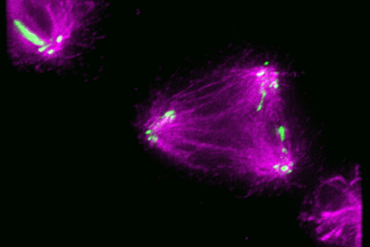
New research from the Cheeseman Lab reveals how cells control the location of the machinery that splits apart chromosomes during cell division.
January 2, 2024
December 20, 2023
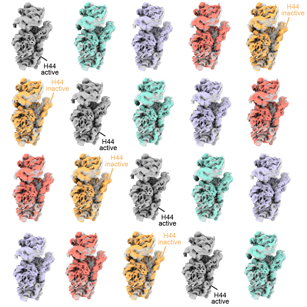
Research from the Drennan Lab in the Department of Biology at MIT explores how a protein called Met18, which is part of a ubiquitous pathway to transfer clusters of iron and sulfur to client proteins in the cytosol and nucleus of cells, can interact with other Met18 units to form intertwined structures
Lillian Eden | Department of Biology
December 18, 2023
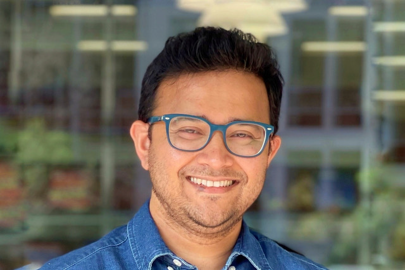
Sunny Das is a postdoc in Whitehead Institute Member Robert Weinberg’s lab studying how breast cancer metastasizes or spreads to other tissues.
December 12, 2023
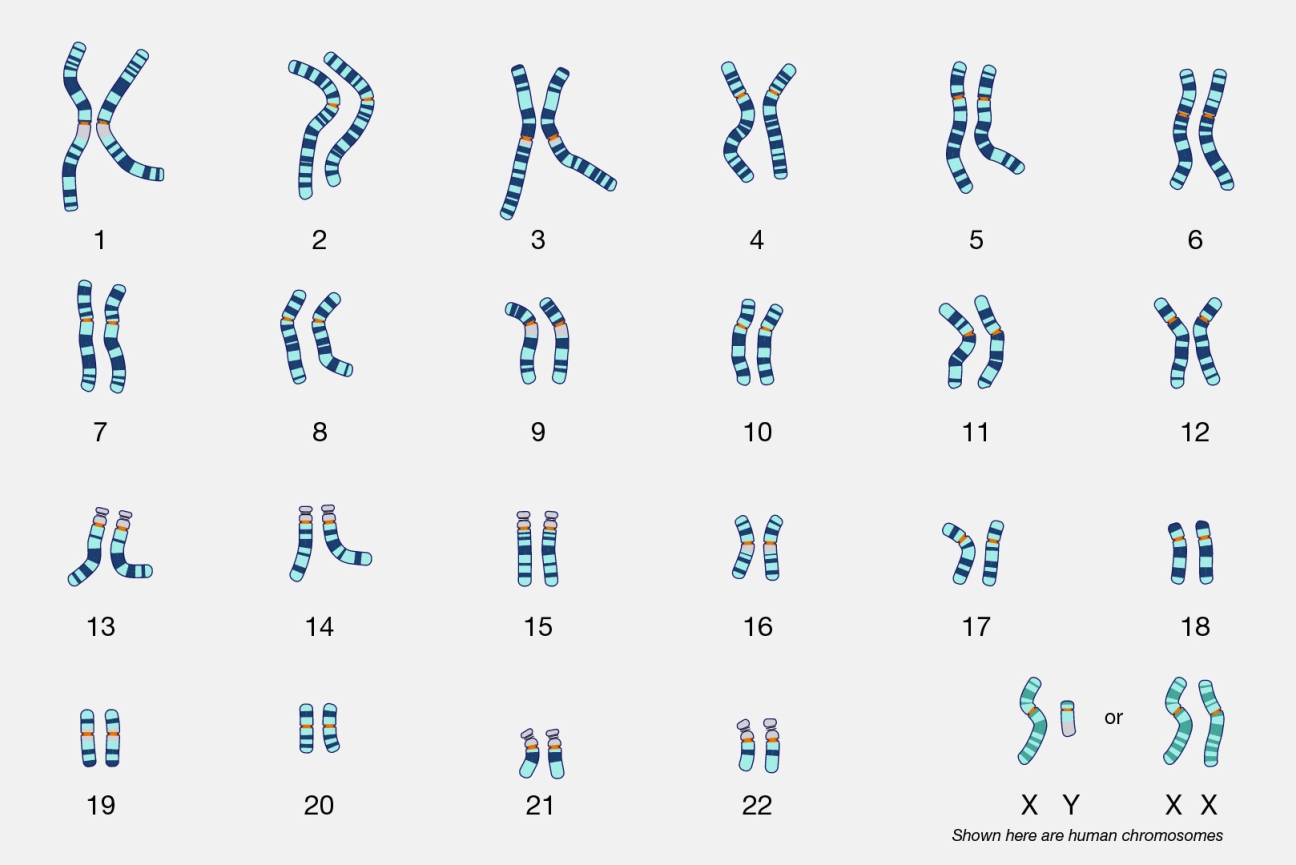
Genes expressed from the X and Y chromosomes impact cells throughout the body—not just in the reproductive system—by dialing up or down the expression of thousands of genes found on other chromosomes.
December 13, 2023
An MIT-based white paper identifies leading questions in the quest to make open-access publications sustainable.
Peter Dizikes | MIT News
November 30, 2023
Davis' support inspired PhD student and protein enthusiast Laurel Kinman to prioritize mentorship in her career
November 30, 2023
Jay Stein, PhD ‘68, was honored with a professorship of Biology at MIT, established by Hologic, at his retirement party last year; the inaugural Jay A. Stein Professor of Biology is Amy Keating, head of the Department of Biology and a noted leader in the field of biological engineering. Stein’s secret of innovation? “Follow the nerd, not the herd.”